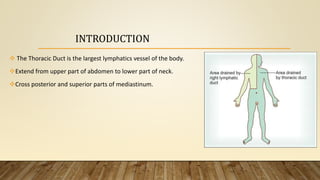
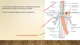
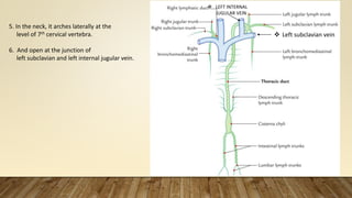
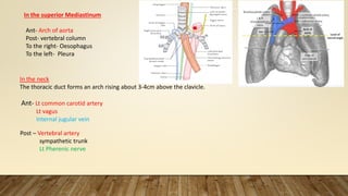
THORACIC DUCT
LEARNING OBJECTIVE
• Introduction
• Course
• Relations
• Tributaries
• Clinical
INTRODUCTION
 The Thoracic Duct is the largest lymphatics vessel of the body.
Extend from upper part of abdomen to lower part of neck.
Cross posterior and superior parts of mediastinum.
1. The thoracic duct begins as a continuation of the upper end
of the cisterna chyli near the lower border of the
12th thoracic vertebra
2. Enters the thorax through the aortic opening of the diaphragm.
COURSES
3. then ascends through the posterior mediastinum from level
of 12th thoracic vertebra to fifth thoracic vertebra.
4. Then it courses through the superior mediastinum.
 DIG- POSTERIOR MEDIASTINUM
5. In the neck, it arches laterally at the
level of 7th cervical vertebra.
6. And open at the junction of
left subclavian and left internal jugular vein.
 LEFT INTERNAL
JUGULAR VEIN
 Left subclavian vein
Relations
At aortic opening of diaphragm
Ant – Diaphragm
Post – vertebral colum
To the right – Azygos vein
To the left - AORTA
In the posterior mediastinum
Ant – (1) Diapragm
(2) Oesophagus
Post - (1) Vertebral column
(2) Rt Posterior intercostal arteries
To the right - AZYGOS VEIN
To the left – Descending Aorta
 DIGRAM- POSTERIOR
MEDIASTINUM
In the superior Mediastinum
Ant- Arch of aorta
Post- vertebral column
To the right- Oesophagus
To the left- Pleura
In the neck
The thoracic duct forms an arch rising about 3-4cm above the clavicle.
Ant- Lt common carotid artery
Lt vagus
Internal jugular vein
Post – Vertebral artery
sympathetic trunk
Lt Pherenic nerve
Tributaries
1.) Thoracic duct recive lymph from both half of the body below the
diaphragm and left half above the abdomen.
2) In thorax thoracic duct receive – lymph vessel from posterior
mediastinal node and small intercostal node.
3) Left jugular trunk, Left subclavian trunk end in thoracic duct.
4) Left bronchiomediastinal trunk – drain lymph from Left half of thorax
And ends in thoracic duct
Rt half above diaphragm
Rt side there is lymphatic duct Into which 
(1) Rt brachiomediastinal
(2) Rt jugular
(3) Rt subclavianlymph drain
Rt lymphatic trunk end in Rt brachiocephalic vein
Applied anatomy
 Thoracic duct is very thin walled and colourless so more prone for injury
during surgery in post mediastinum.
Obstruction of Thoracic duct - Due to mature filarial parasites lymph
vessels get burst chylothorax, chyloperitoneum, chyluria.
 Cervical part of thoracic duct is damaged in block dissection of neck
THORACIC_DUCT.pptx

THORACIC_DUCT.pptx

  • 1.
  • 2.
    LEARNING OBJECTIVE • Introduction •Course • Relations • Tributaries • Clinical
  • 3.
    INTRODUCTION  The ThoracicDuct is the largest lymphatics vessel of the body. Extend from upper part of abdomen to lower part of neck. Cross posterior and superior parts of mediastinum.
  • 4.
    1. The thoracicduct begins as a continuation of the upper end of the cisterna chyli near the lower border of the 12th thoracic vertebra 2. Enters the thorax through the aortic opening of the diaphragm. COURSES
  • 5.
    3. then ascendsthrough the posterior mediastinum from level of 12th thoracic vertebra to fifth thoracic vertebra. 4. Then it courses through the superior mediastinum.  DIG- POSTERIOR MEDIASTINUM
  • 6.
    5. In theneck, it arches laterally at the level of 7th cervical vertebra. 6. And open at the junction of left subclavian and left internal jugular vein.  LEFT INTERNAL JUGULAR VEIN  Left subclavian vein
  • 7.
    Relations At aortic openingof diaphragm Ant – Diaphragm Post – vertebral colum To the right – Azygos vein To the left - AORTA In the posterior mediastinum Ant – (1) Diapragm (2) Oesophagus Post - (1) Vertebral column (2) Rt Posterior intercostal arteries To the right - AZYGOS VEIN To the left – Descending Aorta  DIGRAM- POSTERIOR MEDIASTINUM
  • 8.
    In the superiorMediastinum Ant- Arch of aorta Post- vertebral column To the right- Oesophagus To the left- Pleura In the neck The thoracic duct forms an arch rising about 3-4cm above the clavicle. Ant- Lt common carotid artery Lt vagus Internal jugular vein Post – Vertebral artery sympathetic trunk Lt Pherenic nerve
  • 9.
    Tributaries 1.) Thoracic ductrecive lymph from both half of the body below the diaphragm and left half above the abdomen. 2) In thorax thoracic duct receive – lymph vessel from posterior mediastinal node and small intercostal node. 3) Left jugular trunk, Left subclavian trunk end in thoracic duct. 4) Left bronchiomediastinal trunk – drain lymph from Left half of thorax And ends in thoracic duct Rt half above diaphragm Rt side there is lymphatic duct Into which  (1) Rt brachiomediastinal (2) Rt jugular (3) Rt subclavianlymph drain Rt lymphatic trunk end in Rt brachiocephalic vein
  • 10.
    Applied anatomy  Thoracicduct is very thin walled and colourless so more prone for injury during surgery in post mediastinum. Obstruction of Thoracic duct - Due to mature filarial parasites lymph vessels get burst chylothorax, chyloperitoneum, chyluria.  Cervical part of thoracic duct is damaged in block dissection of neck