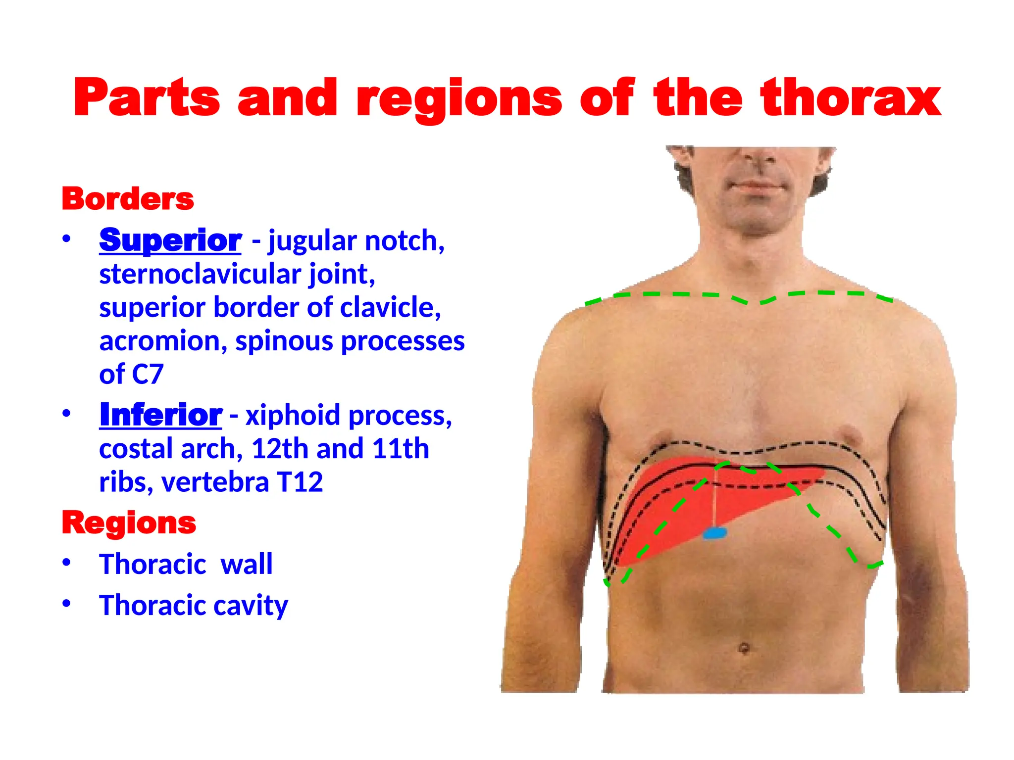
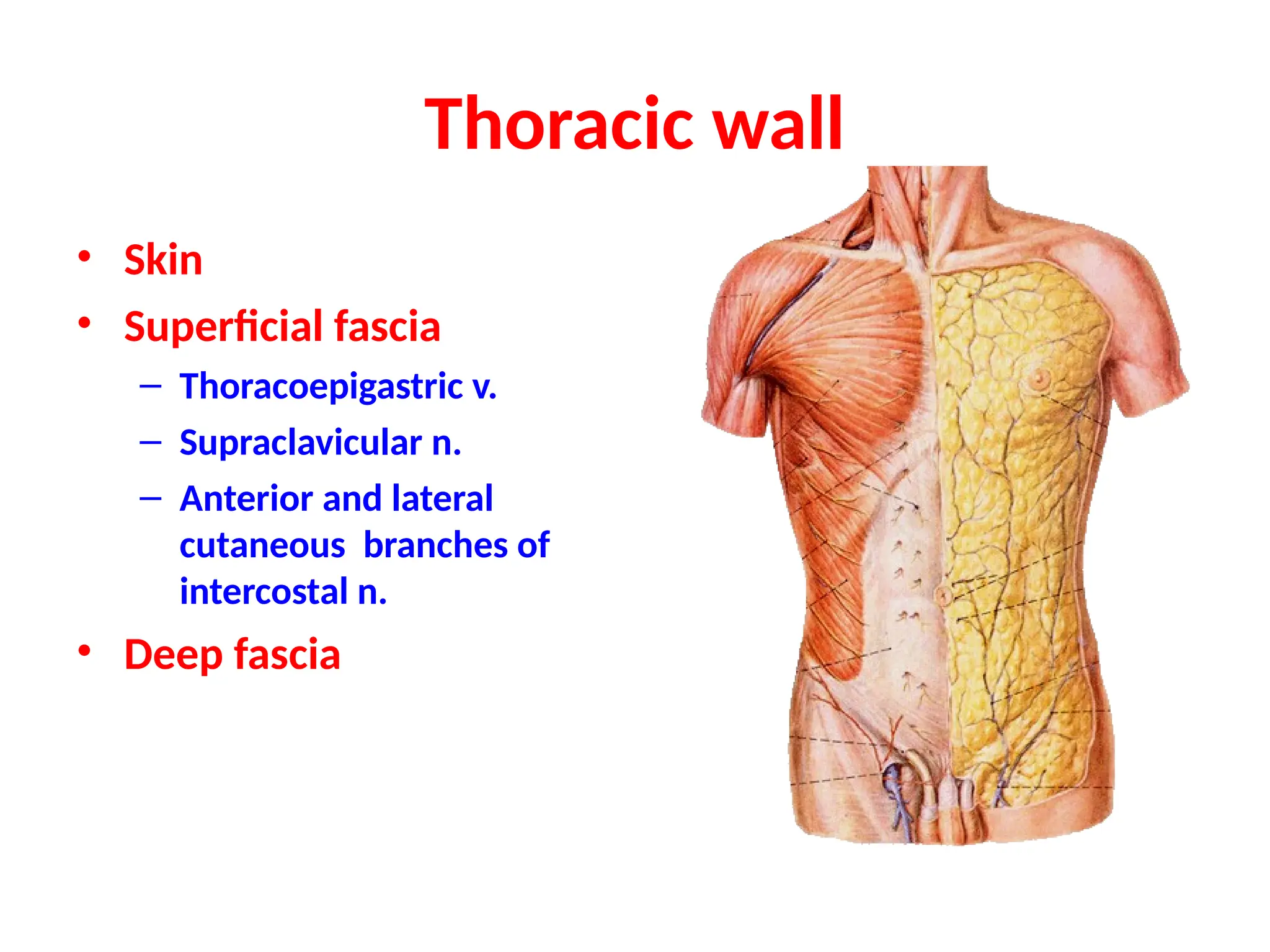
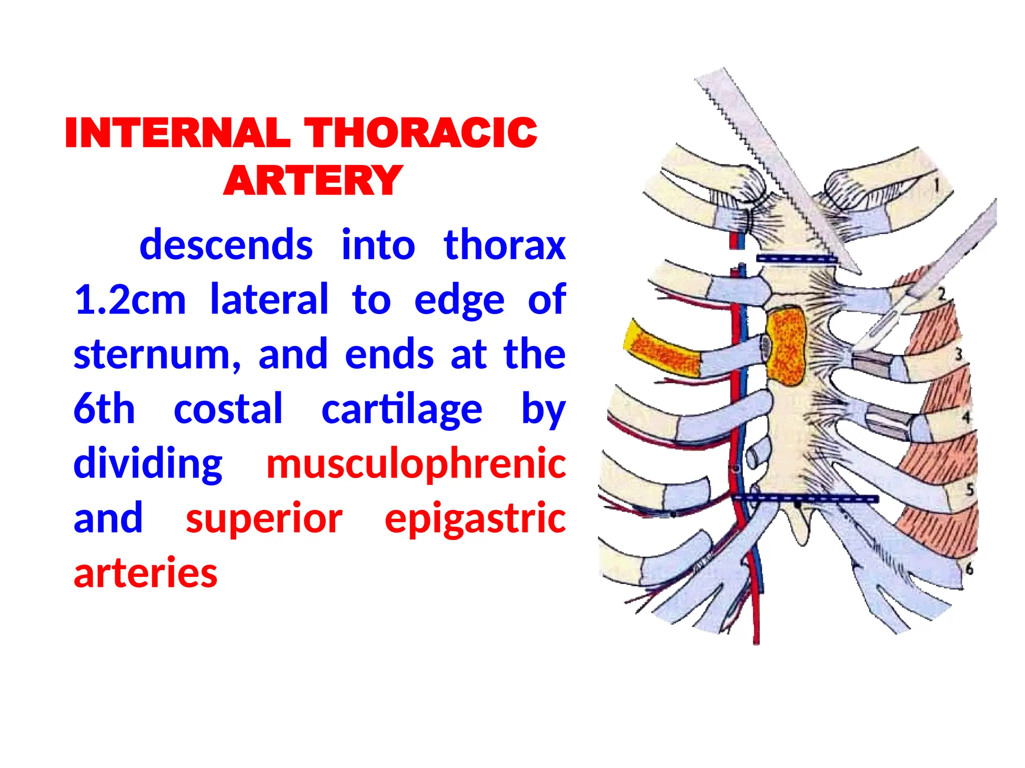
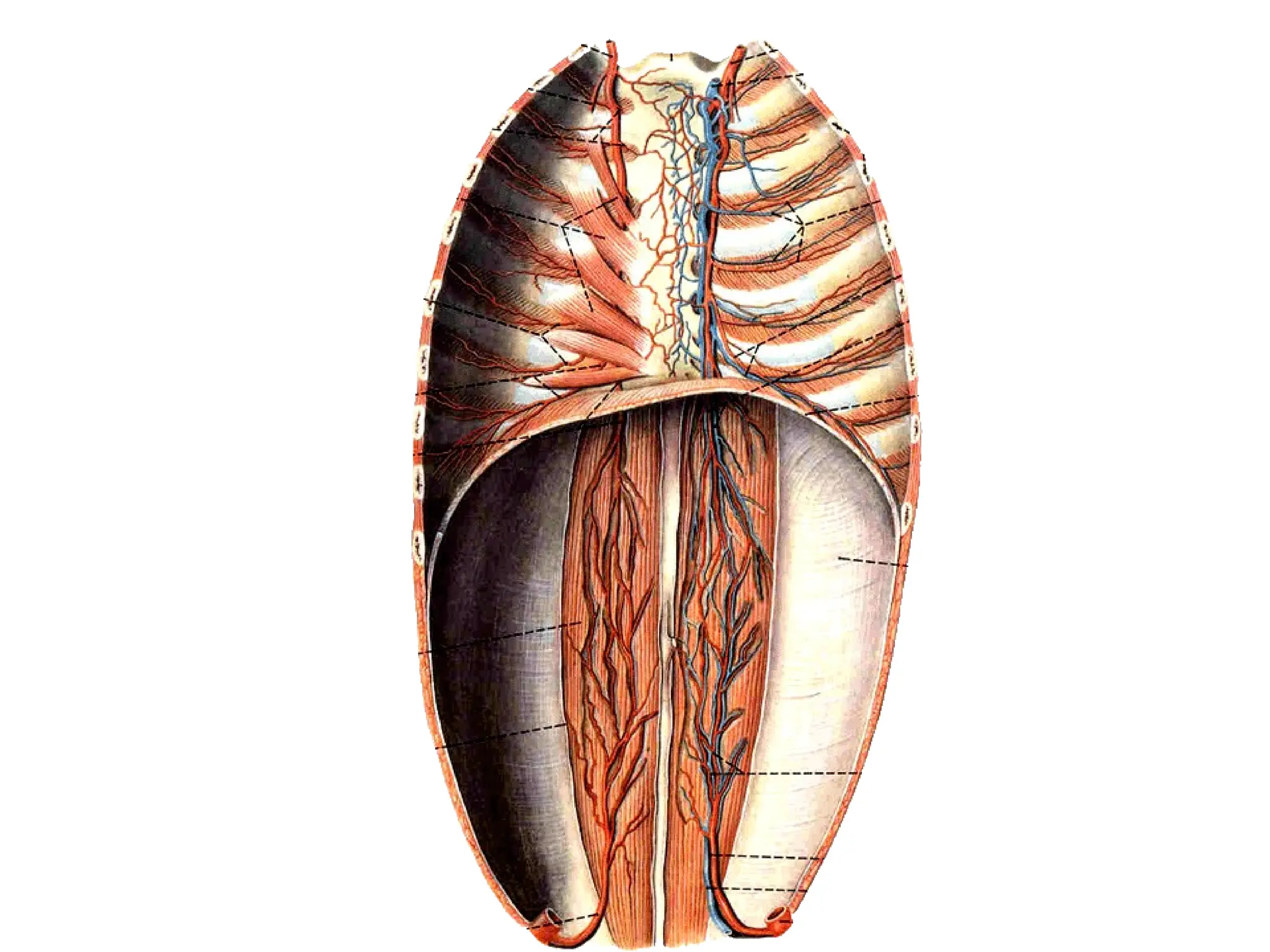
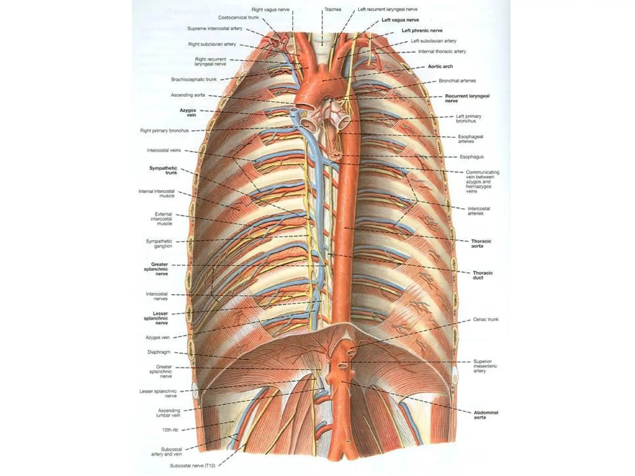
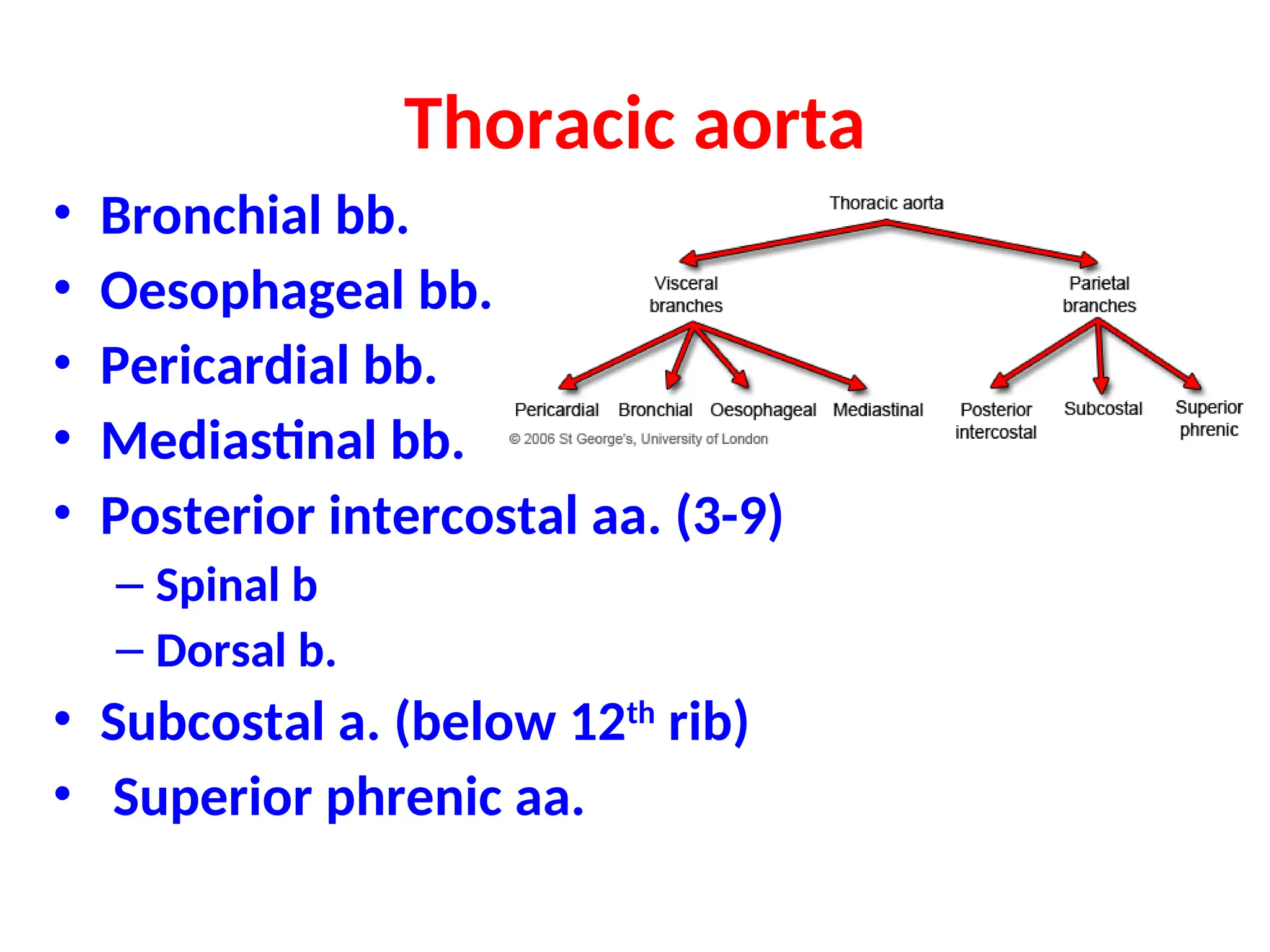
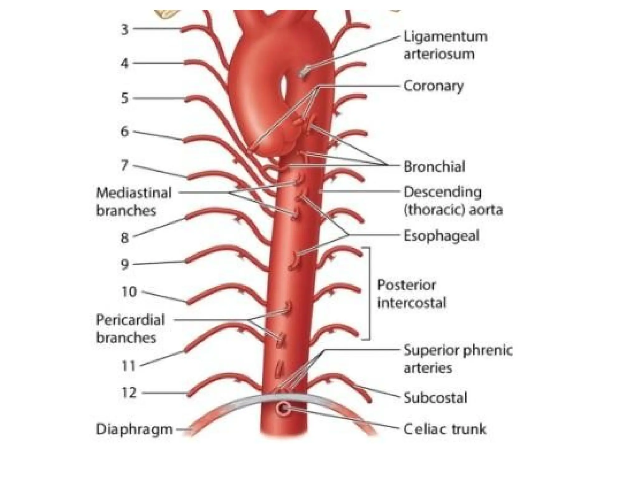
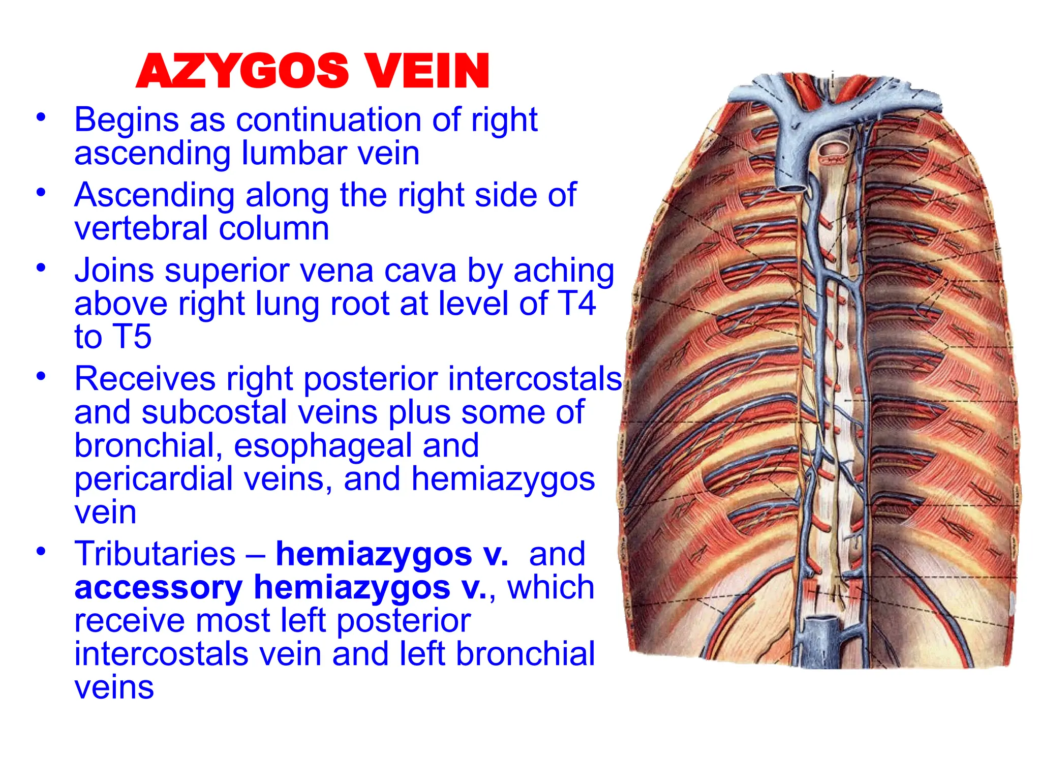
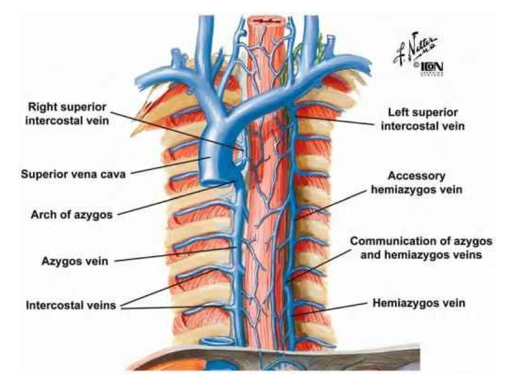
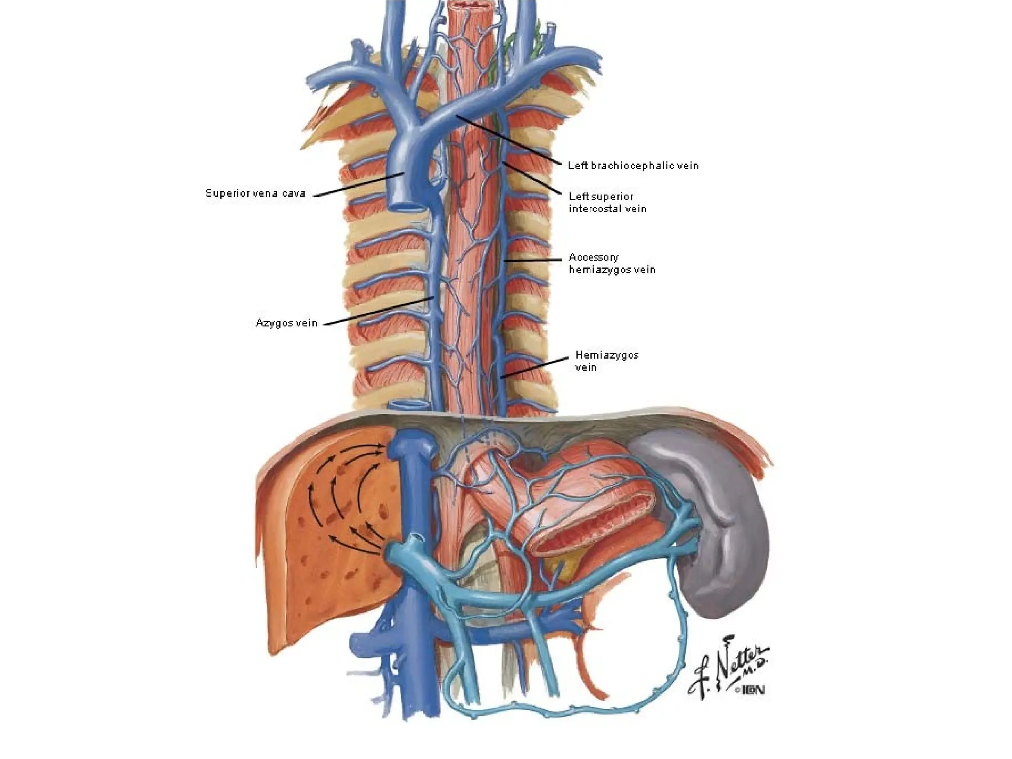
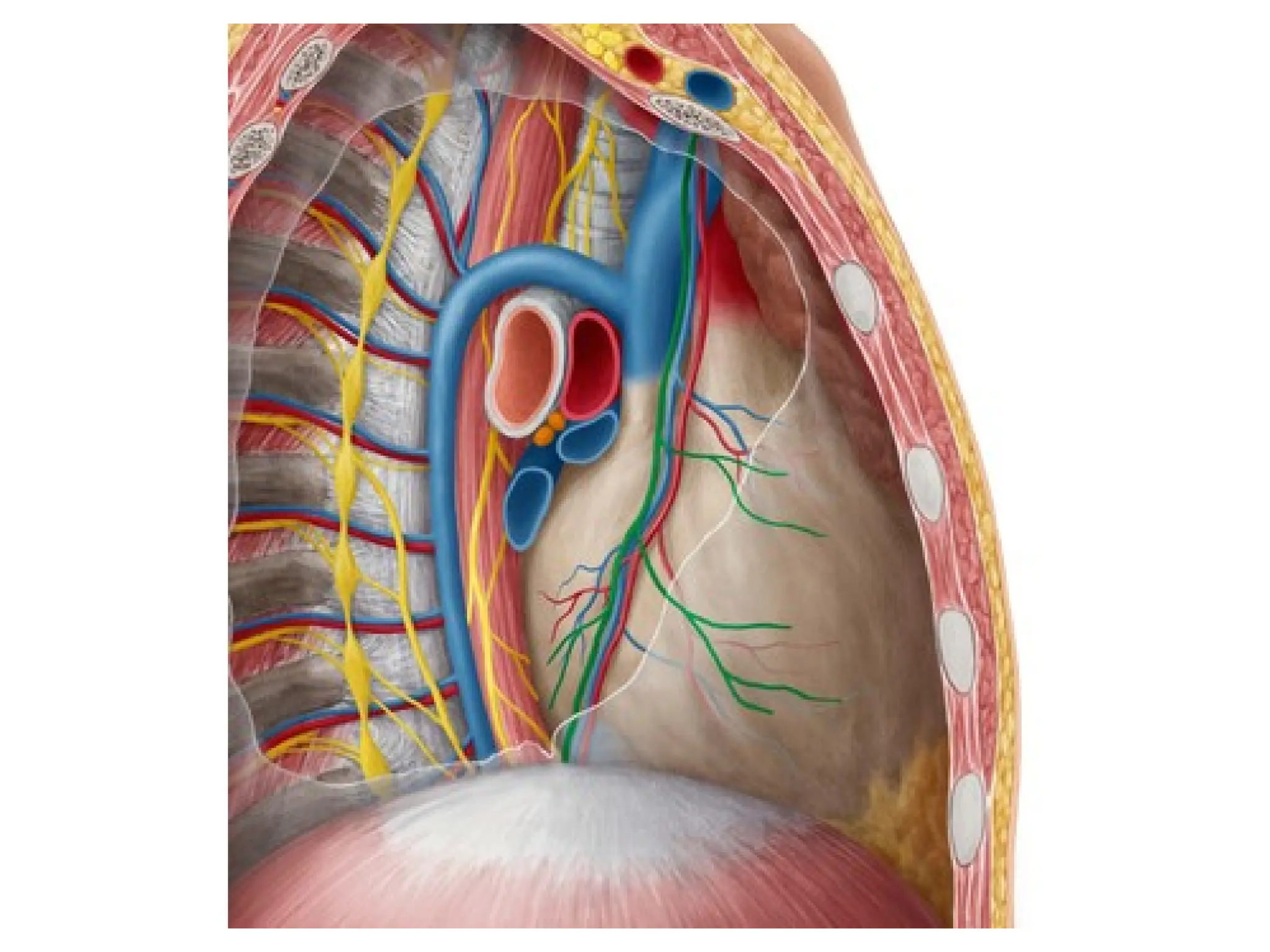
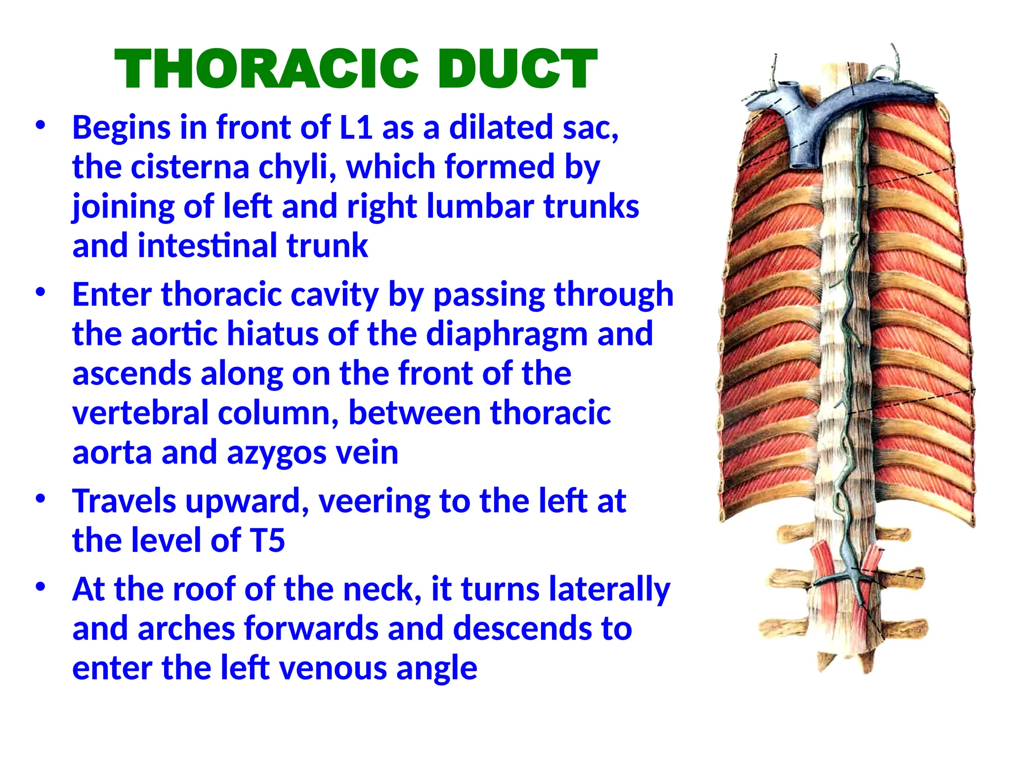
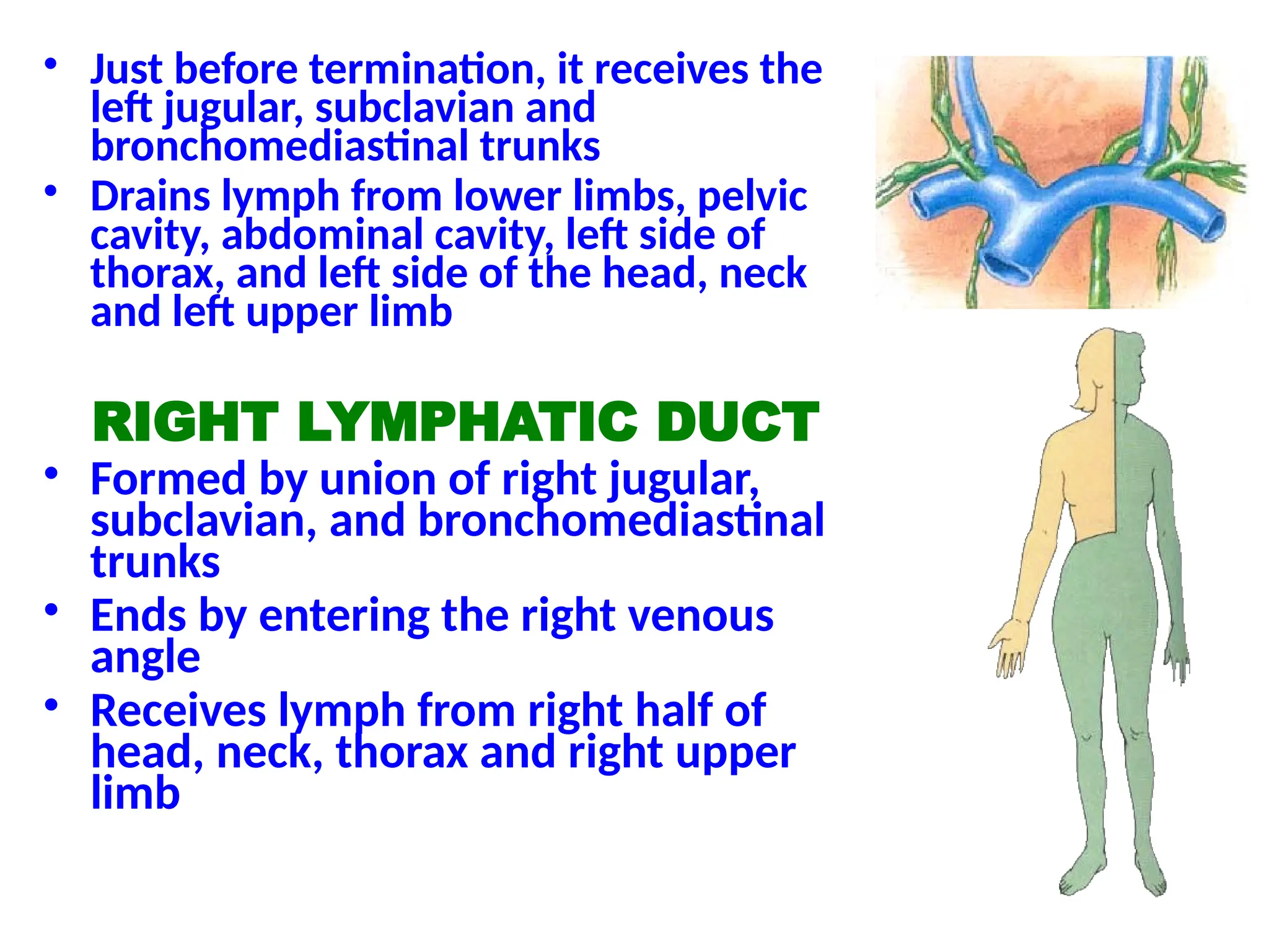
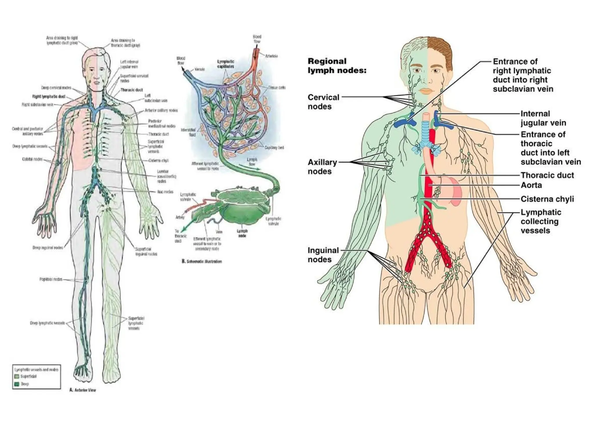
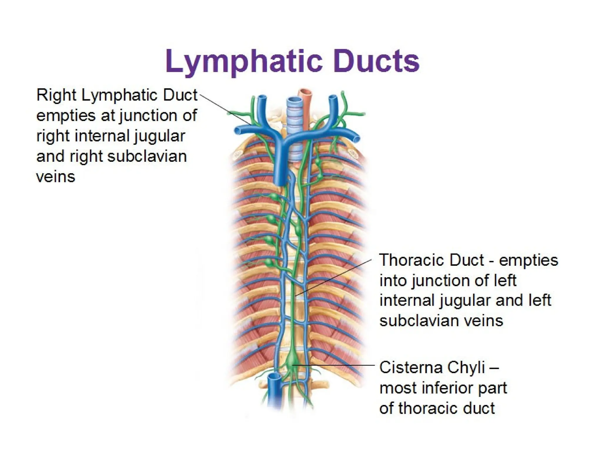
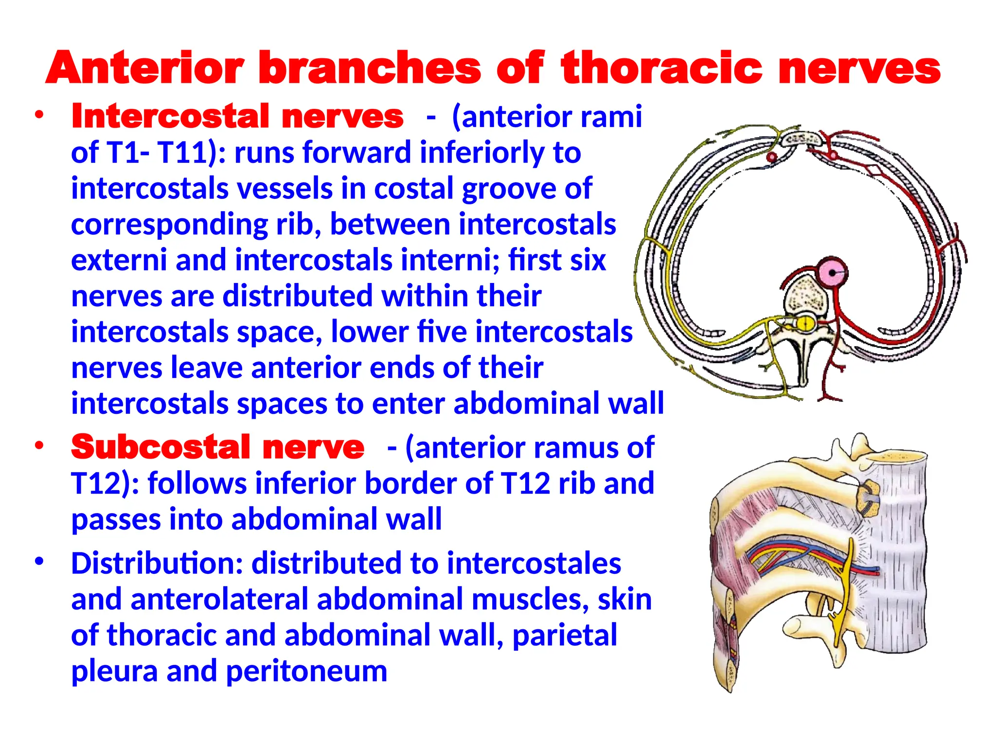
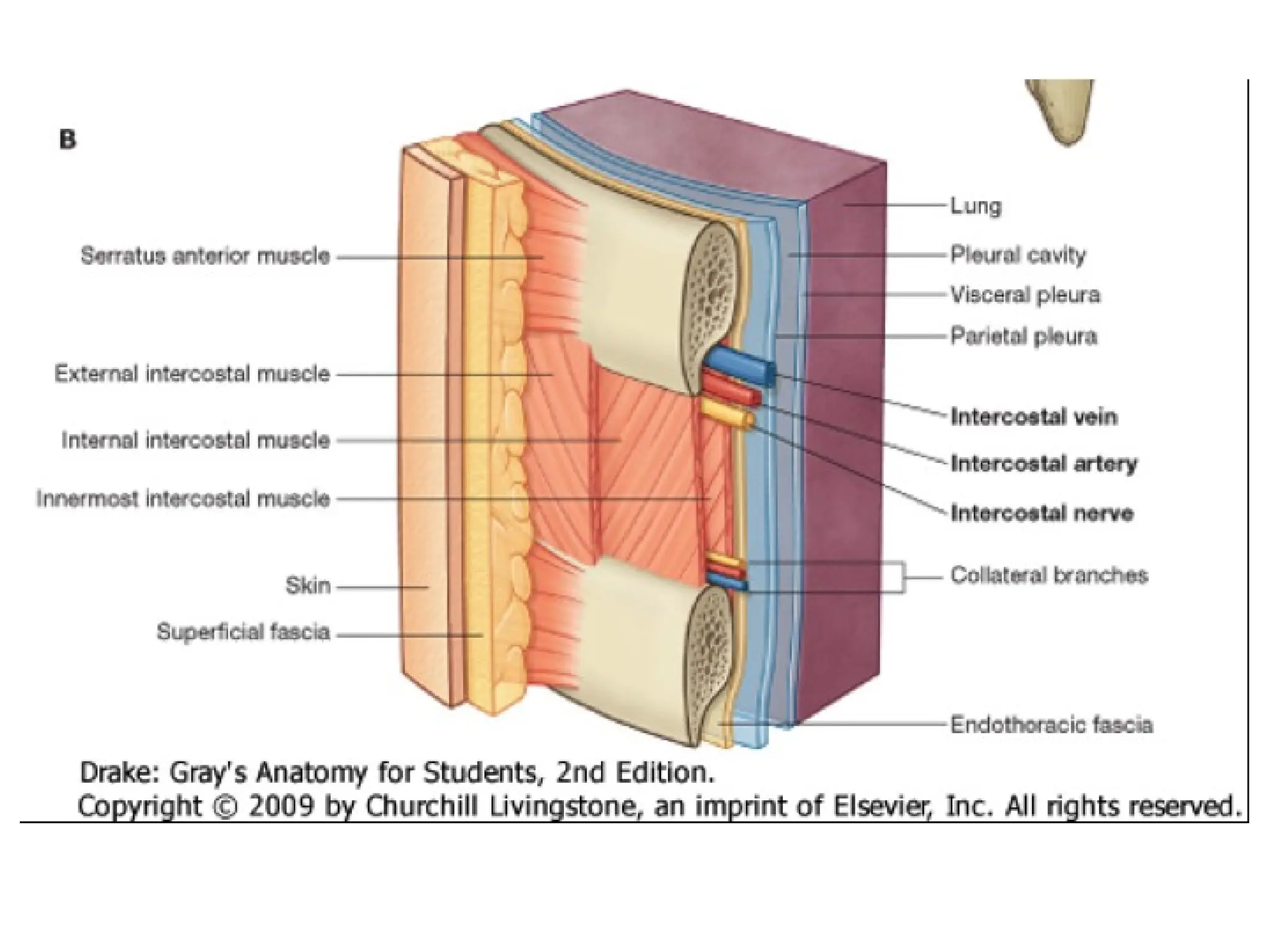
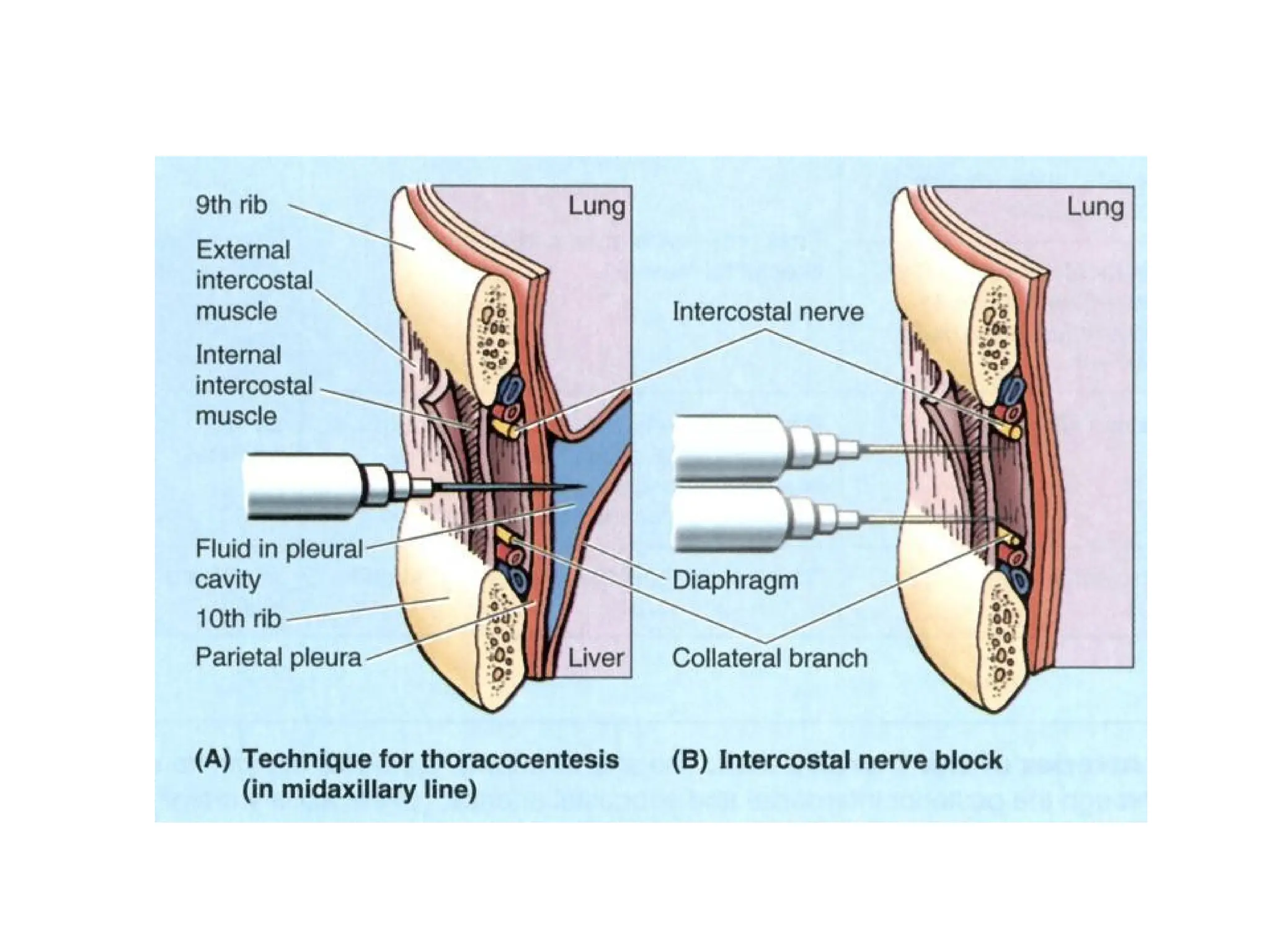
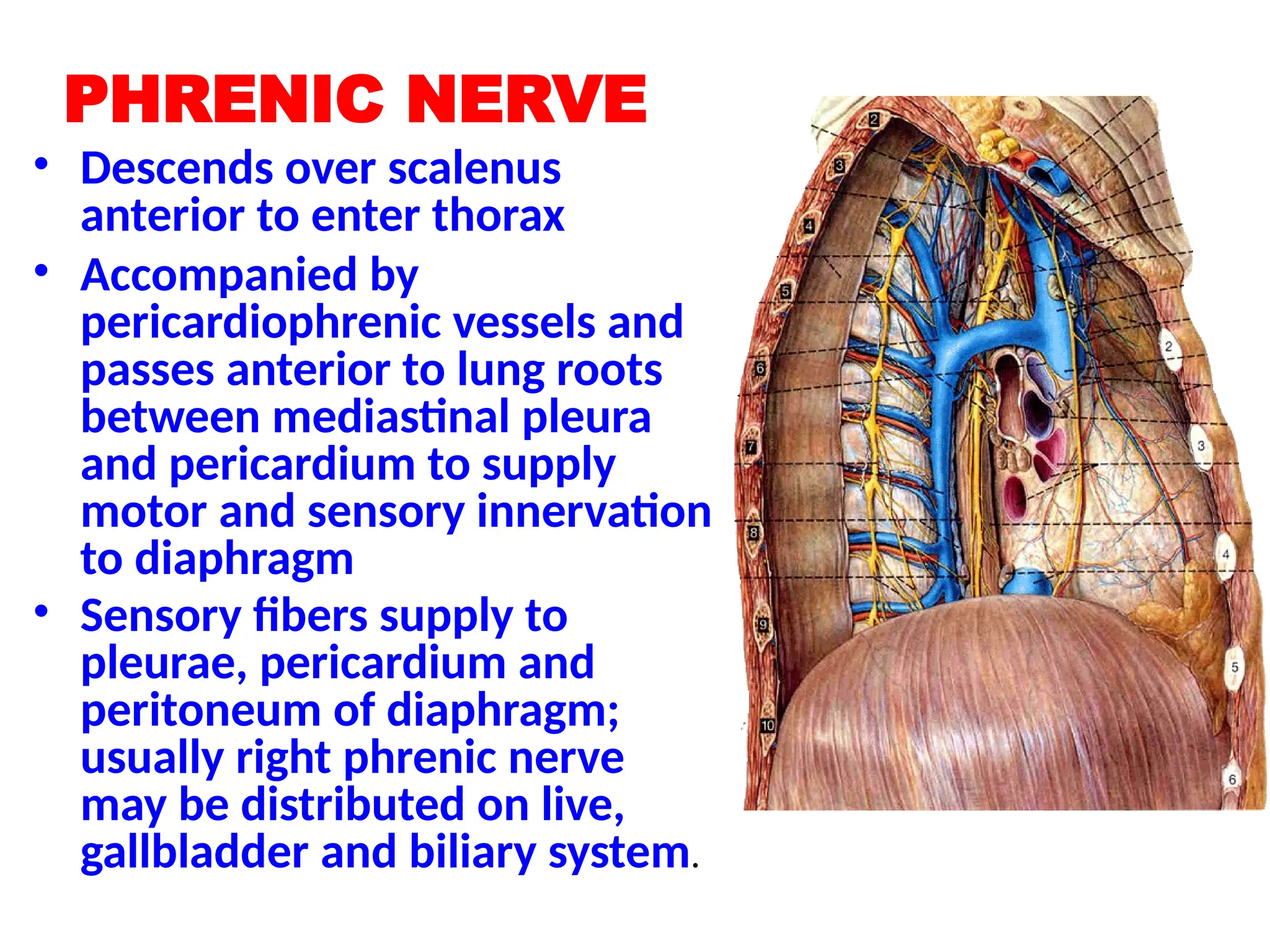
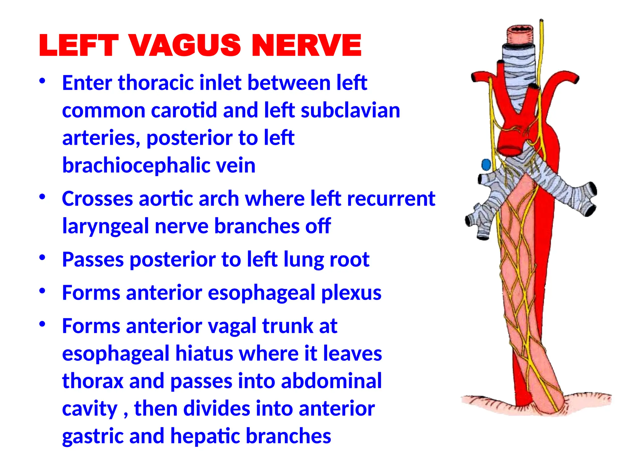
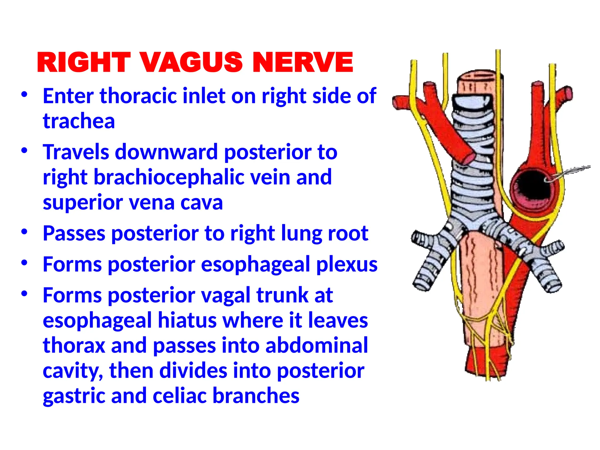
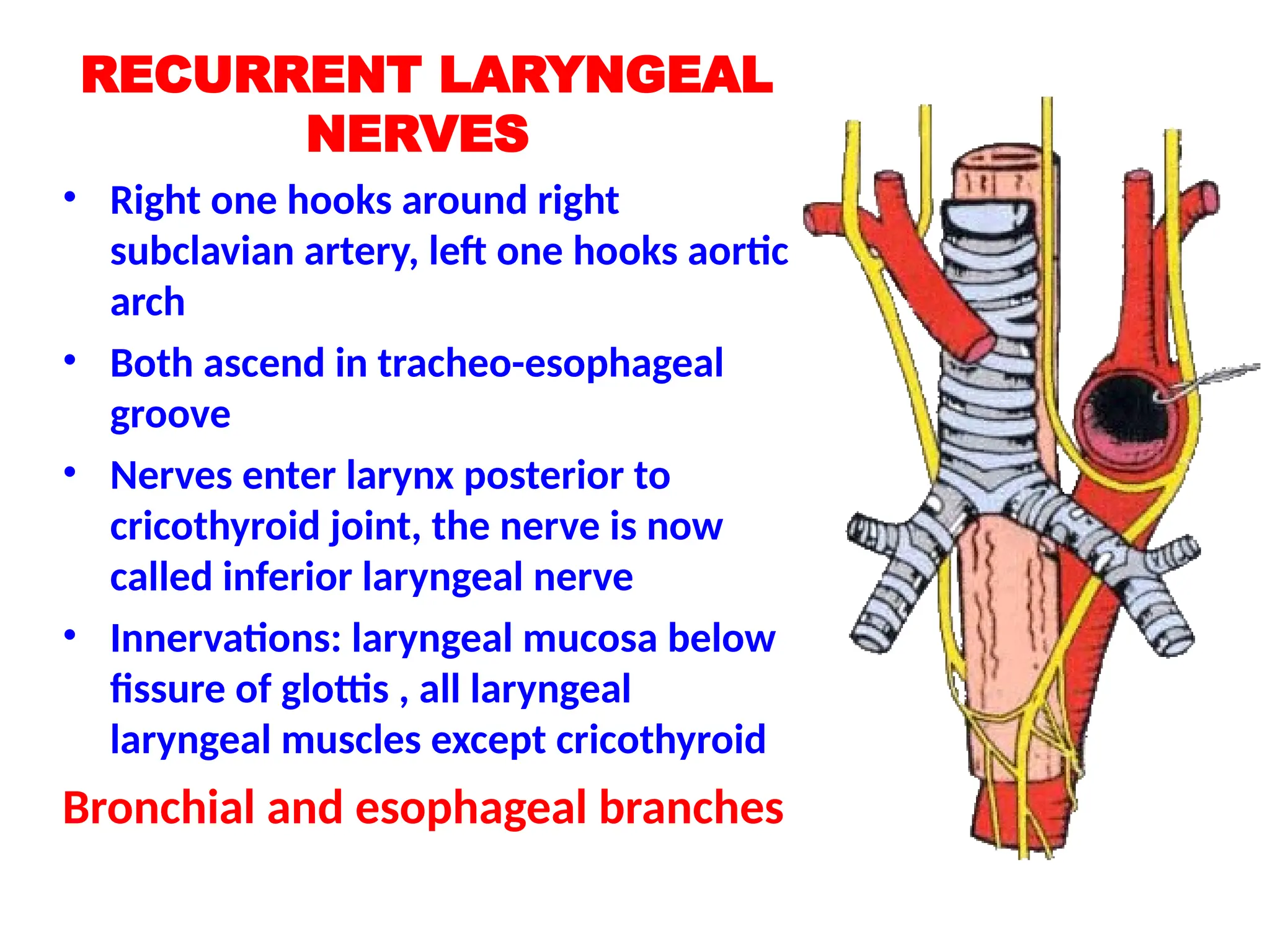
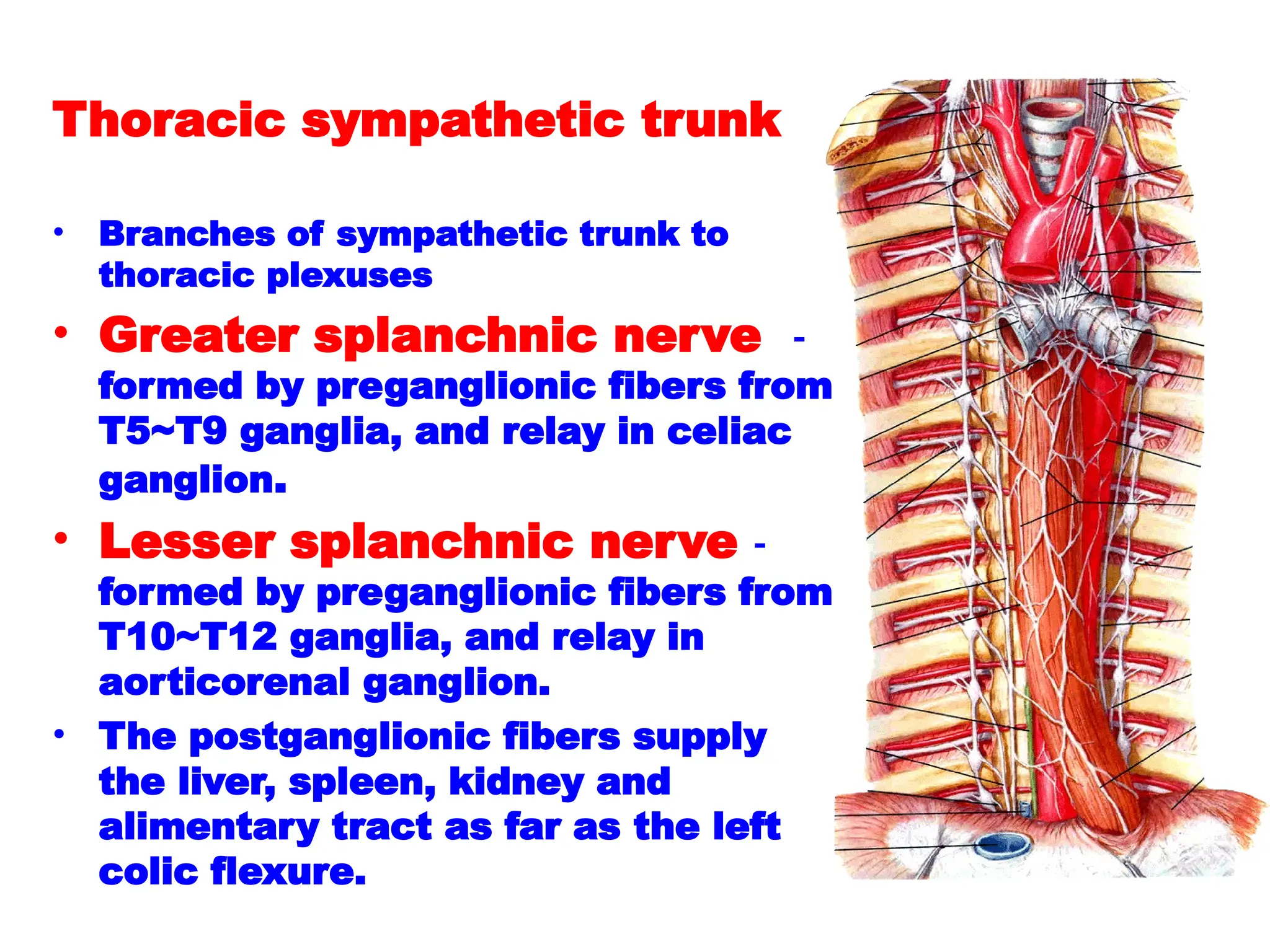
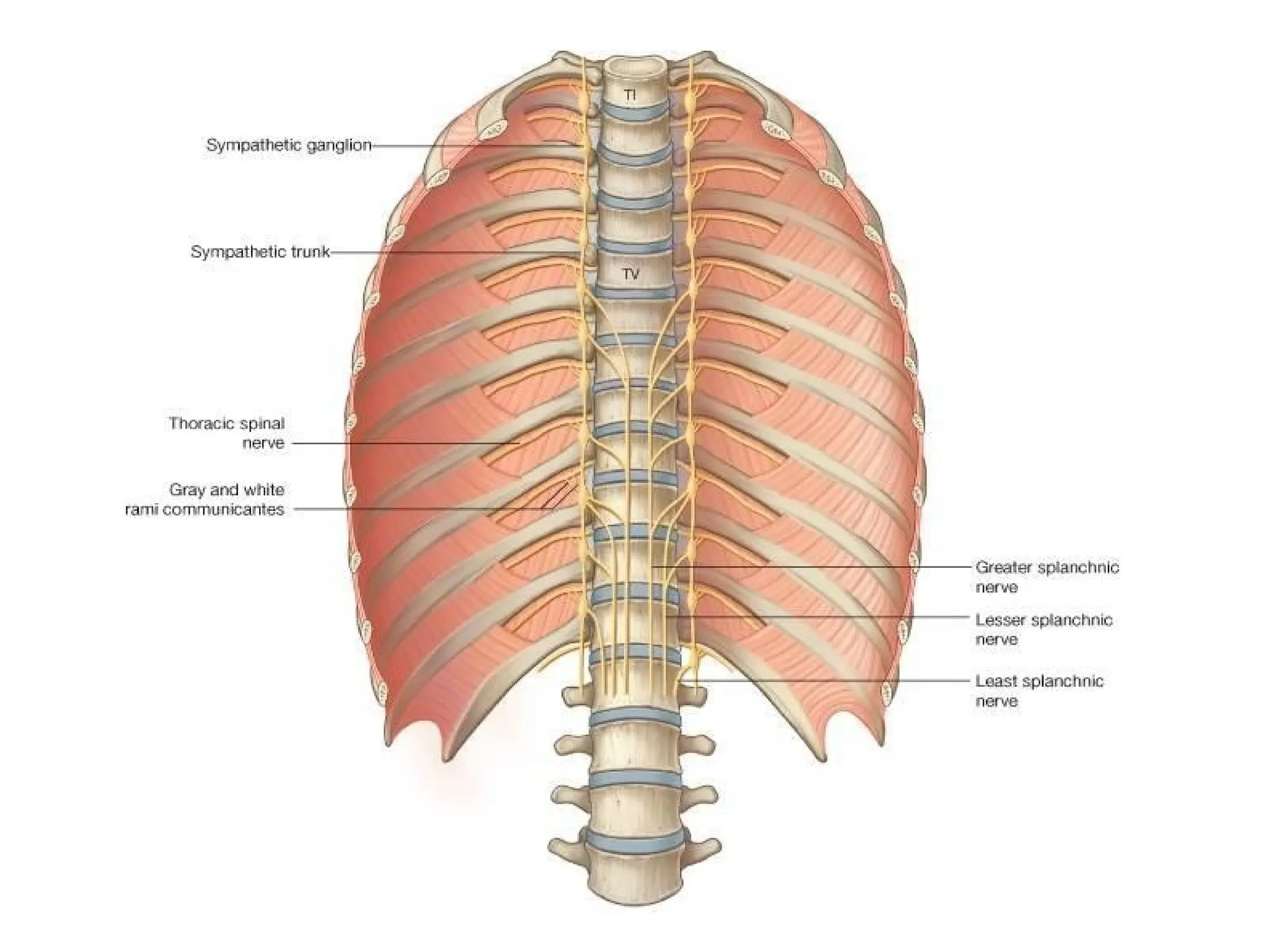
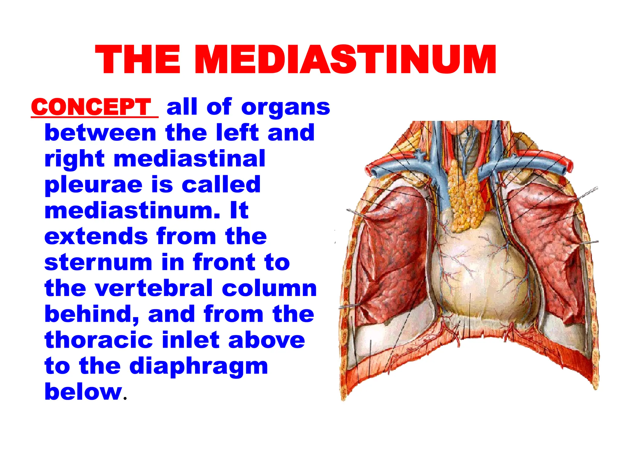
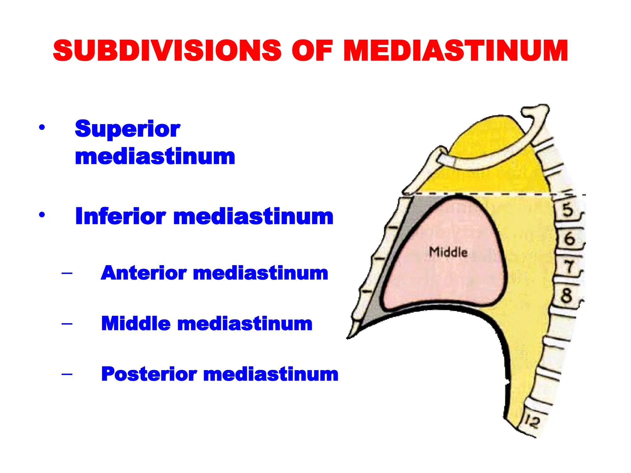
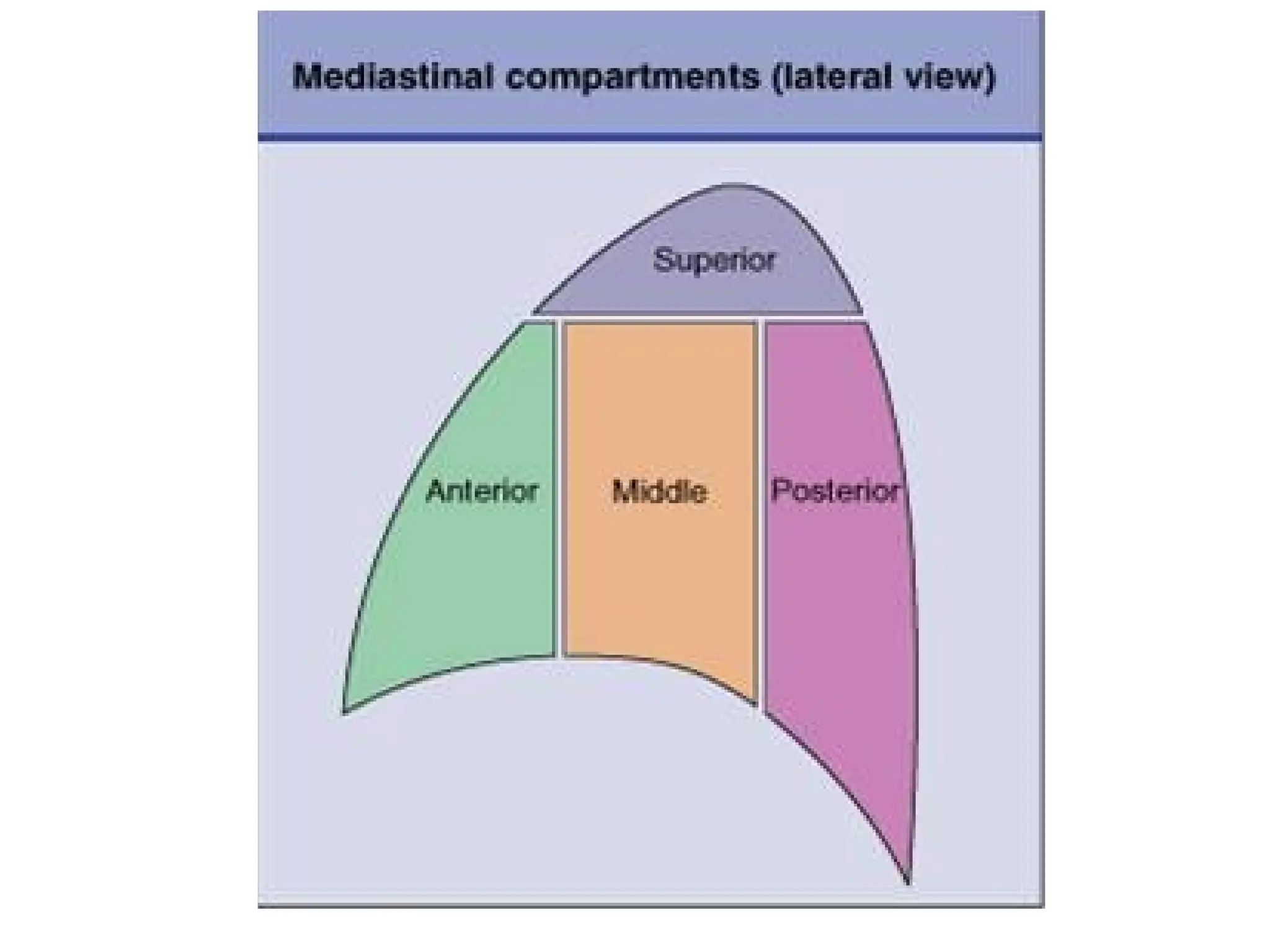
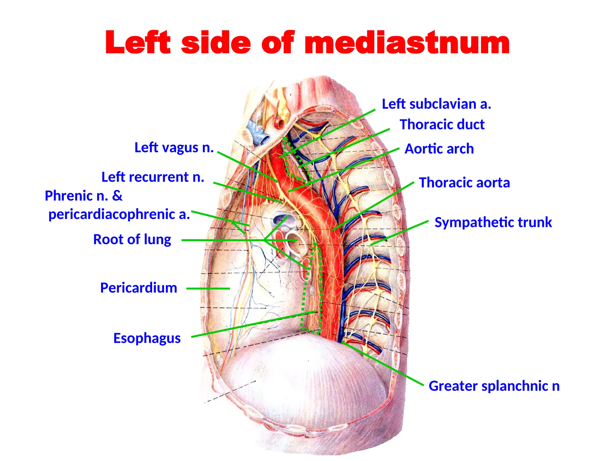
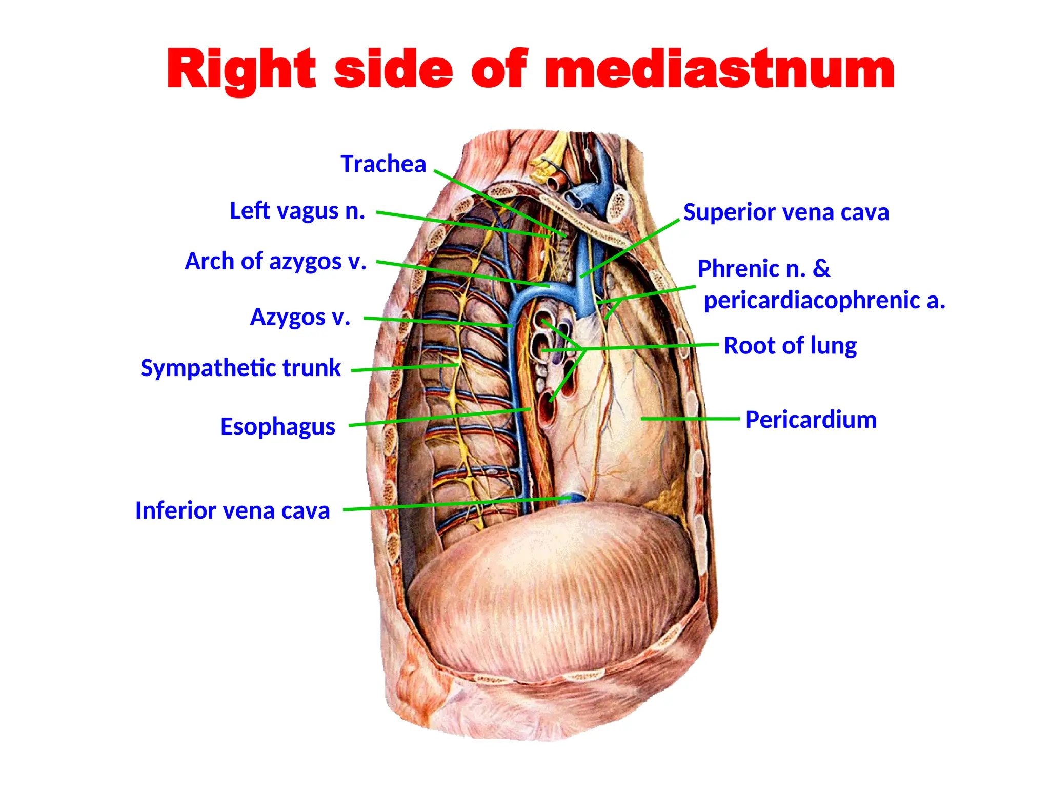
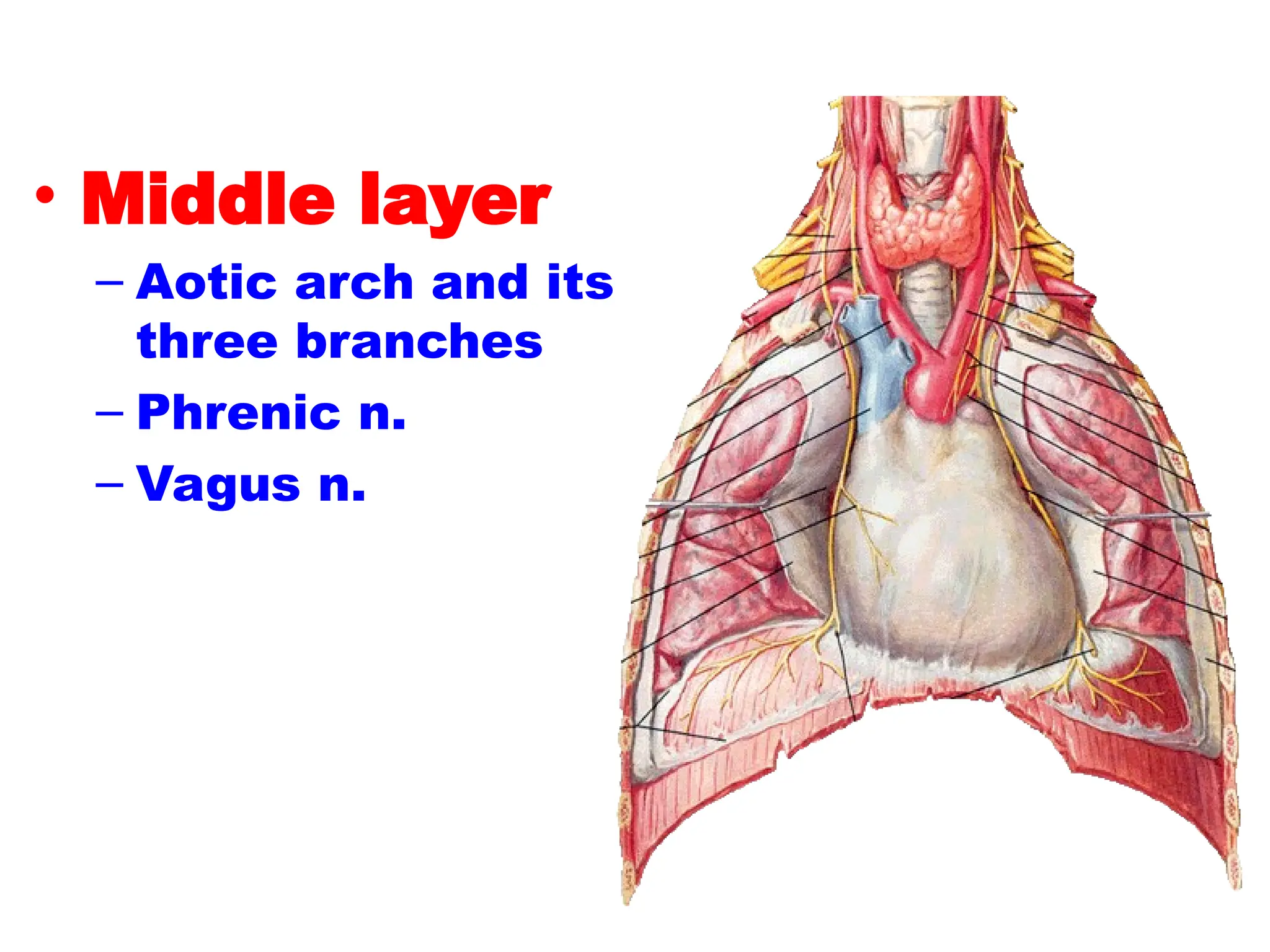
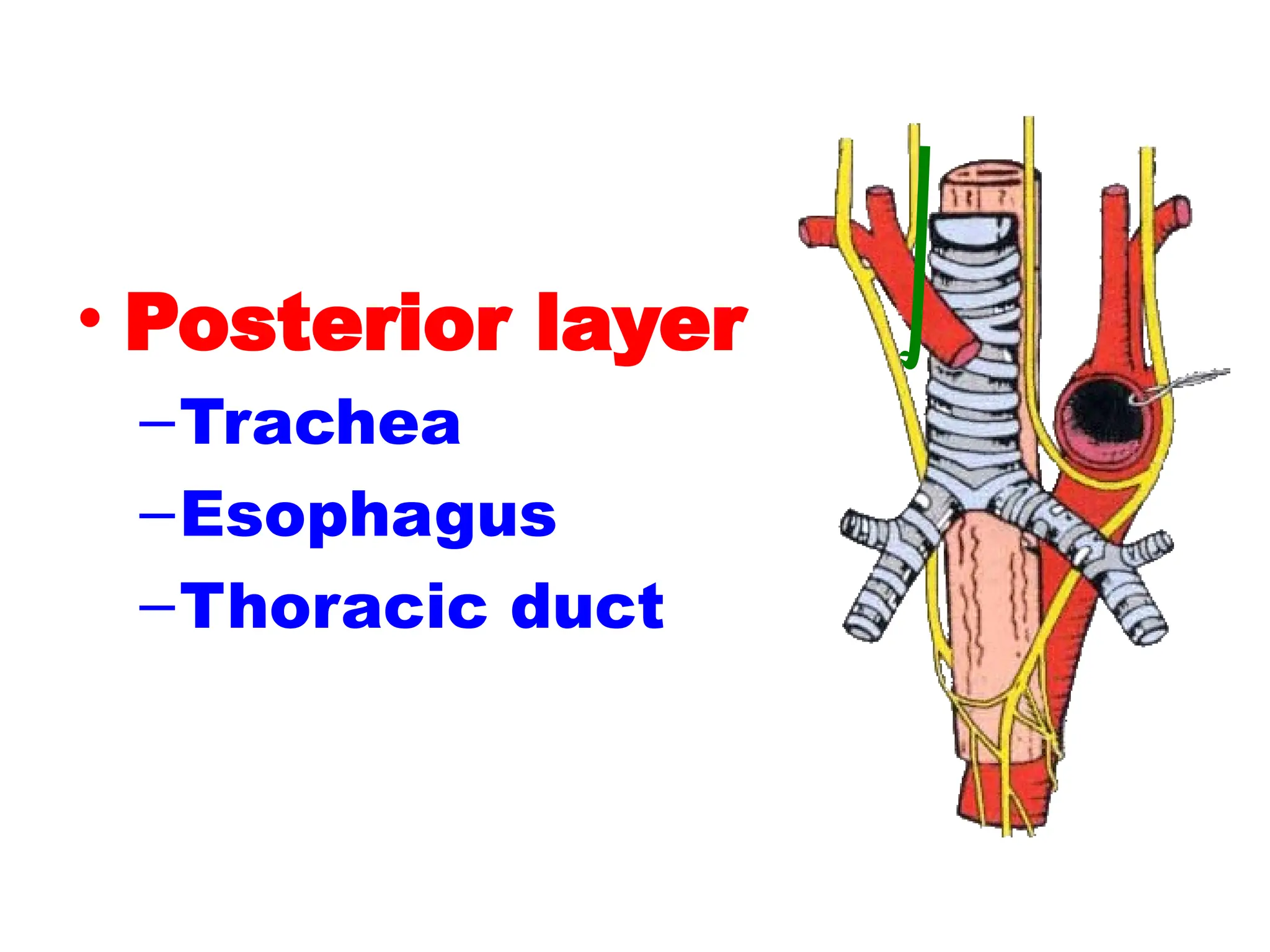
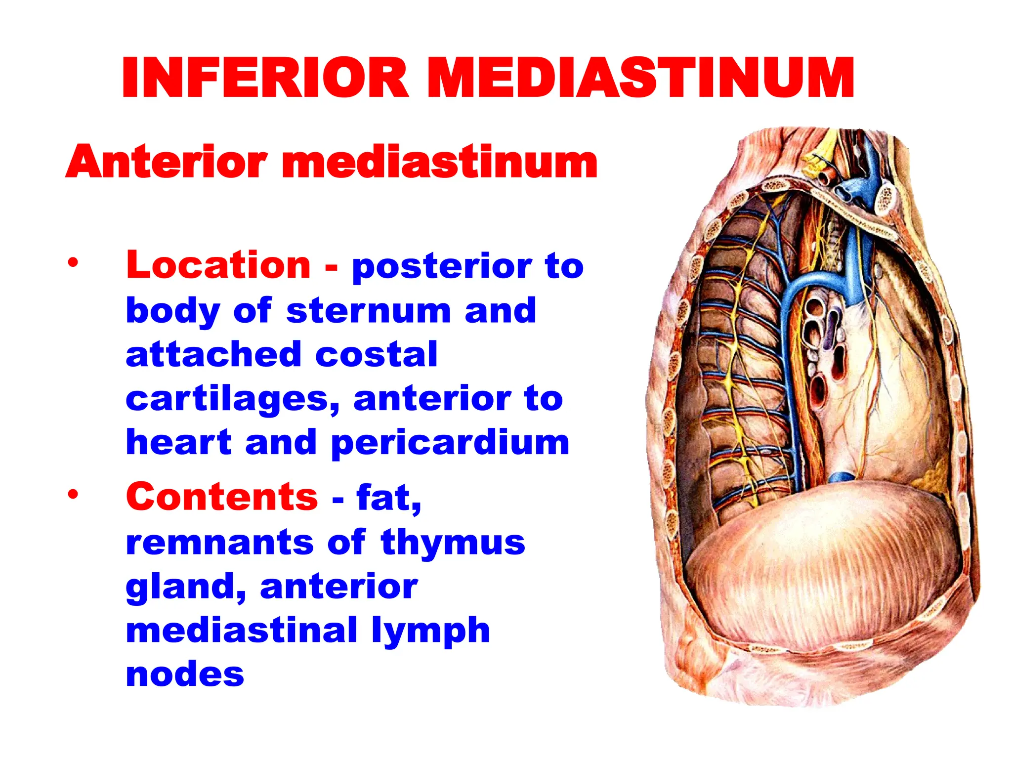
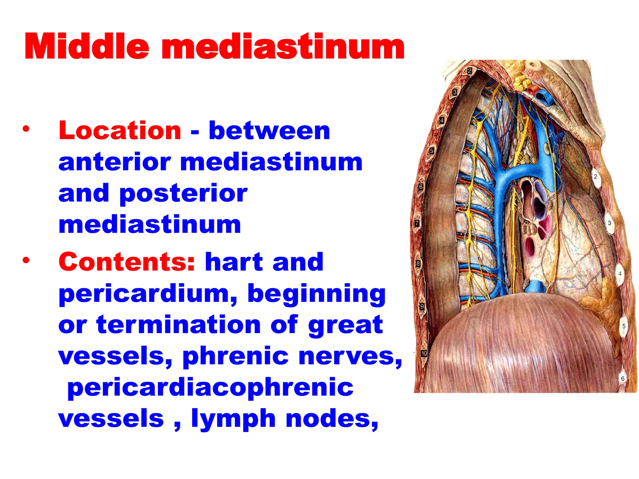
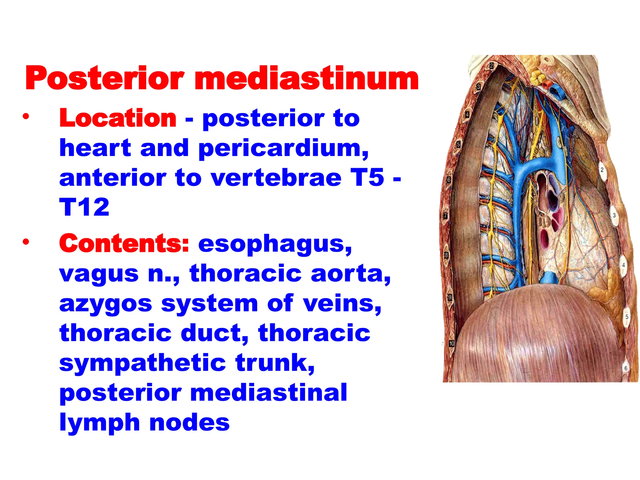
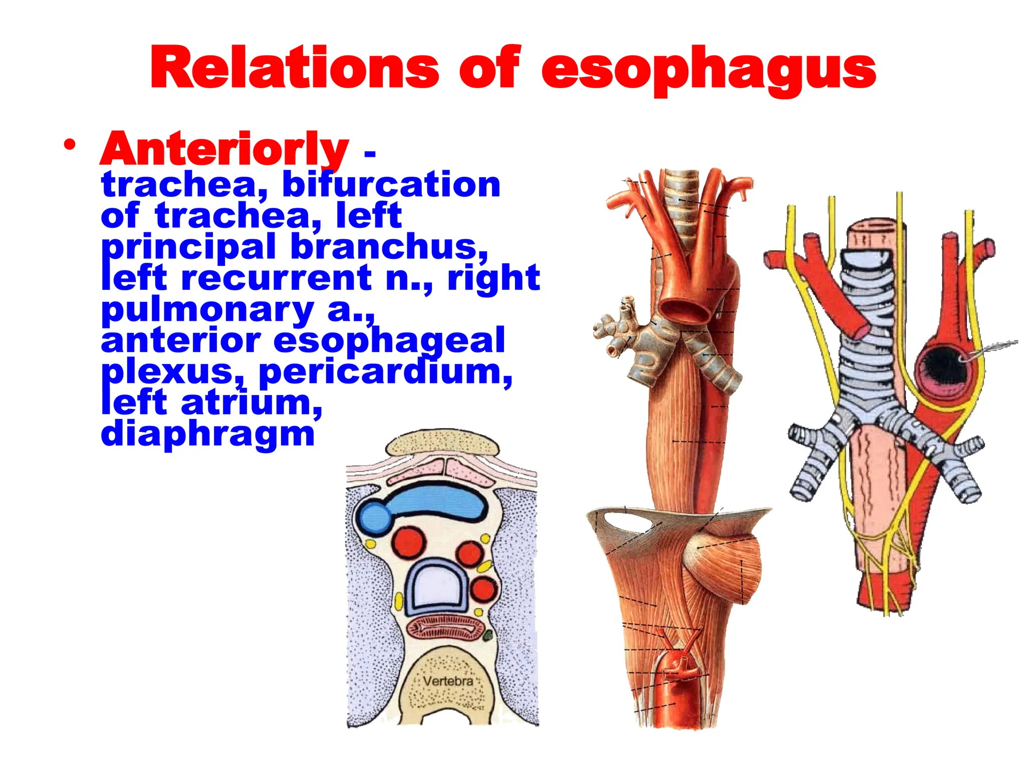
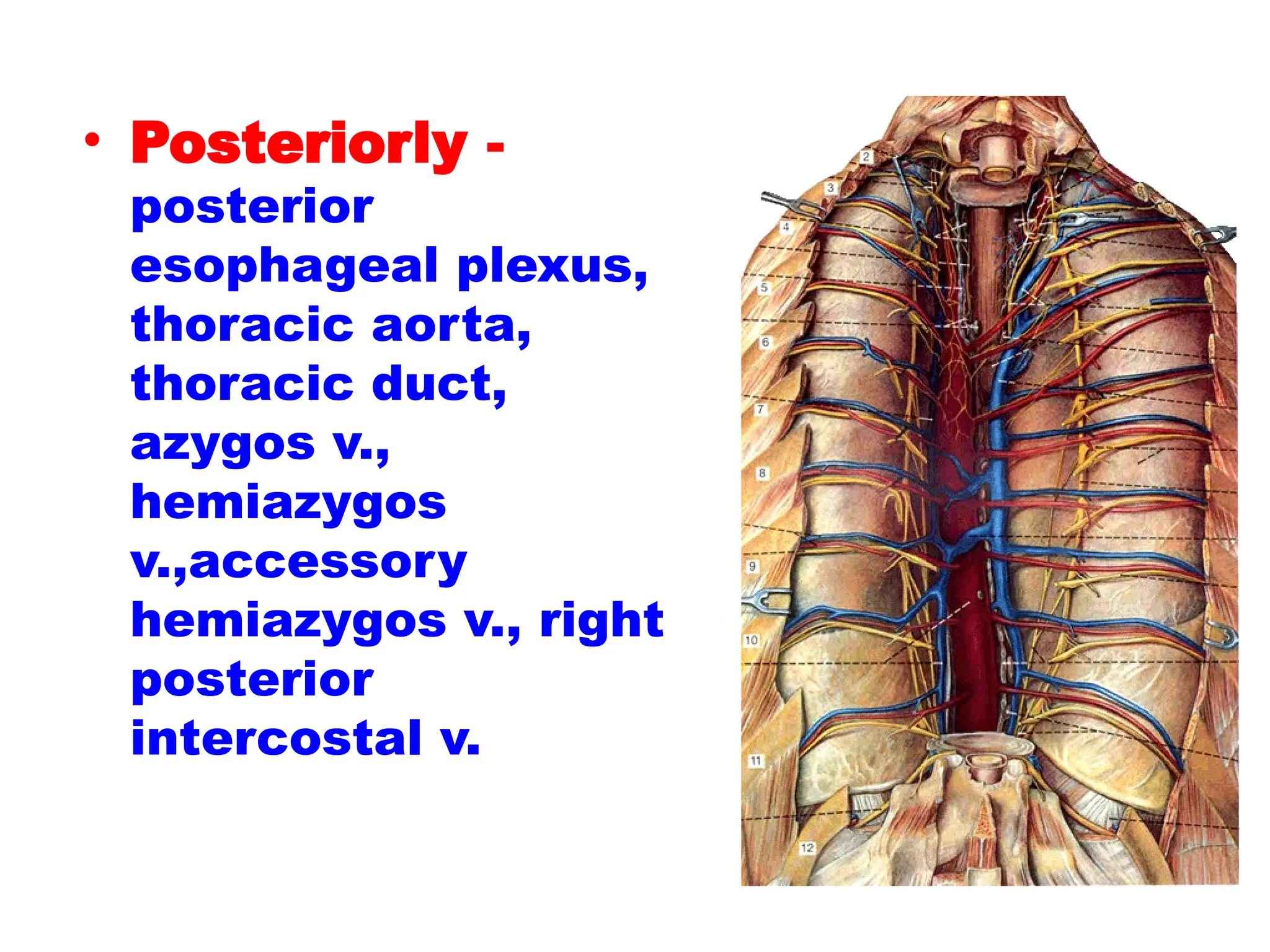
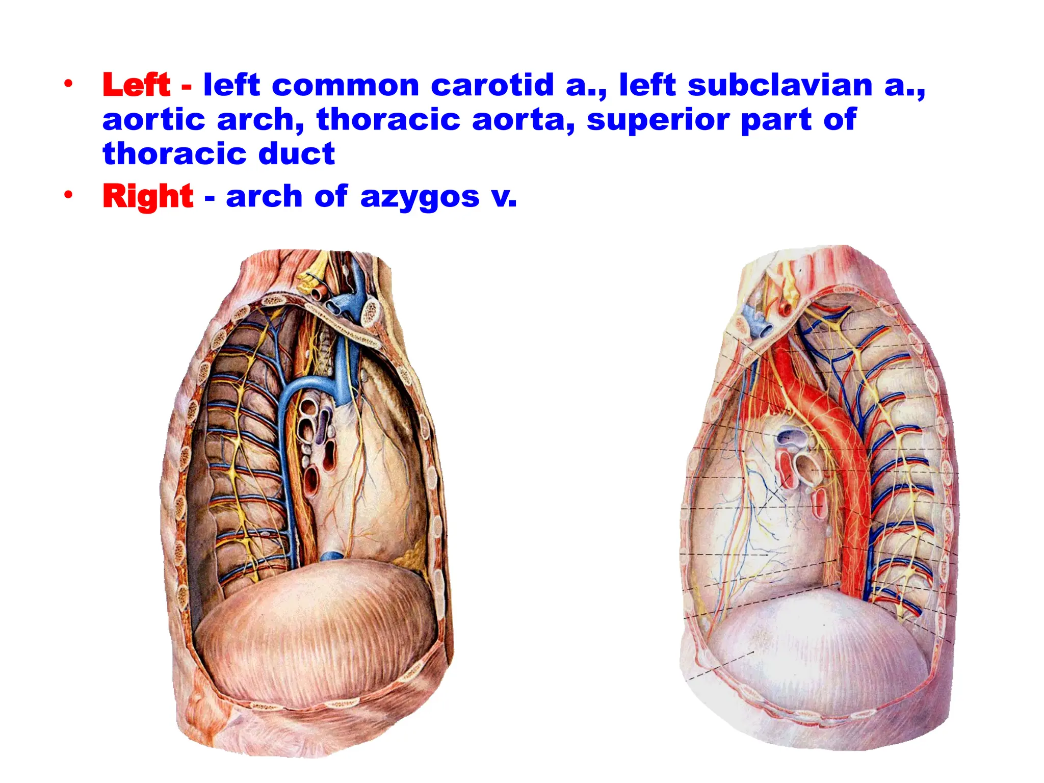
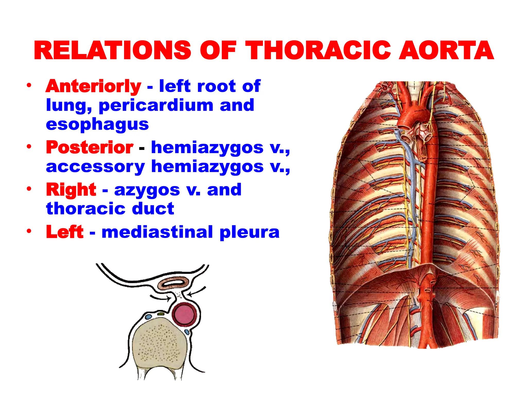
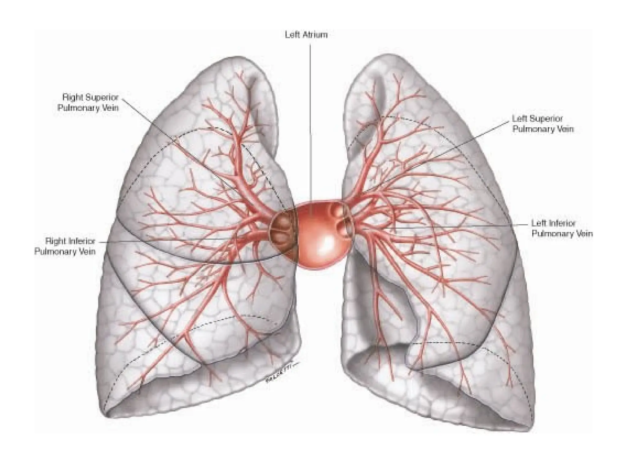
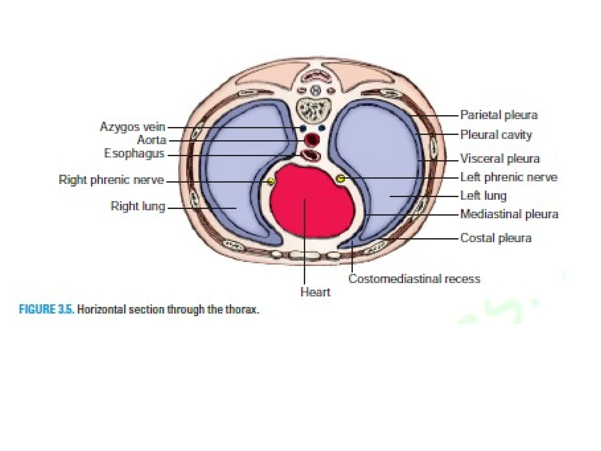
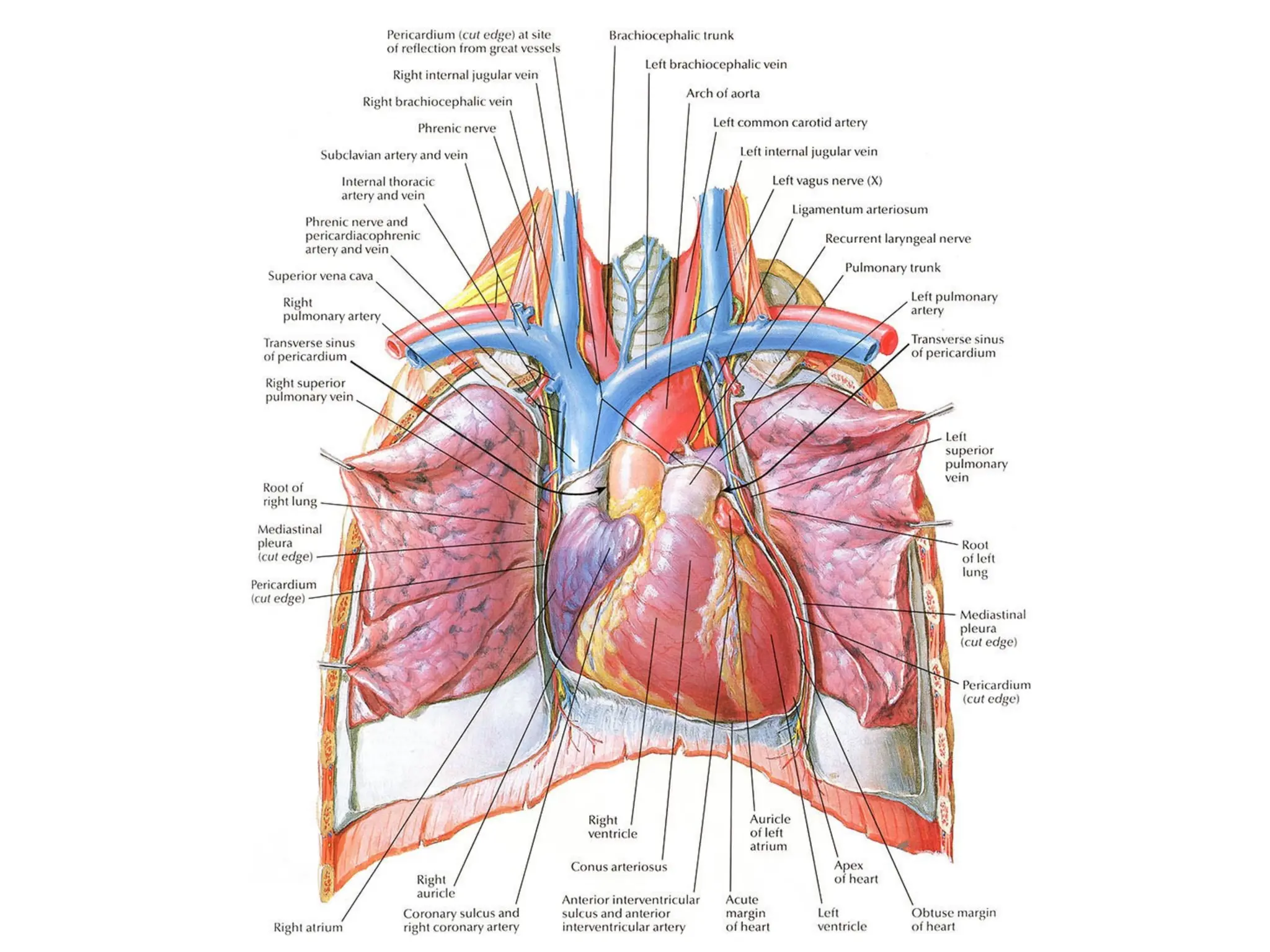
Lecturer: Mirlan Nuruev
TOPOGRAPHY OF THORAX
Parts and regions of the thorax
Borders
• Superior - jugular notch,
sternoclavicular joint,
superior border of clavicle,
acromion, spinous processes
of C7
• Inferior - xiphoid process,
costal arch, 12th and 11th
ribs, vertebra T12
Regions
• Thoracic wall
• Thoracic cavity
Thoracic wall
• Skin
• Superficial fascia
– Thoracoepigastric v.
– Supraclavicular n.
– Anterior and lateral
cutaneous branches of
intercostal n.
• Deep fascia
INTERNAL THORACIC
ARTERY
descends into thorax
1.2cm lateral to edge of
sternum, and ends at the
6th costal cartilage by
dividing musculophrenic
and superior epigastric
arteries
Thoracic aorta
• Bronchial bb.
• Oesophageal bb.
• Pericardial bb.
• Mediastinal bb.
• Posterior intercostal aa. (3-9)
– Spinal b
– Dorsal b.
• Subcostal a. (below 12th
rib)
• Superior phrenic aa.
AZYGOS VEIN
• Begins as continuation of right
ascending lumbar vein
• Ascending along the right side of
vertebral column
• Joins superior vena cava by aching
above right lung root at level of T4
to T5
• Receives right posterior intercostals
and subcostal veins plus some of
bronchial, esophageal and
pericardial veins, and hemiazygos
vein
• Tributaries – hemiazygos v. and
accessory hemiazygos v., which
receive most left posterior
intercostals vein and left bronchial
veins
THORACIC DUCT
• Begins in front of L1 as a dilated sac,
the cisterna chyli, which formed by
joining of left and right lumbar trunks
and intestinal trunk
• Enter thoracic cavity by passing through
the aortic hiatus of the diaphragm and
ascends along on the front of the
vertebral column, between thoracic
aorta and azygos vein
• Travels upward, veering to the left at
the level of T5
• At the roof of the neck, it turns laterally
and arches forwards and descends to
enter the left venous angle
• Just before termination, it receives the
left jugular, subclavian and
bronchomediastinal trunks
• Drains lymph from lower limbs, pelvic
cavity, abdominal cavity, left side of
thorax, and left side of the head, neck
and left upper limb
RIGHT LYMPHATIC DUCT
• Formed by union of right jugular,
subclavian, and bronchomediastinal
trunks
• Ends by entering the right venous
angle
• Receives lymph from right half of
head, neck, thorax and right upper
limb
Anterior branches of thoracic nerves
• Intercostal nerves - (anterior rami
of T1- T11): runs forward inferiorly to
intercostals vessels in costal groove of
corresponding rib, between intercostals
externi and intercostals interni; first six
nerves are distributed within their
intercostals space, lower five intercostals
nerves leave anterior ends of their
intercostals spaces to enter abdominal wall
• Subcostal nerve - (anterior ramus of
T12): follows inferior border of T12 rib and
passes into abdominal wall
• Distribution: distributed to intercostales
and anterolateral abdominal muscles, skin
of thoracic and abdominal wall, parietal
pleura and peritoneum
PHRENIC NERVE
• Descends over scalenus
anterior to enter thorax
• Accompanied by
pericardiophrenic vessels and
passes anterior to lung roots
between mediastinal pleura
and pericardium to supply
motor and sensory innervation
to diaphragm
• Sensory fibers supply to
pleurae, pericardium and
peritoneum of diaphragm;
usually right phrenic nerve
may be distributed on live,
gallbladder and biliary system.
LEFT VAGUS NERVE
• Enter thoracic inlet between left
common carotid and left subclavian
arteries, posterior to left
brachiocephalic vein
• Crosses aortic arch where left recurrent
laryngeal nerve branches off
• Passes posterior to left lung root
• Forms anterior esophageal plexus
• Forms anterior vagal trunk at
esophageal hiatus where it leaves
thorax and passes into abdominal
cavity , then divides into anterior
gastric and hepatic branches
RIGHT VAGUS NERVE
• Enter thoracic inlet on right side of
trachea
• Travels downward posterior to
right brachiocephalic vein and
superior vena cava
• Passes posterior to right lung root
• Forms posterior esophageal plexus
• Forms posterior vagal trunk at
esophageal hiatus where it leaves
thorax and passes into abdominal
cavity, then divides into posterior
gastric and celiac branches
RECURRENT LARYNGEAL
NERVES
• Right one hooks around right
subclavian artery, left one hooks aortic
arch
• Both ascend in tracheo-esophageal
groove
• Nerves enter larynx posterior to
cricothyroid joint, the nerve is now
called inferior laryngeal nerve
• Innervations: laryngeal mucosa below
fissure of glottis , all laryngeal
laryngeal muscles except cricothyroid
Bronchial and esophageal branches
Thoracic sympathetic trunk
• Branches of sympathetic trunk to
thoracic plexuses
• Greater splanchnic nerve -
formed by preganglionic fibers from
T5~T9 ganglia, and relay in celiac
ganglion.
• Lesser splanchnic nerve -
formed by preganglionic fibers from
T10~T12 ganglia, and relay in
aorticorenal ganglion.
• The postganglionic fibers supply
the liver, spleen, kidney and
alimentary tract as far as the left
colic flexure.
THE MEDIASTINUM
CONCEPT all of organs
between the left and
right mediastinal
pleurae is called
mediastinum. It
extends from the
sternum in front to
the vertebral column
behind, and from the
thoracic inlet above
to the diaphragm
below.
SUBDIVISIONS OF MEDIASTINUM
• Superior
mediastinum
• Inferior mediastinum
– Anterior mediastinum
– Middle mediastinum
– Posterior mediastinum
Left side of mediastnum
Root of lung
Pericardium
Phrenic n. &
pericardiacophrenic a.
Left vagus n.
Left recurrent n. Thoracic aorta
Sympathetic trunk
Greater splanchnic n
Aortic arch
Thoracic duct
Left subclavian a.
Esophagus
Right side of mediastnum
Root of lung
Pericardium
Superior vena cava
Phrenic n. &
pericardiacophrenic a.
Left vagus n.
Azygos v.
Sympathetic trunk
Esophagus
Arch of azygos v.
Trachea
Inferior vena cava
• Middle layer
– Aotic arch and its
three branches
– Phrenic n.
– Vagus n.
• Posterior layer
–Trachea
–Esophagus
–Thoracic duct
INFERIOR MEDIASTINUM
Anterior mediastinum
• Location - posterior to
body of sternum and
attached costal
cartilages, anterior to
heart and pericardium
• Contents - fat,
remnants of thymus
gland, anterior
mediastinal lymph
nodes
Middle mediastinum
• Location - between
anterior mediastinum
and posterior
mediastinum
• Contents: hart and
pericardium, beginning
or termination of great
vessels, phrenic nerves,
pericardiacophrenic
vessels , lymph nodes,
Posterior mediastinum
• Location - posterior to
heart and pericardium,
anterior to vertebrae T5 -
T12
• Contents: esophagus,
vagus n., thoracic aorta,
azygos system of veins,
thoracic duct, thoracic
sympathetic trunk,
posterior mediastinal
lymph nodes
Relations of esophagus
• Anteriorly -
trachea, bifurcation
of trachea, left
principal branchus,
left recurrent n., right
pulmonary a.,
anterior esophageal
plexus, pericardium,
left atrium,
diaphragm
• Posteriorly -
posterior
esophageal plexus,
thoracic aorta,
thoracic duct,
azygos v.,
hemiazygos
v.,accessory
hemiazygos v., right
posterior
intercostal v.
• Left - left common carotid a., left subclavian a.,
aortic arch, thoracic aorta, superior part of
thoracic duct
• Right - arch of azygos v.
RELATIONS OF THORACIC AORTA
• Anteriorly - left root of
lung, pericardium and
esophagus
• Posterior - hemiazygos v.,
accessory hemiazygos v.,
• Right - azygos v. and
thoracic duct
• Left - mediastinal pleura
Thank you!!

Lecture 7 OSTA Thorax.pptod ods. n.

  • 1.
  • 2.
    Parts and regionsof the thorax Borders • Superior - jugular notch, sternoclavicular joint, superior border of clavicle, acromion, spinous processes of C7 • Inferior - xiphoid process, costal arch, 12th and 11th ribs, vertebra T12 Regions • Thoracic wall • Thoracic cavity
  • 3.
    Thoracic wall • Skin •Superficial fascia – Thoracoepigastric v. – Supraclavicular n. – Anterior and lateral cutaneous branches of intercostal n. • Deep fascia
  • 4.
    INTERNAL THORACIC ARTERY descends intothorax 1.2cm lateral to edge of sternum, and ends at the 6th costal cartilage by dividing musculophrenic and superior epigastric arteries
  • 7.
    Thoracic aorta • Bronchialbb. • Oesophageal bb. • Pericardial bb. • Mediastinal bb. • Posterior intercostal aa. (3-9) – Spinal b – Dorsal b. • Subcostal a. (below 12th rib) • Superior phrenic aa.
  • 9.
    AZYGOS VEIN • Beginsas continuation of right ascending lumbar vein • Ascending along the right side of vertebral column • Joins superior vena cava by aching above right lung root at level of T4 to T5 • Receives right posterior intercostals and subcostal veins plus some of bronchial, esophageal and pericardial veins, and hemiazygos vein • Tributaries – hemiazygos v. and accessory hemiazygos v., which receive most left posterior intercostals vein and left bronchial veins
  • 13.
    THORACIC DUCT • Beginsin front of L1 as a dilated sac, the cisterna chyli, which formed by joining of left and right lumbar trunks and intestinal trunk • Enter thoracic cavity by passing through the aortic hiatus of the diaphragm and ascends along on the front of the vertebral column, between thoracic aorta and azygos vein • Travels upward, veering to the left at the level of T5 • At the roof of the neck, it turns laterally and arches forwards and descends to enter the left venous angle
  • 14.
    • Just beforetermination, it receives the left jugular, subclavian and bronchomediastinal trunks • Drains lymph from lower limbs, pelvic cavity, abdominal cavity, left side of thorax, and left side of the head, neck and left upper limb RIGHT LYMPHATIC DUCT • Formed by union of right jugular, subclavian, and bronchomediastinal trunks • Ends by entering the right venous angle • Receives lymph from right half of head, neck, thorax and right upper limb
  • 17.
    Anterior branches ofthoracic nerves • Intercostal nerves - (anterior rami of T1- T11): runs forward inferiorly to intercostals vessels in costal groove of corresponding rib, between intercostals externi and intercostals interni; first six nerves are distributed within their intercostals space, lower five intercostals nerves leave anterior ends of their intercostals spaces to enter abdominal wall • Subcostal nerve - (anterior ramus of T12): follows inferior border of T12 rib and passes into abdominal wall • Distribution: distributed to intercostales and anterolateral abdominal muscles, skin of thoracic and abdominal wall, parietal pleura and peritoneum
  • 20.
    PHRENIC NERVE • Descendsover scalenus anterior to enter thorax • Accompanied by pericardiophrenic vessels and passes anterior to lung roots between mediastinal pleura and pericardium to supply motor and sensory innervation to diaphragm • Sensory fibers supply to pleurae, pericardium and peritoneum of diaphragm; usually right phrenic nerve may be distributed on live, gallbladder and biliary system.
  • 21.
    LEFT VAGUS NERVE •Enter thoracic inlet between left common carotid and left subclavian arteries, posterior to left brachiocephalic vein • Crosses aortic arch where left recurrent laryngeal nerve branches off • Passes posterior to left lung root • Forms anterior esophageal plexus • Forms anterior vagal trunk at esophageal hiatus where it leaves thorax and passes into abdominal cavity , then divides into anterior gastric and hepatic branches
  • 22.
    RIGHT VAGUS NERVE •Enter thoracic inlet on right side of trachea • Travels downward posterior to right brachiocephalic vein and superior vena cava • Passes posterior to right lung root • Forms posterior esophageal plexus • Forms posterior vagal trunk at esophageal hiatus where it leaves thorax and passes into abdominal cavity, then divides into posterior gastric and celiac branches
  • 23.
    RECURRENT LARYNGEAL NERVES • Rightone hooks around right subclavian artery, left one hooks aortic arch • Both ascend in tracheo-esophageal groove • Nerves enter larynx posterior to cricothyroid joint, the nerve is now called inferior laryngeal nerve • Innervations: laryngeal mucosa below fissure of glottis , all laryngeal laryngeal muscles except cricothyroid Bronchial and esophageal branches
  • 24.
    Thoracic sympathetic trunk •Branches of sympathetic trunk to thoracic plexuses • Greater splanchnic nerve - formed by preganglionic fibers from T5~T9 ganglia, and relay in celiac ganglion. • Lesser splanchnic nerve - formed by preganglionic fibers from T10~T12 ganglia, and relay in aorticorenal ganglion. • The postganglionic fibers supply the liver, spleen, kidney and alimentary tract as far as the left colic flexure.
  • 26.
    THE MEDIASTINUM CONCEPT allof organs between the left and right mediastinal pleurae is called mediastinum. It extends from the sternum in front to the vertebral column behind, and from the thoracic inlet above to the diaphragm below.
  • 27.
    SUBDIVISIONS OF MEDIASTINUM •Superior mediastinum • Inferior mediastinum – Anterior mediastinum – Middle mediastinum – Posterior mediastinum
  • 29.
    Left side ofmediastnum Root of lung Pericardium Phrenic n. & pericardiacophrenic a. Left vagus n. Left recurrent n. Thoracic aorta Sympathetic trunk Greater splanchnic n Aortic arch Thoracic duct Left subclavian a. Esophagus
  • 30.
    Right side ofmediastnum Root of lung Pericardium Superior vena cava Phrenic n. & pericardiacophrenic a. Left vagus n. Azygos v. Sympathetic trunk Esophagus Arch of azygos v. Trachea Inferior vena cava
  • 31.
    • Middle layer –Aotic arch and its three branches – Phrenic n. – Vagus n.
  • 32.
  • 33.
    INFERIOR MEDIASTINUM Anterior mediastinum •Location - posterior to body of sternum and attached costal cartilages, anterior to heart and pericardium • Contents - fat, remnants of thymus gland, anterior mediastinal lymph nodes
  • 34.
    Middle mediastinum • Location- between anterior mediastinum and posterior mediastinum • Contents: hart and pericardium, beginning or termination of great vessels, phrenic nerves, pericardiacophrenic vessels , lymph nodes,
  • 35.
    Posterior mediastinum • Location- posterior to heart and pericardium, anterior to vertebrae T5 - T12 • Contents: esophagus, vagus n., thoracic aorta, azygos system of veins, thoracic duct, thoracic sympathetic trunk, posterior mediastinal lymph nodes
  • 36.
    Relations of esophagus •Anteriorly - trachea, bifurcation of trachea, left principal branchus, left recurrent n., right pulmonary a., anterior esophageal plexus, pericardium, left atrium, diaphragm
  • 37.
    • Posteriorly - posterior esophagealplexus, thoracic aorta, thoracic duct, azygos v., hemiazygos v.,accessory hemiazygos v., right posterior intercostal v.
  • 38.
    • Left -left common carotid a., left subclavian a., aortic arch, thoracic aorta, superior part of thoracic duct • Right - arch of azygos v.
  • 39.
    RELATIONS OF THORACICAORTA • Anteriorly - left root of lung, pericardium and esophagus • Posterior - hemiazygos v., accessory hemiazygos v., • Right - azygos v. and thoracic duct • Left - mediastinal pleura
  • 45.