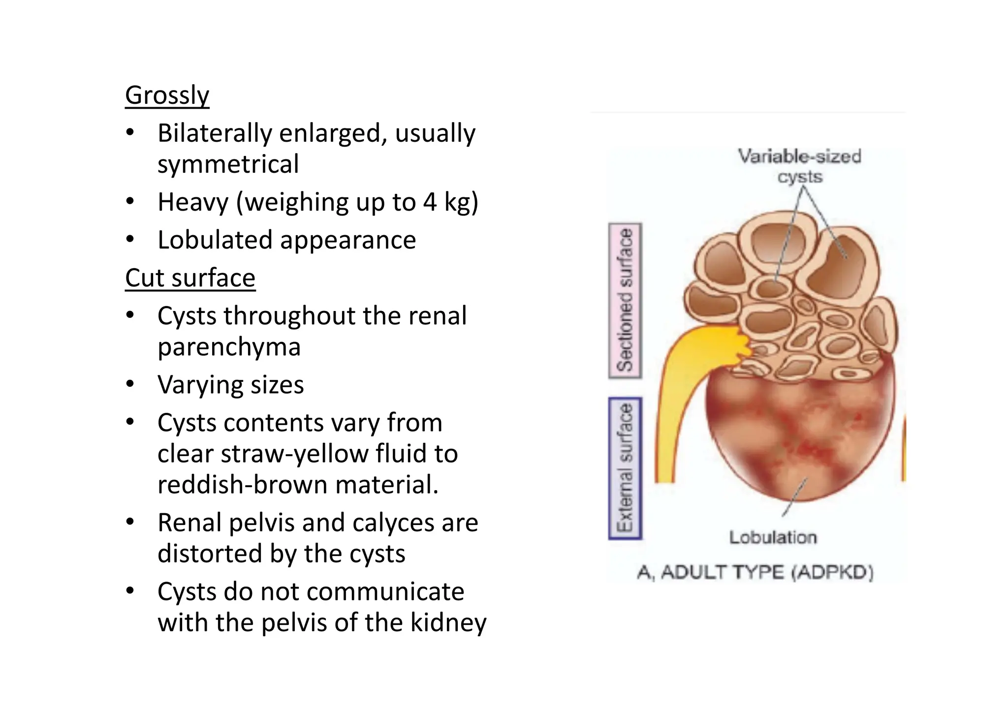
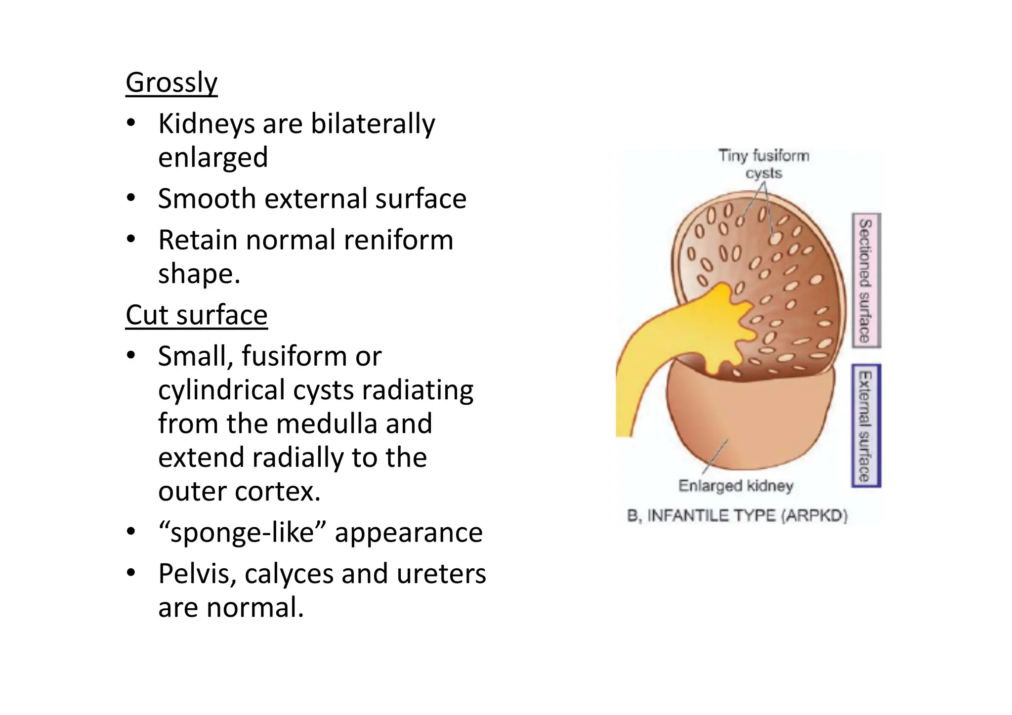
The Kidney and
Lower Urinary Tract - I
Dr A. Rapsang
Concentration test
• An artificial fluid deprivation is induced in the patient
for more than 20 hours.
• If the nephron is normal, water is selectively
reabsorbed resulting in excretion of urine of high
solute concentration
• However, if the tubular cells are nonfunctional, the
solute concentration of the urine will remain
constant regardless of stress of water deprivation.
Dilution test
• An excess of fluid is given to the patient.
• Normally, renal compensation should result in
excretion of urine with high water content and lower
• If the renal tubules are diseased, the concentration
of solutes in the urine will remain constant
irrespective of the excess water intake.
Blood chemistry.
• Impairment of renal function results in elevation of
end-products of protein metabolism.
• Urea - normal range 20-40 mg/dl
• Blood urea nitrogen (BUN) - normal range 10-20
mg/dl
• Creatinine - normal range 0.6-1.2 mg/dl
• An increase of these end-products in the blood is
called azotaemia.
• ‘uraemia’ is defined as association of these
biochemical abnormalities with clinical signs and
symptoms.
Creatinine clearance test
• The clearance of creatinine is determined by
collecting urine over 24-hour period
• A blood sample is withdrawn during the day.
• This test is routinely employed method of estimating
GFR.
Diseases of the kidneys are divided into 4 major groups
• Glomerular diseases: Often immunologically-
mediated
– Acute
– Chronic.
• Tubular diseases: Often caused by toxic or infectious
agents and are often acute.
• Interstitial diseases: Commonly due to toxic or
infectious agents
• Vascular diseases
• Others
– Congenital anomalies
– Obstructive uropathy (including urolithiasis)
– Tumours of the kidneys.
Acute Renal Failure (ARF)
• Acute renal failure (ARF) is a syndrome characterised
by
– Rapid onset of renal dysfunction
• Oliguria or anuria
– Sudden increase in metabolic waste-products in
the blood
• Urea
• Creatinine
– Development of uraemia.
Etiopathogenesis.
• Pre-renal causes. Pre-renal diseases are those which
cause sudden decrease in blood flow to the nephron.
– Inadequate cardiac output
– Hypovolaemia
– Vascular disease causing reduced perfusion of the kidneys.
• Intra-renal causes. Disease of renal tissue itself.
• Post-renal causes. Caused by obstruction to the flow
of urine anywhere along the renal tract distal to the
opening of the collecting ducts.
– Mass within the lumen/wall of the tract
– External compression anywhere along the lower urinary
tract—ureter, bladder neck or urethra.
Clinical features.
Depend to a large extent on the underlying cause of
ARF and on the stage of the disease at which the
patient presents.
• Syndrome of acute nephritis.
• Extensive proliferation of epithelial cells in the
glomeruli → decrease in GFR.
– Mild proteinuria
– Haematuria
– Edema
– Mild hypertension.
• E.g., Acute post-streptococcal glomerulonephritis
• Syndrome accompanying tubular pathology.
• Destruction of the tubular cells of the nephron
• E.g., Acute tubular necrosis
• The disease typically progresses through 3 characteristic
stages from oliguria → diuresis → recovery.
• Oliguric phase:
– First 7 to 10 days
– Urinary output < 400 ml/day.
– Accumulation of waste products
• Azotaemia
• Metabolic acidosis
• Hyperkalaemia
• Hypernatraemia
• Hypervolaemia
• Pulmonary oedema.
• Diuretic phase:
• With the onset of healing of tubules, there is
improvement in urinary output.
• Urine is of low specific gravity.
• Phase of recovery:
• Full recovery with healing of tubular epithelial cells
• Death occurs in 50% cases
• Pre-renal syndrome.
• ARF occurring secondary to disorders in which
neither the glomerulus nor the tubules are damaged
• E.g., Renal arterial obstruction
– Hypovolaemia
– Hypotension
– Cardiac insufficiency.
– Oliguria
– Azotaemia
– Fluid retention and oedema.
Chronic Renal Failure (CRF)
• Chronic renal failure is a syndrome characterised by
progressive and irreversible deterioration of renal
function due to slow destruction of renal
parenchyma
Etiopathogenesis.
• Diseases causing glomerular pathology.
– Primary glomerular pathology
• Chronic glomerulonephritis
– Systemic glomerular pathology
• SLE
• Diabetic nephropathy.
• Diseases causing tubulointerstitial pathology.
• Vascular causes
– Long-standing hypertension
• Infectious causes
– Chronic pyelonephritis.
• Toxic causes:
– Intake of high doses of analgesics
– Other substances -lead, cadmium and uranium.
• Obstructive causes
– Stones, blood clots, tumours, strictures and enlarged
prostate.
CRF evolves progressively through 4 stages:
• Decreased renal reserve.
– Damage to renal parenchyma is marginal and the kidneys
remain functional.
– The GFR is about 50% of normal
– BUN and creatinine are normal
– Patients are usually asymptomatic
• Renal insufficiency.
– 75% of functional renal parenchyma has been destroyed.
– The GFR is about 25% of normal
– Elevation in BUN and serum creatinine.
– Polyuria and nocturia
• Renal failure.
– 90% of functional renal tissue has been destroyed.
– The GFR is approximately 10% of normal.
– Regulation of sodium and water is lost - oedema,
metabolic acidosis, hypocalcaemia, and signs and
symptoms of uraemia.
• End-stage kidney.
– The GFR at this stage is less than 5% of normal
– Uraemic syndrome
– Progressive primary (renal) and secondary systemic (extra-
renal) symptoms.
Clinical features.
Uraemic syndrome
• Primary uraemic (renal) manifestations.
– Metabolic acidosis - Kussmaul breathing,
hyperkalaemia and hypercalcaemia.
– Hyperkalaemia - cardiac arrhythmias, weakness,
nausea, intestinal colic, diarrhoea, muscular
irritability and flaccid paralysis.
– Sodium and water imbalance -hypervolaemia and
circulatory overload with congestive heart failure.
– Hyperuricaemia - gout.
– Azotaemia.
• Secondary uraemic (extra-renal) manifestations.
– Anaemia – due to decreased production of
erythropoietin
– Integumentary system
• Deposit of urinary pigment such as urochrome in the skin
causes sallow-yellow colour.
• The urea content in the sweat rises. On perspiration, urea
remains on the facial skin as powdery ‘uraemic frost’.
– Cardiovascular system
• Fluid retention
– Congestive heart failure.
– Respiratory system
• Pulmonary congestion
• Pulmonary oedema
– Digestive system.
• Mucosal ulcerations
• Gastrointestinal irritation - nausea, vomiting and
diarrhoea.
– Skeletal system -renal osteodystrophy
• Osteomalacia - deficiency of vitamin D (which is
normally activated by the kidney)
• Osteitis fibrosa - due to elevated levels of
parathormone → mobilises calcium from bone →
hypercalcaemia → deposits of excess calcium salts in
joints and soft tissues and weakening of bones (renal
osteodystrophy).
CONGENITAL MALFORMATIONS
Malformations of the kidneys are classified into 3 broad
groups:
• Abnormalities in amount of renal tissue.
– Renal hypoplasia
– Renomegaly
• Anomalies of position, form and orientation.
– Renal ectopia (pelvic kidney)
– Renal fusion (horseshoe kidney)
– Persistent foetal lobation.
• Anomalies of differentiation.
– Cystic diseases of the kidney
CYSTIC DISEASES OF KIDNEY
• Cystic lesions of the kidney may be
– Congenital or acquired,
– Non-neoplastic or neoplastic.
• Majority of these lesions are congenital non-
neoplastic.
• Cystic lesions can occur at any age
• Their usual clinical presentation may include:
– Abdominal mass
– Infection
– Respiratory distress (due to accompanied pulmonary
hypoplasia)
– Haemorrhage
– Neoplastic transformation.
Polycystic Kidney Disease
• Major portion of the renal parenchyma is
converted into cysts of varying size.
• The disease occurs in two forms:
• An adult type - inherited as an autosomal
dominant disease
• An infantile type - inherited as an autosomal
recessive disorder.
• Adult polycystic kidney disease
• Always bilateral and diffuse.
• Though the kidneys are abnormal at birth, renal
function is retained
• Symptoms appear in adult life, mostly between
the age of 30 and 50 years.
Grossly
• Bilaterally enlarged, usually
symmetrical
• Heavy (weighing up to 4 kg)
• Lobulated appearance
Cut surface
• Cysts throughout the renal
parenchyma
• Varying sizes
• Cysts contents vary from
clear straw-yellow fluid to
reddish-brown material.
• Renal pelvis and calyces are
distorted by the cysts
• Cysts do not communicate
with the pelvis of the kidney
Histologically
• Some cysts contain glomerular tufts
• Some cysts have epithelial lining
• The intervening tissue between the cysts shows
some normal renal parenchyma.
• There can be fibrosis and chronic inflammation
Clinical features.
• Commonly manifests in 3rd to 5th decades of life.
• Dull-ache in the lumbar region
• Haematuria
• Renal colic
• Hypertension
• Urinary tract infections
• Progressive CRF
– Polyuria
– Proteinuria.
• Other associated congenital anomalies seen less
frequently are cysts in the pancreas, spleen, lungs
and other organs.
Infantile polycystic kidney disease
• Less common
• Bilateral.
• The age at presentation may be perinatal, neonatal,
infantile or juvenile
Grossly
• Kidneys are bilaterally
enlarged
• Smooth external surface
• Retain normal reniform
shape.
Cut surface
• Small, fusiform or
cylindrical cysts radiating
from the medulla and
extend radially to the
outer cortex.
• “sponge-like” appearance
• Pelvis, calyces and ureters
are normal.
Histologically
• The total number of nephrons is normal.
• Since the cysts are formed from dilatation of
collecting tubules, all the collecting tubules show
cylindrical or saccular dilatations and are lined by
cuboidal to low columnar epithelium.
• Many of the glomeruli are also cystically dilated.
Clinical features.
• Depend on age of the child.
• In severe form, the gross bilateral cystic renal
enlargement - interfere with delivery.
• In infancy, renal failure may manifest early.
• Almost all cases of infantile polycystic kidney disease
have associated multiple epithelium-lined cysts in
the liver or proliferation of portal bile ductules.
• In older children - congenital hepatic fibrosis
– Portal hypertension
– Splenomegaly.
Assignment
• Short notes on
– Kidney function tests
– Dilution and concentration tests
– Creatinine clearance test
– Uraemic syndrome
• Define
– Azotemia
– Uraemia
• Definition, etiopathogenesis and clinical features of
– ARF
– CRF
• Pathological and clinical features of Polycystic kidney
disease

The Kidney and anatomy and pathophysiology

  • 1.
    The Kidney and LowerUrinary Tract - I Dr A. Rapsang
  • 3.
    Concentration test • Anartificial fluid deprivation is induced in the patient for more than 20 hours. • If the nephron is normal, water is selectively reabsorbed resulting in excretion of urine of high solute concentration • However, if the tubular cells are nonfunctional, the solute concentration of the urine will remain constant regardless of stress of water deprivation.
  • 4.
    Dilution test • Anexcess of fluid is given to the patient. • Normally, renal compensation should result in excretion of urine with high water content and lower • If the renal tubules are diseased, the concentration of solutes in the urine will remain constant irrespective of the excess water intake.
  • 5.
    Blood chemistry. • Impairmentof renal function results in elevation of end-products of protein metabolism. • Urea - normal range 20-40 mg/dl • Blood urea nitrogen (BUN) - normal range 10-20 mg/dl • Creatinine - normal range 0.6-1.2 mg/dl • An increase of these end-products in the blood is called azotaemia. • ‘uraemia’ is defined as association of these biochemical abnormalities with clinical signs and symptoms.
  • 6.
    Creatinine clearance test •The clearance of creatinine is determined by collecting urine over 24-hour period • A blood sample is withdrawn during the day. • This test is routinely employed method of estimating GFR.
  • 7.
    Diseases of thekidneys are divided into 4 major groups • Glomerular diseases: Often immunologically- mediated – Acute – Chronic. • Tubular diseases: Often caused by toxic or infectious agents and are often acute. • Interstitial diseases: Commonly due to toxic or infectious agents • Vascular diseases • Others – Congenital anomalies – Obstructive uropathy (including urolithiasis) – Tumours of the kidneys.
  • 8.
  • 9.
    • Acute renalfailure (ARF) is a syndrome characterised by – Rapid onset of renal dysfunction • Oliguria or anuria – Sudden increase in metabolic waste-products in the blood • Urea • Creatinine – Development of uraemia.
  • 10.
    Etiopathogenesis. • Pre-renal causes.Pre-renal diseases are those which cause sudden decrease in blood flow to the nephron. – Inadequate cardiac output – Hypovolaemia – Vascular disease causing reduced perfusion of the kidneys. • Intra-renal causes. Disease of renal tissue itself. • Post-renal causes. Caused by obstruction to the flow of urine anywhere along the renal tract distal to the opening of the collecting ducts. – Mass within the lumen/wall of the tract – External compression anywhere along the lower urinary tract—ureter, bladder neck or urethra.
  • 11.
    Clinical features. Depend toa large extent on the underlying cause of ARF and on the stage of the disease at which the patient presents. • Syndrome of acute nephritis. • Extensive proliferation of epithelial cells in the glomeruli → decrease in GFR. – Mild proteinuria – Haematuria – Edema – Mild hypertension. • E.g., Acute post-streptococcal glomerulonephritis
  • 12.
    • Syndrome accompanyingtubular pathology. • Destruction of the tubular cells of the nephron • E.g., Acute tubular necrosis • The disease typically progresses through 3 characteristic stages from oliguria → diuresis → recovery. • Oliguric phase: – First 7 to 10 days – Urinary output < 400 ml/day. – Accumulation of waste products • Azotaemia • Metabolic acidosis • Hyperkalaemia • Hypernatraemia • Hypervolaemia • Pulmonary oedema.
  • 13.
    • Diuretic phase: •With the onset of healing of tubules, there is improvement in urinary output. • Urine is of low specific gravity. • Phase of recovery: • Full recovery with healing of tubular epithelial cells • Death occurs in 50% cases
  • 14.
    • Pre-renal syndrome. •ARF occurring secondary to disorders in which neither the glomerulus nor the tubules are damaged • E.g., Renal arterial obstruction – Hypovolaemia – Hypotension – Cardiac insufficiency. – Oliguria – Azotaemia – Fluid retention and oedema.
  • 15.
  • 16.
    • Chronic renalfailure is a syndrome characterised by progressive and irreversible deterioration of renal function due to slow destruction of renal parenchyma Etiopathogenesis. • Diseases causing glomerular pathology. – Primary glomerular pathology • Chronic glomerulonephritis – Systemic glomerular pathology • SLE • Diabetic nephropathy.
  • 17.
    • Diseases causingtubulointerstitial pathology. • Vascular causes – Long-standing hypertension • Infectious causes – Chronic pyelonephritis. • Toxic causes: – Intake of high doses of analgesics – Other substances -lead, cadmium and uranium. • Obstructive causes – Stones, blood clots, tumours, strictures and enlarged prostate.
  • 18.
    CRF evolves progressivelythrough 4 stages: • Decreased renal reserve. – Damage to renal parenchyma is marginal and the kidneys remain functional. – The GFR is about 50% of normal – BUN and creatinine are normal – Patients are usually asymptomatic • Renal insufficiency. – 75% of functional renal parenchyma has been destroyed. – The GFR is about 25% of normal – Elevation in BUN and serum creatinine. – Polyuria and nocturia
  • 19.
    • Renal failure. –90% of functional renal tissue has been destroyed. – The GFR is approximately 10% of normal. – Regulation of sodium and water is lost - oedema, metabolic acidosis, hypocalcaemia, and signs and symptoms of uraemia. • End-stage kidney. – The GFR at this stage is less than 5% of normal – Uraemic syndrome – Progressive primary (renal) and secondary systemic (extra- renal) symptoms.
  • 20.
    Clinical features. Uraemic syndrome •Primary uraemic (renal) manifestations. – Metabolic acidosis - Kussmaul breathing, hyperkalaemia and hypercalcaemia. – Hyperkalaemia - cardiac arrhythmias, weakness, nausea, intestinal colic, diarrhoea, muscular irritability and flaccid paralysis. – Sodium and water imbalance -hypervolaemia and circulatory overload with congestive heart failure. – Hyperuricaemia - gout. – Azotaemia.
  • 21.
    • Secondary uraemic(extra-renal) manifestations. – Anaemia – due to decreased production of erythropoietin – Integumentary system • Deposit of urinary pigment such as urochrome in the skin causes sallow-yellow colour. • The urea content in the sweat rises. On perspiration, urea remains on the facial skin as powdery ‘uraemic frost’. – Cardiovascular system • Fluid retention – Congestive heart failure. – Respiratory system • Pulmonary congestion • Pulmonary oedema
  • 22.
    – Digestive system. •Mucosal ulcerations • Gastrointestinal irritation - nausea, vomiting and diarrhoea. – Skeletal system -renal osteodystrophy • Osteomalacia - deficiency of vitamin D (which is normally activated by the kidney) • Osteitis fibrosa - due to elevated levels of parathormone → mobilises calcium from bone → hypercalcaemia → deposits of excess calcium salts in joints and soft tissues and weakening of bones (renal osteodystrophy).
  • 23.
  • 24.
    Malformations of thekidneys are classified into 3 broad groups: • Abnormalities in amount of renal tissue. – Renal hypoplasia – Renomegaly • Anomalies of position, form and orientation. – Renal ectopia (pelvic kidney) – Renal fusion (horseshoe kidney) – Persistent foetal lobation. • Anomalies of differentiation. – Cystic diseases of the kidney
  • 25.
    CYSTIC DISEASES OFKIDNEY • Cystic lesions of the kidney may be – Congenital or acquired, – Non-neoplastic or neoplastic. • Majority of these lesions are congenital non- neoplastic. • Cystic lesions can occur at any age • Their usual clinical presentation may include: – Abdominal mass – Infection – Respiratory distress (due to accompanied pulmonary hypoplasia) – Haemorrhage – Neoplastic transformation.
  • 27.
    Polycystic Kidney Disease •Major portion of the renal parenchyma is converted into cysts of varying size. • The disease occurs in two forms: • An adult type - inherited as an autosomal dominant disease • An infantile type - inherited as an autosomal recessive disorder. • Adult polycystic kidney disease • Always bilateral and diffuse. • Though the kidneys are abnormal at birth, renal function is retained • Symptoms appear in adult life, mostly between the age of 30 and 50 years.
  • 28.
    Grossly • Bilaterally enlarged,usually symmetrical • Heavy (weighing up to 4 kg) • Lobulated appearance Cut surface • Cysts throughout the renal parenchyma • Varying sizes • Cysts contents vary from clear straw-yellow fluid to reddish-brown material. • Renal pelvis and calyces are distorted by the cysts • Cysts do not communicate with the pelvis of the kidney
  • 29.
    Histologically • Some cystscontain glomerular tufts • Some cysts have epithelial lining • The intervening tissue between the cysts shows some normal renal parenchyma. • There can be fibrosis and chronic inflammation
  • 30.
    Clinical features. • Commonlymanifests in 3rd to 5th decades of life. • Dull-ache in the lumbar region • Haematuria • Renal colic • Hypertension • Urinary tract infections • Progressive CRF – Polyuria – Proteinuria. • Other associated congenital anomalies seen less frequently are cysts in the pancreas, spleen, lungs and other organs.
  • 31.
    Infantile polycystic kidneydisease • Less common • Bilateral. • The age at presentation may be perinatal, neonatal, infantile or juvenile
  • 32.
    Grossly • Kidneys arebilaterally enlarged • Smooth external surface • Retain normal reniform shape. Cut surface • Small, fusiform or cylindrical cysts radiating from the medulla and extend radially to the outer cortex. • “sponge-like” appearance • Pelvis, calyces and ureters are normal.
  • 33.
    Histologically • The totalnumber of nephrons is normal. • Since the cysts are formed from dilatation of collecting tubules, all the collecting tubules show cylindrical or saccular dilatations and are lined by cuboidal to low columnar epithelium. • Many of the glomeruli are also cystically dilated.
  • 34.
    Clinical features. • Dependon age of the child. • In severe form, the gross bilateral cystic renal enlargement - interfere with delivery. • In infancy, renal failure may manifest early. • Almost all cases of infantile polycystic kidney disease have associated multiple epithelium-lined cysts in the liver or proliferation of portal bile ductules. • In older children - congenital hepatic fibrosis – Portal hypertension – Splenomegaly.
  • 35.
    Assignment • Short noteson – Kidney function tests – Dilution and concentration tests – Creatinine clearance test – Uraemic syndrome • Define – Azotemia – Uraemia • Definition, etiopathogenesis and clinical features of – ARF – CRF • Pathological and clinical features of Polycystic kidney disease