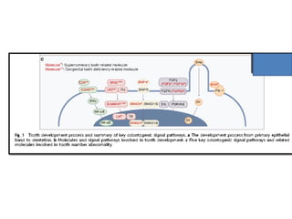
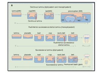
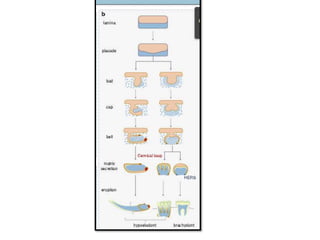
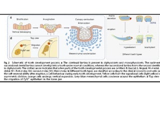
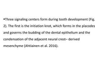
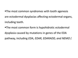
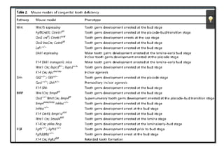
This document discusses genetic factors that influence tooth development abnormalities. It begins by describing the normal development of primary and permanent dentition, regulated by signaling centers. Tooth agenesis and supernumerary tooth formation can result from mutations affecting genes in the Wnt, BMP, Shh, and FGF signaling pathways. Specific syndromes associated with tooth number anomalies like ectodermal dysplasia and cleidocranial dysplasia are also discussed. The document concludes by covering variations in tooth size and shape, including double teeth formed by fusion or gemination, taurodontism, and other dental anomalies.

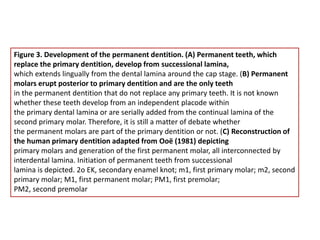


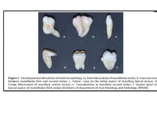

![Concrescence is defined as the cemental union of two
adjacent teeth without confluence of the underlying dentin
showing independent pulpchambers and root canals [14, 18].
It may occur during or after the completion of root
formation.
If the condition occurs during development, it is called
true/developmental concrescence and acquired/post
inflammatory concrescence if after root formation](https://image.slidesharecdn.com/teethabnormalities-230502181043-2fcae6ba/85/dental-abnormalities-on-genetic-background-new-vision-pptx-66-320.jpg)
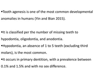
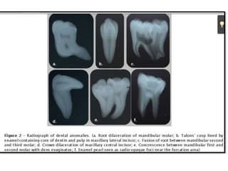
![Dilacerations usually occur in the apical third of
the root when the anterior teeth are involved, middle
third when first molars are involved and coronal
third when third molars are involved [39].
Root dilacerations are common than crown
dilacerations and occur usually in the posterior
region of permanent dentition [](https://image.slidesharecdn.com/teethabnormalities-230502181043-2fcae6ba/85/dental-abnormalities-on-genetic-background-new-vision-pptx-67-320.jpg)
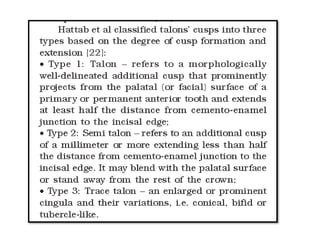
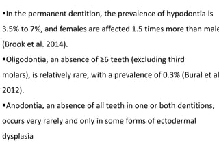
![Dens evaginatus (DE) is a developmental aberration of a
tooth resulting in formation of an accessory cusp whose
morphology has been described as abnormal tubercle,
elevation, protuberance, excrescence, extrusion, or a bulge
[36].
It is also referred to as tuberculated cusp, accessory
tubercle, occlusal tuberculated premolar, Leong’s
premolar, evaginatus odontoma, and occlusal pearl](https://image.slidesharecdn.com/teethabnormalities-230502181043-2fcae6ba/85/dental-abnormalities-on-genetic-background-new-vision-pptx-70-320.jpg)