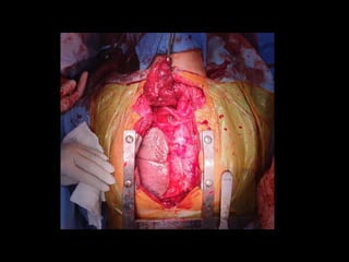
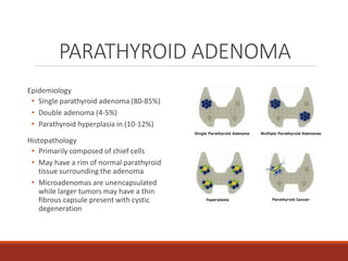
TCVS
CONFERENCE
OCTOBER 10, 2019
CLINICAL CLERK ALEXANDER XERXES MALICSE
CLINICAL CLERK JESSICA MARTINEZ
OSG 54/F
Married, Filipino
Roman Catholic
CC: Difficulty in Walking
o Enlarging non-tender anterior
neck mass
o No dyspnea, no hoarseness
o No consult was done
3 YEARS PTA
2 YEARS PTA
2 MONTHS
PTA
1 WEEK PTA
ADMISSION
o Intermittent Joint Pains on both
elbows and knees
o 7/10
o Bilateral flank pain
o No fever, no hematuria, no dysuria
o No consult done
3 YEARS PTA
2 YEARS PTA
2 MONTHS
PTA
1 WEEK PTA
ADMISSION
o Progressive enlargement of the
anterior neck mass and worsening of
the joint pains
3 YEARS PTA
2 YEARS PTA
2 MONTHS
PTA
1 WEEK PTA
ADMISSION
o Joint Pain 8/10
o 5 pounds unintentional weight loss
o Persistence of flank pain
o Sought consult with a urologist
o STAGHORN CALCULI
o s/p DJ Stent Insertion, Bilateral
3 YEARS PTA
2 YEARS PTA
2 MONTHS
PTA
1 WEEK PTA
ADMISSION
o Joint Pain 8/10
o 5 pounds unintentional weight loss
o Persistence of flank pain
o Sought consult with a urologist
o STAGHORN CALCULI
o s/p DJ Stent Insertion, Bilateral
o Advised to consult an endocrinologist
3 YEARS PTA
2 YEARS PTA
2 MONTHS
PTA
1 WEEK PTA
ADMISSION
o Difficulty walking
o Unable to stand for a long period
3 YEARS PTA
2 YEARS PTA
2 MONTHS
PTA
1 WEEK PTA
ADMISSION
o Consulted a private endocrinologist
o THYROID FUNCTION TEST
o Normal
o SERUM IONIZED CALCIUM
o Elevated
o BONE DEXA SCAN
o Osteoporosis
3 YEARS PTA
2 YEARS PTA
2 MONTHS
PTA
1 WEEK PTA
ADMISSION
o SESTAMIBI SCAN
o Enlarged right parathyroid with
substernal extension
o Hyperfunctioning
3 YEARS PTA
2 YEARS PTA
2 MONTHS
PTA
1 WEEK PTA
ADMISSION
o SESTAMIBI SCAN
o Enlarged right parathyroid with
substernal extension
o Hyperfunctioning
o CT SCAN
o Large soft tissue mass
o 11.8 x 5.9 x 5 cm
o right thoracic inlet and superior and
middle mediastinum
3 YEARS PTA
2 YEARS PTA
2 MONTHS
PTA
1 WEEK PTA
ADMISSION
o SESTAMIBI SCAN
o Enlarged right parathyroid with
substernal extension
o Hyperfunctioning
o CT SCAN
o Large soft tissue mass
o 11.8 x 5.9 x 5 cm
o right thoracic inlet and superior and
middle mediastinum
o She was advised surgery, hence,
admission
3 YEARS PTA
2 YEARS PTA
2 MONTHS
PTA
1 WEEK PTA
ADMISSION
REVIEW OF SYSTEMS
General Survey: (+) weight loss, (+) weakness, (-) loss of appetite
Cutaneous: (+) pallor, (-) itchiness, (-) jaundice, (-) abnormal hair or
nail growth, (-) erythematous
Eye: (-) blurring of vision, (-) lacrimation, (-) pain
Ear: (-) tinnitus, (-) discharge, (-) deafness, (-) otalgia
Nose: (-) epistaxis, (-) discharge, (-) obstruction, (-) abnormal sense of
smell, (-) postnasal drip, (-) sinus pain
Mouth: (-) gum bleeding, (-) mouth sores, (-) fissures
Throat: (-) soreness, (-) tonsillar pain, (-) dysphagia, (-) odynophagia
Neck: (-) neck stiffness, (-) limitation of motion
REVIEW OF SYSTEMS
Cardiology: (-) easy fatigability, (-) orthopnea, (-) PND, (-) palpitations
Respiratory: (+) cough, (-) shortness of breath, (-) increase in respiration
Gastrointestinal: (+) dark stool, (-) nausea, (-) vomiting, (-) hematemesis,
(-) hematochezia, (-) abdominal pain, (-) diarrhea
Endocrine: (+) polyuria, (-) polydipsia, (-) polyphagia
Genitourinary: (+) nocturia, (-) frequency, (-) urgency, (-) hesitancy
Neurologic: (-) limb numbness, (-) dizziness, (-) paresthesia, (-) seizures, (-)
paralysis
Psychiatry: (-) anxiety, (-) depression, (-) hallucination, (-) paranoia
Musculoskeletal: See HPI
Hematologic: (-) abnormal bleeding, (-) bruising
PAST MEDICAL HISTORY
Previous Illnesses/hospitalizations:
(+) Leptospirosis (1989)
(+) Multinodular nontoxic goiter
(-) HTN
(-) DM
(-) MI/Stroke
(-) Asthma
(-) PTB
(-) Pneumonia
Known allergies: none
Previous accidents/surgeries:
DJ stent insertion (2019)
Current Medications:
Ferrous Sulfate + Folic acid
FAMILY HISTORY
(+) Hypertension- Father
(+) DM- Sister, aunt
(+) Goiter- Cousin
(+) Emphysema- Father
(-) pneumonia, TB
(-) cirrhosis
(-) stroke
SOCIAL HISTORY
• Non-smoker; exposed to second
hand smoking
• Non-alcoholic beverage drinker
• Denies Illicit Drug Use
PHYSICAL EXAM
GENERAL SURVEY: Conscious, coherent, not in respiratory distress,
ambulatory, GCS 15 (E4V5M6)
VITAL SIGNS: BP:120/80 HR: 72 RR:22 Temperature: 36.8 O2 sat: 99%
Ht: 146cm Wt: 110 lbs
SKIN/CUTANEOUS: (+) pallor, (-) cyanosis, (-) jaundice
HEAD: evenly distributed hair, (-) facial deformities, (-) characteristic facies
EYES: (+) pale palpebral conjunctiva, (-) periorbital edema, (-) opacity of lens
EARS: (-) tragal tenderness, non-hyperemic EAC
NOSE: No gross deformities, midline septum, (-) discharge, (-) epistaxis
MOUTH: Dry lips, moist buccal mucosa, (-) oral ulcers, (-) oral plaques
PHYSICAL EXAM
NECK: (+) multinodular anterior neck mass, thyroid is enlarged, no palpable
cervical lymphadenopathies, no distended neck veins, (-) carotid bruit
RESPIRATORY: Symmetrical chest expansion, equal tactile and vocal fremiti, (-)
crackles
CARDIOVASCULAR: Adynamic precordium, apex beat at 5th LICS MCL, (-)
heaves, (-) thrills, (-) lifts, (-) murmur, soft S1 followed by loud S2 at base, loud
S1 followed by soft S2 at apex, pulses regular
GASTROINTESTINAL: soft, nontender abdomen, normoactive bowel sounds, (-)
abdominal bruit, tympanitic on all quadrants, (-) direct and rebound
tenderness
GENITOURINARY: (-) CVA tenderness
EXTREMITIES: (-) bipedal edema, ++ radial pulse, equal, CRT <2 sec
Primary Hyperparathyroidism Secondary
to Parathyroid Adenoma
Multinodular Nontoxic Goiter
CKD Stage 3B Secondary to
Hypercalcemia Secondary to Primary
Hyperparathyroidism
ADMITTING DIAGNOSIS
Cystoscopy, Retrograde Pyelography,
DJ Stent Replacement, Bilateral
Total Thyroidectomy, Parathyroidectomy,
Sternotomy,
PLAN
Cystoscopy, Retrograde Pyelography,
DJ Stent Replacement, Bilateral
Total Thyroidectomy; Sternotomy;
Parathyroidectomy, Right;
Extended Thymectomy
OR DONE
DISCUSSION
PARATHYROID GLAND
• Usually four parathyroid glands
• Ovoid in shape, measuring 5-7 mm, and weighing
40-50 mg
• Superior glands more consistent in position
(dorsal to RLN)
• Inferior glands more variable in position
(ventral to RLN)
• Supplied by superior and inferior thyroid arteries
• Yellow-brown tissue similar to surrounding fatty
tissue
• Contains mainly chief cells which produce
parathyroid hormone (PTH)
PARATHYROID HORMONE (PTH)
Synthesized by the parathyroid glands
Bone
• Stimulates osteoclasts
• Inhibits osteoblasts
Kidney
• Increases reabsorption of calcium
• Increases phosphate excretion
GI tract
• Stimulates hydroxylation of 25-OH D 
1,25 OH D
• 1,25 OH D increases the intestinal
absorption of dietary calcium and
phosphate
DISORDERS OF THE
PARATHYROID GLANDS
Pathophysiology Clinical Manifestations Management
Primary Hyperparathyroidism
Increased PTH from abnormal parathyroid
glands
Results from enlargement of a single gland or
parathyroid adenoma (80%), multiple
adenomas or hyperplasia (15-20%), and
parathyroid carcinoma (1%)
Classic Pentad
• Kidney stones
• Painful bones
• Abdominal groans
• Psychic moans
• Fatigue overtones
Symptomatic: parathyroidectomy
Secondary Hyperparathyroidism
Increased response to hypocalcemic states Calciphylaxis: painful, violaceous
lesions on the extremities
Medical: cinacalcet (calcimimetic)
Parathyroidectomy: if PTH remains high
Tertiary Hyperparathyroidism
Autonomously functioning parathyroid glands
after correction of underlying disorder in
secondary hyperparathyroidism
Similar to primary
hyperparathyroidism
Medical: cinacalcet (calcimimetic)
Parathyroidectomy: if symptomatic or if with
persistence of elevated PTH >1 year after kidney
transplantation
Hypoparathyroidism
Most common: surgically induced Chvostek sign, Trousseau sign Medical: calcium and vitamin D supplementation
PARATHYROID ADENOMA
Epidemiology
• Single parathyroid adenoma (80-85%)
• Double adenoma (4-5%)
• Parathyroid hyperplasia in (10-12%)
Histopathology
• Primarily composed of chief cells
• May have a rim of normal parathyroid
tissue surrounding the adenoma
• Microadenomas are unencapsulated
while larger tumors may have a thin
fibrous capsule present with cystic
degeneration
PARATHYROID ADENOMA
Clinical presentation
• Asymptomatic with hypercalcemia
incidentally discovered on routine lab
work
• Symptomatic commonly manifesting as
symptoms of hypercalcemia such as bone
pain, fatigue, polyuria, nephrolithiasis,
constipation, and neuropsychiatric
disturbance
Diagnostics
• Elevated serum calcium
• Ultrasound
• Technetium (Tc) Sestamibi
PARATHYROID ADENOMA
Management
• Bilateral 4-gland exploration
• Minimally invasive parathyroidectomy
Complications
• Untreated parathyroid adenoma:
parathyroid crisis (hypercalcemia >15
mg/dL)
• Resection of the parathyroid adenoma:
injury to the recurrent laryngeal nerve
BACKGROUND
• Pre-operative localization has allowed for minimally invasive targeted surgical
approaches in cases of a single adenoma
• Patients with pre-operatively localized glands benefited from a targeted open
approach – Open Minimally Invasive Parathyroidectomy (OMIP)
• Endoscopic adaptation allowed for even smaller incisions – Minimally Invasive Video
Assisted Parathyroidectomy (MIVAP)
METHODS
GROUP B
GROUP A
Traditional parathyroidectomy with
BNE MIVAP with ioPTH
Exclusion Criteria
• Gland >3cm in size
• Family history of parathyroid disease
• Previous neck surgery
• Clinical suspicion for a parathyroid carcinoma or inflammatory thyroid condition
METHODS
• Pre and post-operative video laryngostroboscopy (VLS) for any voice changes
• Operative time: from incision to placement of dressing
• Pain evaluation: visual analogue scale (1 hour, 24 hours)
• Evaluation for cure
• 6 month follow up of Ca and PTH values
• Recurrence: Ca >10.5mg/dL and/or PTH >72 pg/mL
RESULTS
Operative times, post-operative pain levels, and complication rates compared
between group A and group B
CONCLUSION
MIVAP with ioPTH demonstrated significantly improved operative times and
post-operative pain levels, while maintaining equivalent recurrence rates
Thank you!

TCVS CON.pptx

  • 1.
    TCVS CONFERENCE OCTOBER 10, 2019 CLINICALCLERK ALEXANDER XERXES MALICSE CLINICAL CLERK JESSICA MARTINEZ
  • 2.
    OSG 54/F Married, Filipino RomanCatholic CC: Difficulty in Walking
  • 3.
    o Enlarging non-tenderanterior neck mass o No dyspnea, no hoarseness o No consult was done 3 YEARS PTA 2 YEARS PTA 2 MONTHS PTA 1 WEEK PTA ADMISSION
  • 4.
    o Intermittent JointPains on both elbows and knees o 7/10 o Bilateral flank pain o No fever, no hematuria, no dysuria o No consult done 3 YEARS PTA 2 YEARS PTA 2 MONTHS PTA 1 WEEK PTA ADMISSION
  • 5.
    o Progressive enlargementof the anterior neck mass and worsening of the joint pains 3 YEARS PTA 2 YEARS PTA 2 MONTHS PTA 1 WEEK PTA ADMISSION
  • 6.
    o Joint Pain8/10 o 5 pounds unintentional weight loss o Persistence of flank pain o Sought consult with a urologist o STAGHORN CALCULI o s/p DJ Stent Insertion, Bilateral 3 YEARS PTA 2 YEARS PTA 2 MONTHS PTA 1 WEEK PTA ADMISSION
  • 8.
    o Joint Pain8/10 o 5 pounds unintentional weight loss o Persistence of flank pain o Sought consult with a urologist o STAGHORN CALCULI o s/p DJ Stent Insertion, Bilateral o Advised to consult an endocrinologist 3 YEARS PTA 2 YEARS PTA 2 MONTHS PTA 1 WEEK PTA ADMISSION
  • 9.
    o Difficulty walking oUnable to stand for a long period 3 YEARS PTA 2 YEARS PTA 2 MONTHS PTA 1 WEEK PTA ADMISSION
  • 10.
    o Consulted aprivate endocrinologist o THYROID FUNCTION TEST o Normal o SERUM IONIZED CALCIUM o Elevated o BONE DEXA SCAN o Osteoporosis 3 YEARS PTA 2 YEARS PTA 2 MONTHS PTA 1 WEEK PTA ADMISSION
  • 12.
    o SESTAMIBI SCAN oEnlarged right parathyroid with substernal extension o Hyperfunctioning 3 YEARS PTA 2 YEARS PTA 2 MONTHS PTA 1 WEEK PTA ADMISSION
  • 16.
    o SESTAMIBI SCAN oEnlarged right parathyroid with substernal extension o Hyperfunctioning o CT SCAN o Large soft tissue mass o 11.8 x 5.9 x 5 cm o right thoracic inlet and superior and middle mediastinum 3 YEARS PTA 2 YEARS PTA 2 MONTHS PTA 1 WEEK PTA ADMISSION
  • 17.
    o SESTAMIBI SCAN oEnlarged right parathyroid with substernal extension o Hyperfunctioning o CT SCAN o Large soft tissue mass o 11.8 x 5.9 x 5 cm o right thoracic inlet and superior and middle mediastinum o She was advised surgery, hence, admission 3 YEARS PTA 2 YEARS PTA 2 MONTHS PTA 1 WEEK PTA ADMISSION
  • 18.
    REVIEW OF SYSTEMS GeneralSurvey: (+) weight loss, (+) weakness, (-) loss of appetite Cutaneous: (+) pallor, (-) itchiness, (-) jaundice, (-) abnormal hair or nail growth, (-) erythematous Eye: (-) blurring of vision, (-) lacrimation, (-) pain Ear: (-) tinnitus, (-) discharge, (-) deafness, (-) otalgia Nose: (-) epistaxis, (-) discharge, (-) obstruction, (-) abnormal sense of smell, (-) postnasal drip, (-) sinus pain Mouth: (-) gum bleeding, (-) mouth sores, (-) fissures Throat: (-) soreness, (-) tonsillar pain, (-) dysphagia, (-) odynophagia Neck: (-) neck stiffness, (-) limitation of motion
  • 19.
    REVIEW OF SYSTEMS Cardiology:(-) easy fatigability, (-) orthopnea, (-) PND, (-) palpitations Respiratory: (+) cough, (-) shortness of breath, (-) increase in respiration Gastrointestinal: (+) dark stool, (-) nausea, (-) vomiting, (-) hematemesis, (-) hematochezia, (-) abdominal pain, (-) diarrhea Endocrine: (+) polyuria, (-) polydipsia, (-) polyphagia Genitourinary: (+) nocturia, (-) frequency, (-) urgency, (-) hesitancy Neurologic: (-) limb numbness, (-) dizziness, (-) paresthesia, (-) seizures, (-) paralysis Psychiatry: (-) anxiety, (-) depression, (-) hallucination, (-) paranoia Musculoskeletal: See HPI Hematologic: (-) abnormal bleeding, (-) bruising
  • 20.
    PAST MEDICAL HISTORY PreviousIllnesses/hospitalizations: (+) Leptospirosis (1989) (+) Multinodular nontoxic goiter (-) HTN (-) DM (-) MI/Stroke (-) Asthma (-) PTB (-) Pneumonia Known allergies: none Previous accidents/surgeries: DJ stent insertion (2019) Current Medications: Ferrous Sulfate + Folic acid
  • 21.
    FAMILY HISTORY (+) Hypertension-Father (+) DM- Sister, aunt (+) Goiter- Cousin (+) Emphysema- Father (-) pneumonia, TB (-) cirrhosis (-) stroke SOCIAL HISTORY • Non-smoker; exposed to second hand smoking • Non-alcoholic beverage drinker • Denies Illicit Drug Use
  • 22.
    PHYSICAL EXAM GENERAL SURVEY:Conscious, coherent, not in respiratory distress, ambulatory, GCS 15 (E4V5M6) VITAL SIGNS: BP:120/80 HR: 72 RR:22 Temperature: 36.8 O2 sat: 99% Ht: 146cm Wt: 110 lbs SKIN/CUTANEOUS: (+) pallor, (-) cyanosis, (-) jaundice HEAD: evenly distributed hair, (-) facial deformities, (-) characteristic facies EYES: (+) pale palpebral conjunctiva, (-) periorbital edema, (-) opacity of lens EARS: (-) tragal tenderness, non-hyperemic EAC NOSE: No gross deformities, midline septum, (-) discharge, (-) epistaxis MOUTH: Dry lips, moist buccal mucosa, (-) oral ulcers, (-) oral plaques
  • 24.
    PHYSICAL EXAM NECK: (+)multinodular anterior neck mass, thyroid is enlarged, no palpable cervical lymphadenopathies, no distended neck veins, (-) carotid bruit RESPIRATORY: Symmetrical chest expansion, equal tactile and vocal fremiti, (-) crackles CARDIOVASCULAR: Adynamic precordium, apex beat at 5th LICS MCL, (-) heaves, (-) thrills, (-) lifts, (-) murmur, soft S1 followed by loud S2 at base, loud S1 followed by soft S2 at apex, pulses regular GASTROINTESTINAL: soft, nontender abdomen, normoactive bowel sounds, (-) abdominal bruit, tympanitic on all quadrants, (-) direct and rebound tenderness GENITOURINARY: (-) CVA tenderness EXTREMITIES: (-) bipedal edema, ++ radial pulse, equal, CRT <2 sec
  • 25.
    Primary Hyperparathyroidism Secondary toParathyroid Adenoma Multinodular Nontoxic Goiter CKD Stage 3B Secondary to Hypercalcemia Secondary to Primary Hyperparathyroidism ADMITTING DIAGNOSIS
  • 26.
    Cystoscopy, Retrograde Pyelography, DJStent Replacement, Bilateral Total Thyroidectomy, Parathyroidectomy, Sternotomy, PLAN
  • 30.
    Cystoscopy, Retrograde Pyelography, DJStent Replacement, Bilateral Total Thyroidectomy; Sternotomy; Parathyroidectomy, Right; Extended Thymectomy OR DONE
  • 31.
  • 32.
    PARATHYROID GLAND • Usuallyfour parathyroid glands • Ovoid in shape, measuring 5-7 mm, and weighing 40-50 mg • Superior glands more consistent in position (dorsal to RLN) • Inferior glands more variable in position (ventral to RLN) • Supplied by superior and inferior thyroid arteries • Yellow-brown tissue similar to surrounding fatty tissue • Contains mainly chief cells which produce parathyroid hormone (PTH)
  • 33.
    PARATHYROID HORMONE (PTH) Synthesizedby the parathyroid glands Bone • Stimulates osteoclasts • Inhibits osteoblasts Kidney • Increases reabsorption of calcium • Increases phosphate excretion GI tract • Stimulates hydroxylation of 25-OH D  1,25 OH D • 1,25 OH D increases the intestinal absorption of dietary calcium and phosphate
  • 34.
    DISORDERS OF THE PARATHYROIDGLANDS Pathophysiology Clinical Manifestations Management Primary Hyperparathyroidism Increased PTH from abnormal parathyroid glands Results from enlargement of a single gland or parathyroid adenoma (80%), multiple adenomas or hyperplasia (15-20%), and parathyroid carcinoma (1%) Classic Pentad • Kidney stones • Painful bones • Abdominal groans • Psychic moans • Fatigue overtones Symptomatic: parathyroidectomy Secondary Hyperparathyroidism Increased response to hypocalcemic states Calciphylaxis: painful, violaceous lesions on the extremities Medical: cinacalcet (calcimimetic) Parathyroidectomy: if PTH remains high Tertiary Hyperparathyroidism Autonomously functioning parathyroid glands after correction of underlying disorder in secondary hyperparathyroidism Similar to primary hyperparathyroidism Medical: cinacalcet (calcimimetic) Parathyroidectomy: if symptomatic or if with persistence of elevated PTH >1 year after kidney transplantation Hypoparathyroidism Most common: surgically induced Chvostek sign, Trousseau sign Medical: calcium and vitamin D supplementation
  • 35.
    PARATHYROID ADENOMA Epidemiology • Singleparathyroid adenoma (80-85%) • Double adenoma (4-5%) • Parathyroid hyperplasia in (10-12%) Histopathology • Primarily composed of chief cells • May have a rim of normal parathyroid tissue surrounding the adenoma • Microadenomas are unencapsulated while larger tumors may have a thin fibrous capsule present with cystic degeneration
  • 36.
    PARATHYROID ADENOMA Clinical presentation •Asymptomatic with hypercalcemia incidentally discovered on routine lab work • Symptomatic commonly manifesting as symptoms of hypercalcemia such as bone pain, fatigue, polyuria, nephrolithiasis, constipation, and neuropsychiatric disturbance Diagnostics • Elevated serum calcium • Ultrasound • Technetium (Tc) Sestamibi
  • 37.
    PARATHYROID ADENOMA Management • Bilateral4-gland exploration • Minimally invasive parathyroidectomy Complications • Untreated parathyroid adenoma: parathyroid crisis (hypercalcemia >15 mg/dL) • Resection of the parathyroid adenoma: injury to the recurrent laryngeal nerve
  • 39.
    BACKGROUND • Pre-operative localizationhas allowed for minimally invasive targeted surgical approaches in cases of a single adenoma • Patients with pre-operatively localized glands benefited from a targeted open approach – Open Minimally Invasive Parathyroidectomy (OMIP) • Endoscopic adaptation allowed for even smaller incisions – Minimally Invasive Video Assisted Parathyroidectomy (MIVAP)
  • 40.
    METHODS GROUP B GROUP A Traditionalparathyroidectomy with BNE MIVAP with ioPTH Exclusion Criteria • Gland >3cm in size • Family history of parathyroid disease • Previous neck surgery • Clinical suspicion for a parathyroid carcinoma or inflammatory thyroid condition
  • 41.
    METHODS • Pre andpost-operative video laryngostroboscopy (VLS) for any voice changes • Operative time: from incision to placement of dressing • Pain evaluation: visual analogue scale (1 hour, 24 hours) • Evaluation for cure • 6 month follow up of Ca and PTH values • Recurrence: Ca >10.5mg/dL and/or PTH >72 pg/mL
  • 42.
    RESULTS Operative times, post-operativepain levels, and complication rates compared between group A and group B
  • 43.
    CONCLUSION MIVAP with ioPTHdemonstrated significantly improved operative times and post-operative pain levels, while maintaining equivalent recurrence rates
  • 44.