Download to read offline
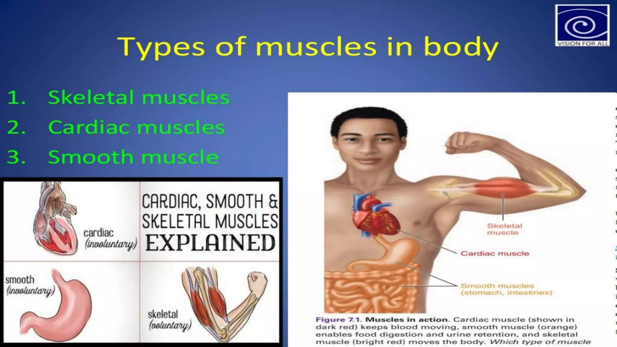
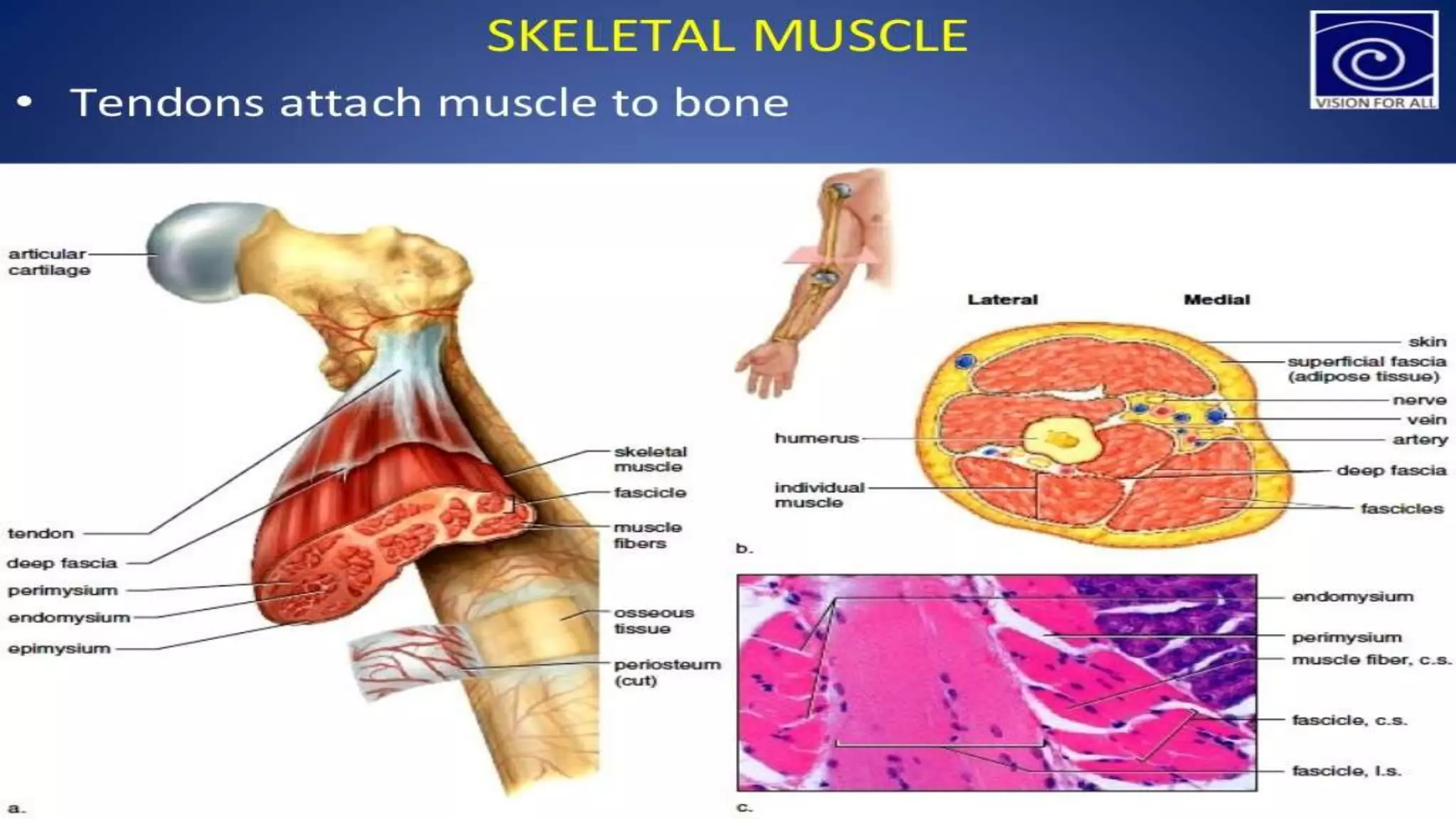

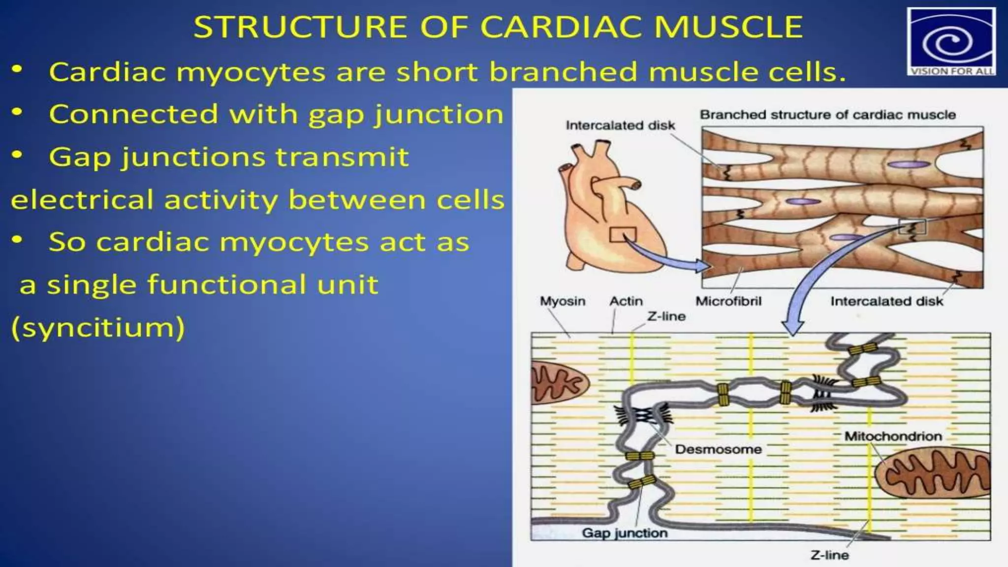
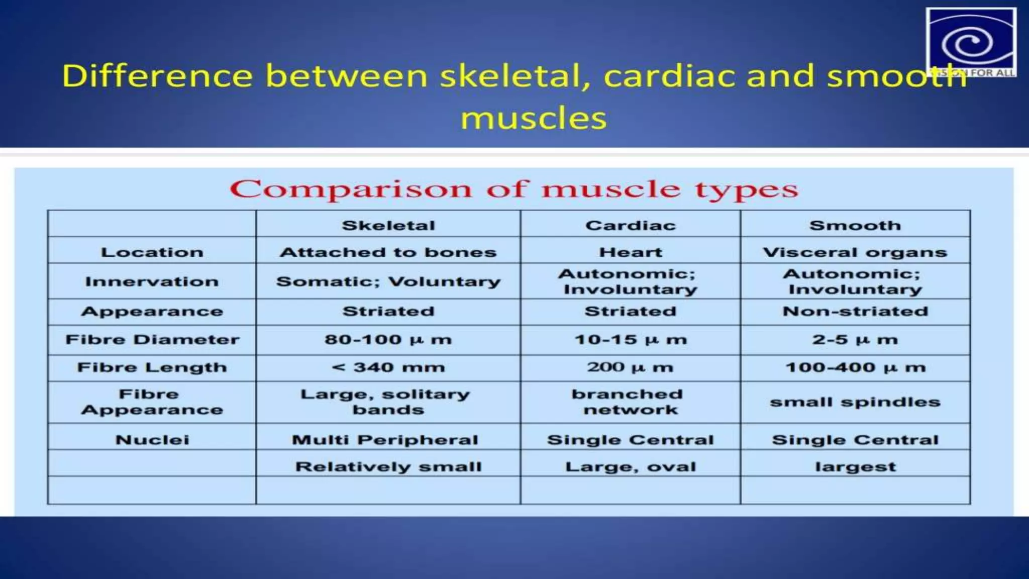


Skeletal muscle is voluntary muscle that is attached to bones and controls movement. It comprises 40-50% of body weight and there are approximately 650 muscles. Skeletal muscle contracts and relaxes in alternating fashion using ATP to generate movement. Cardiac muscle is involuntary muscle found only in the heart. It has a striated appearance and coordinates contractions to pump blood through the circulatory system. Cardiac muscle cells connect through intercalated discs to contract in a wave-like pattern. Smooth muscle is involuntary muscle found in organs and passageways like the stomach, intestines, arteries and veins. It controls involuntary functions like digestion and regulates blood flow. Smooth muscle lacks striations and responds to chemical and neural stimuli for


