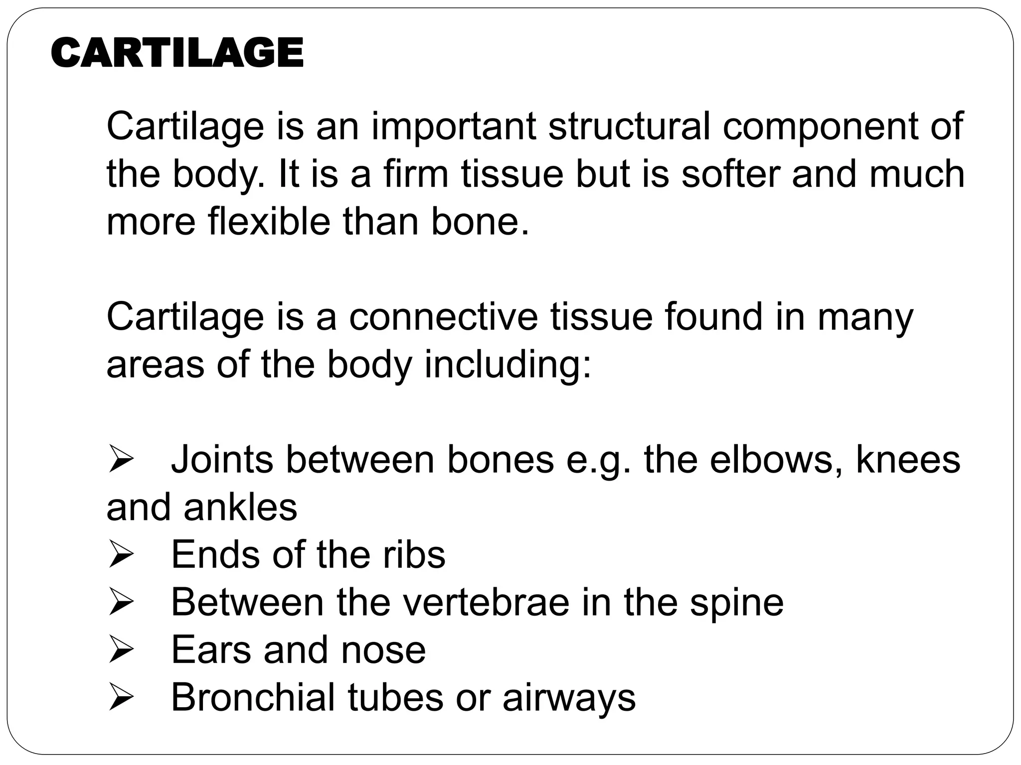
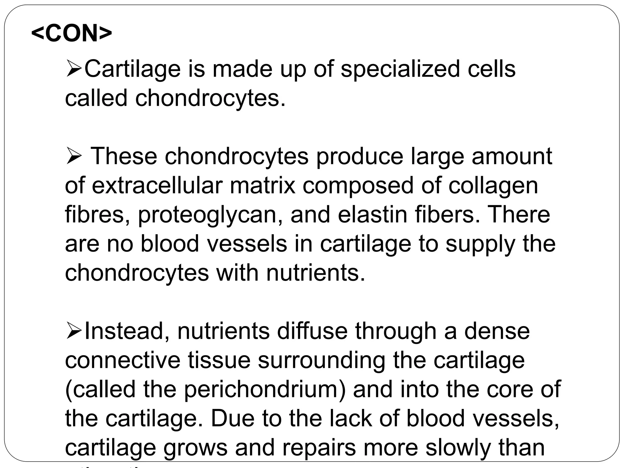
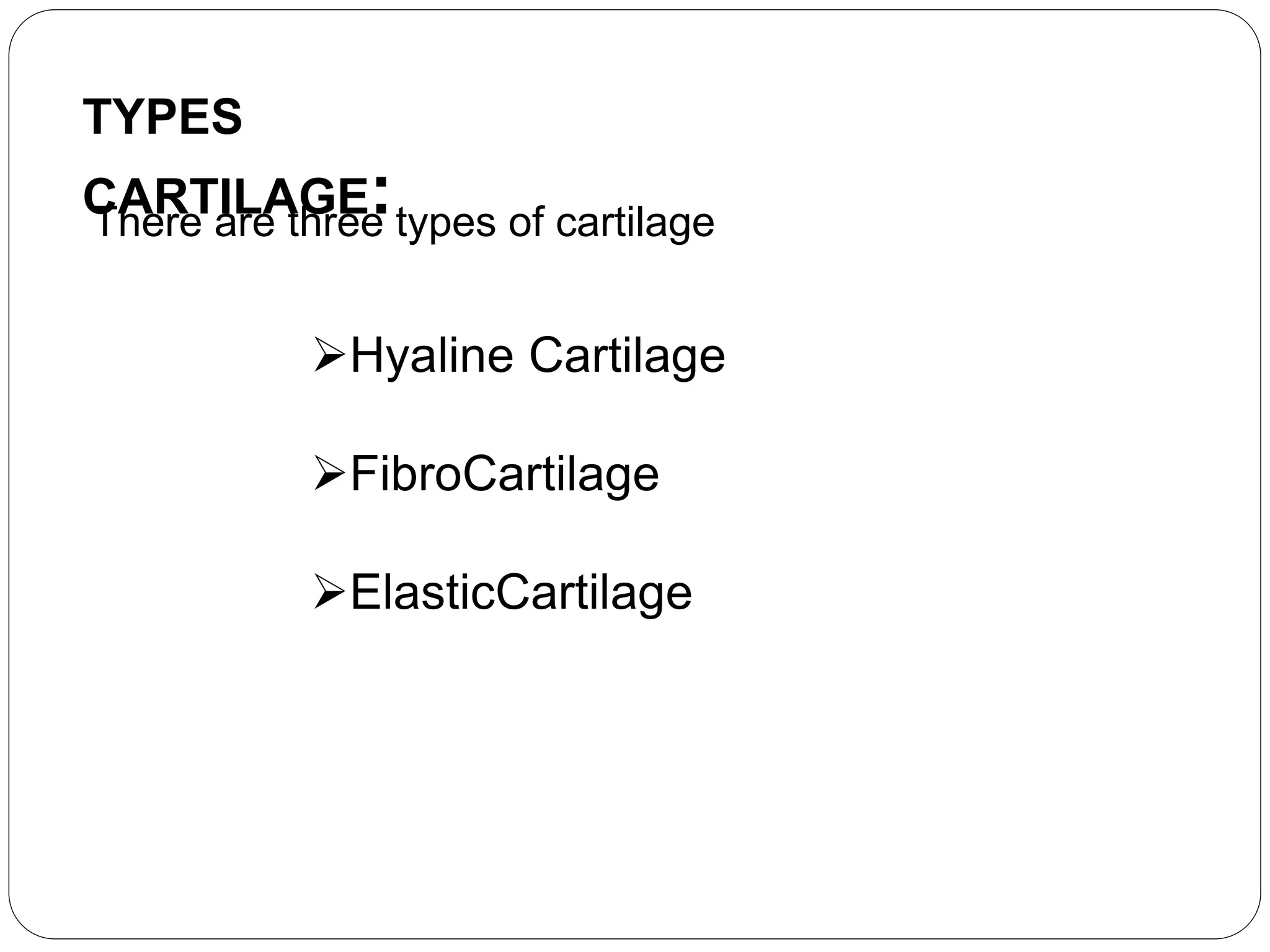
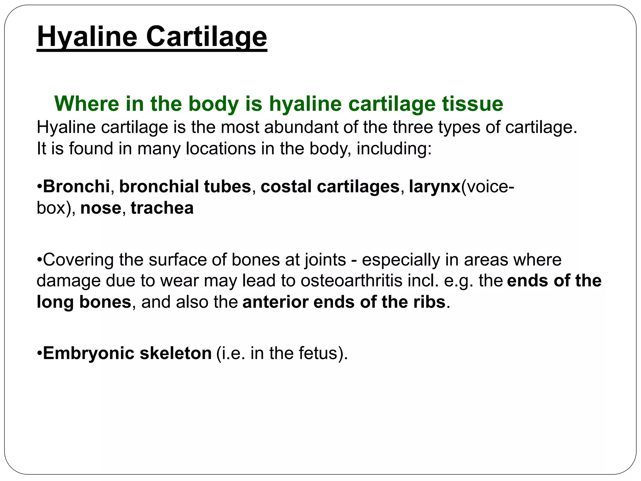
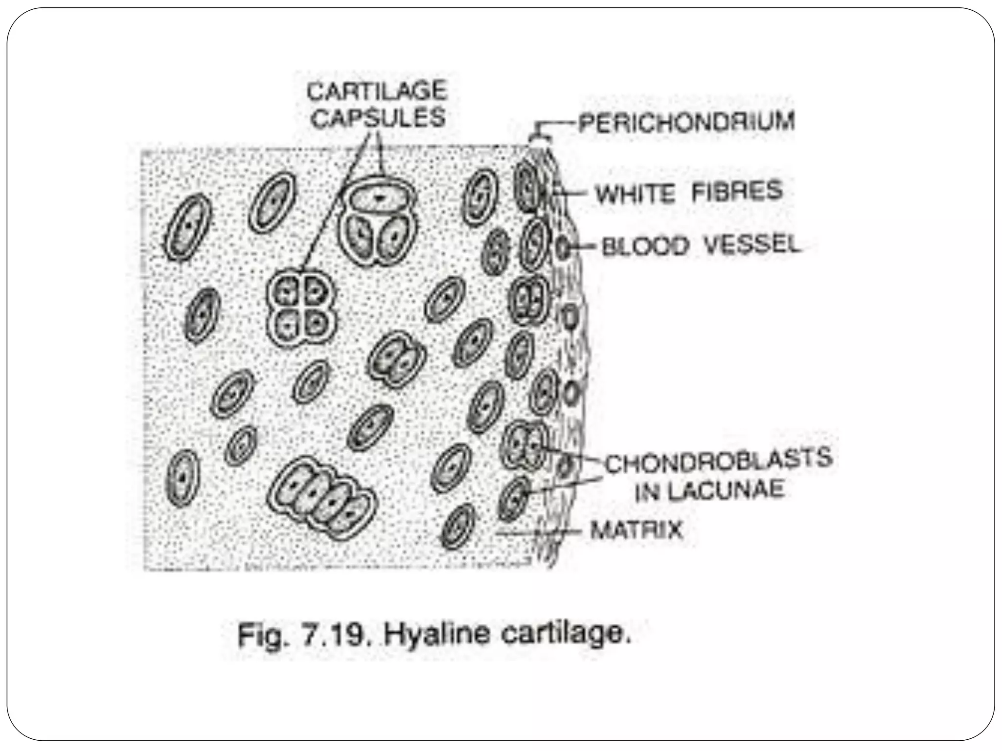
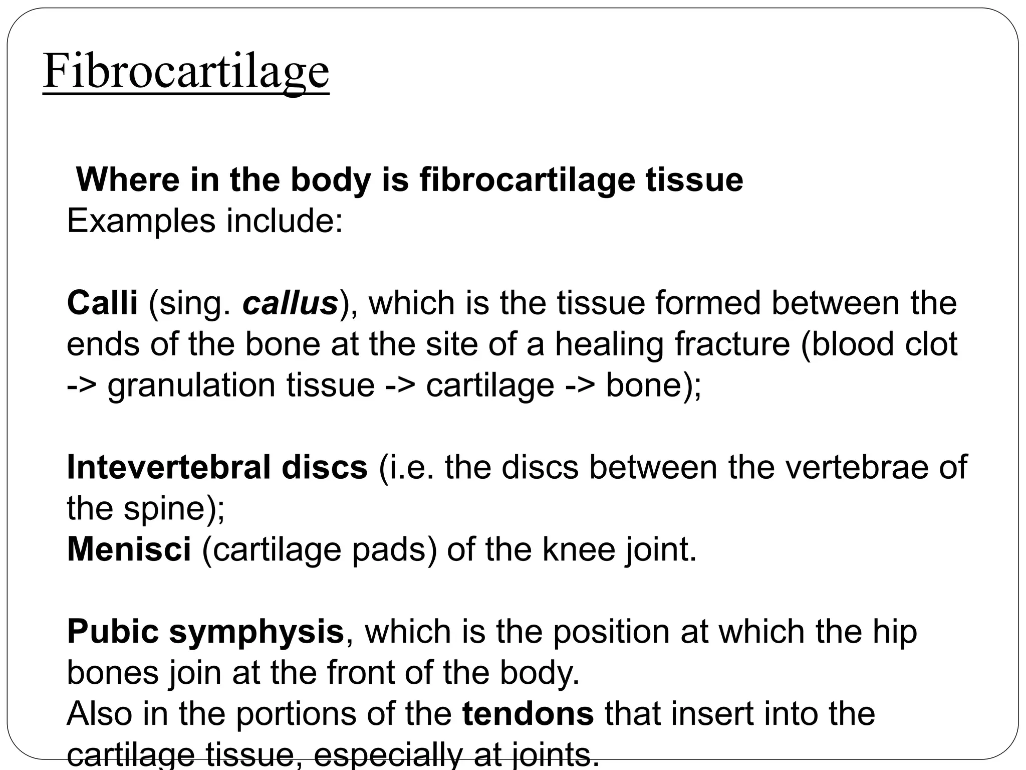
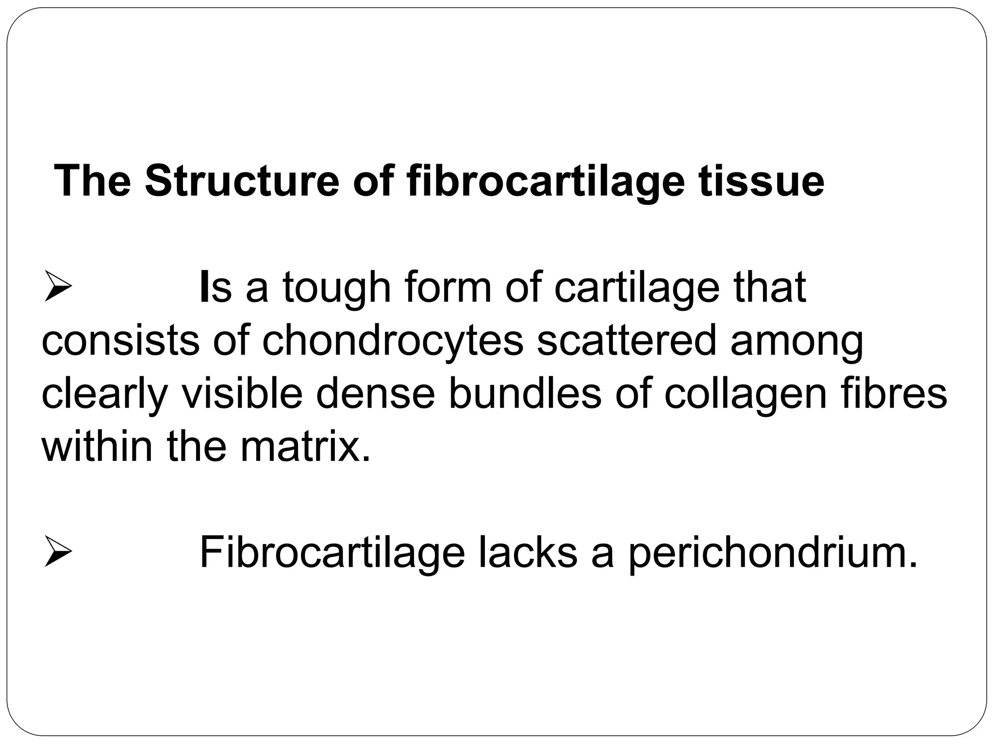
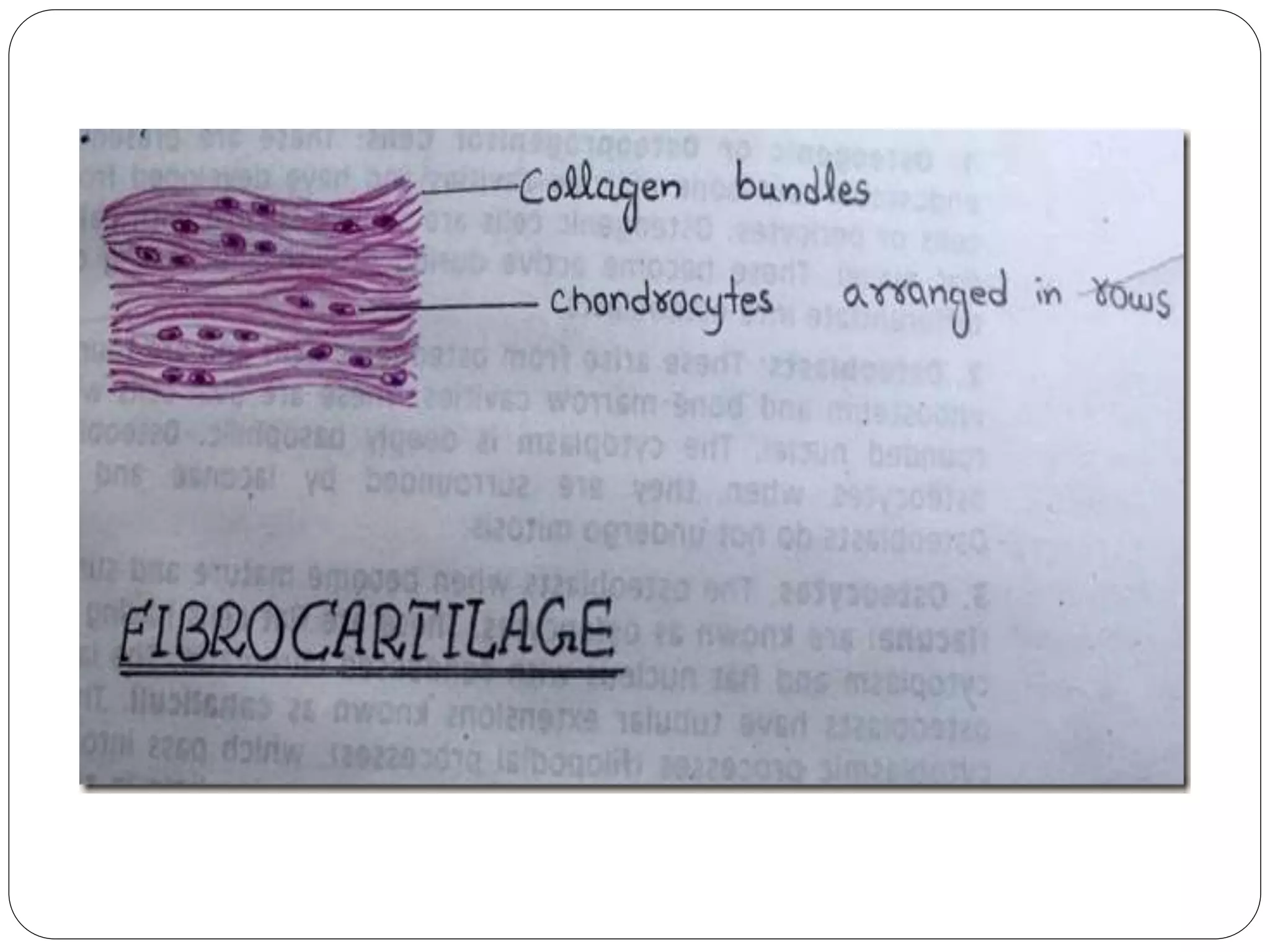
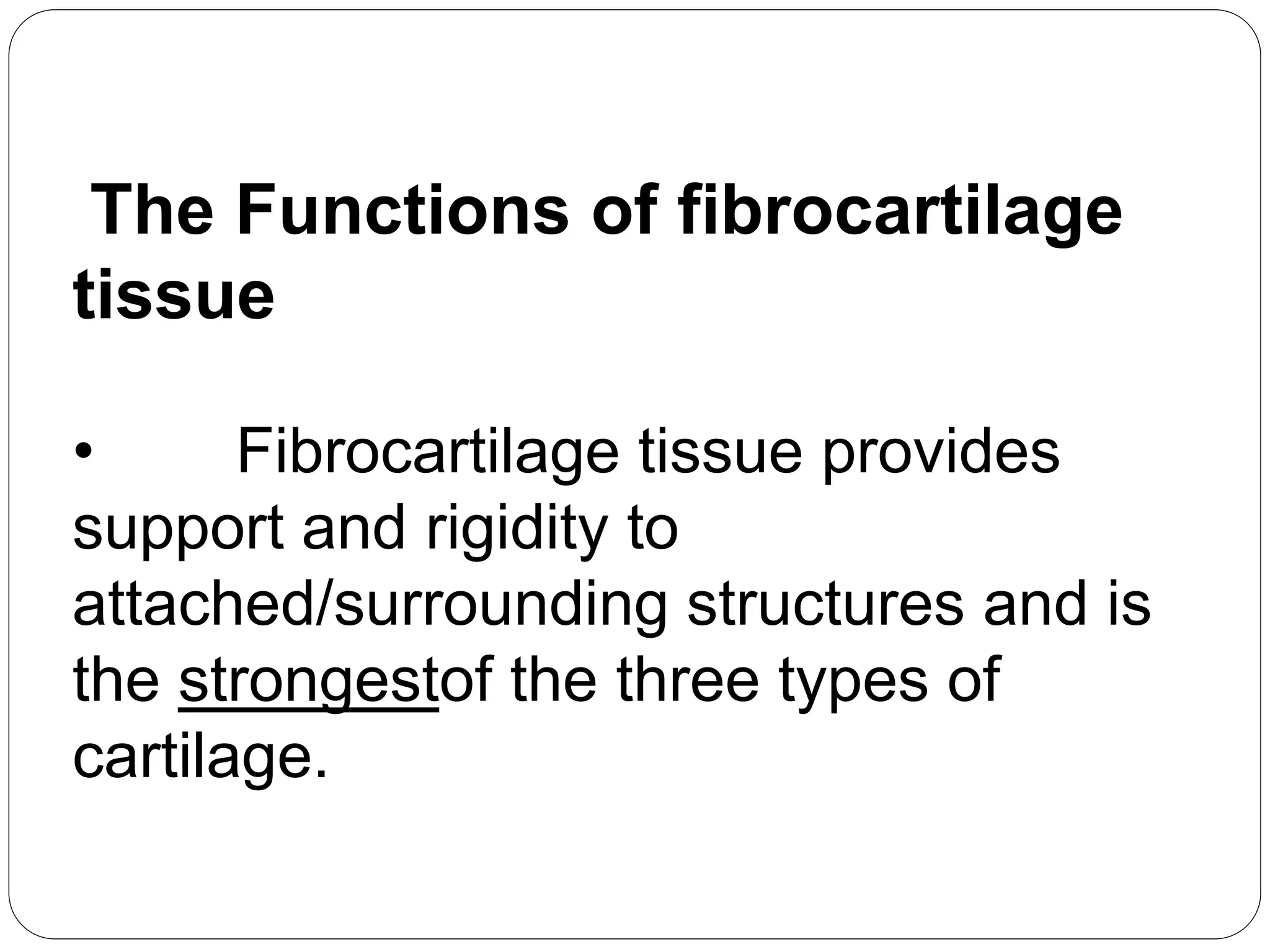
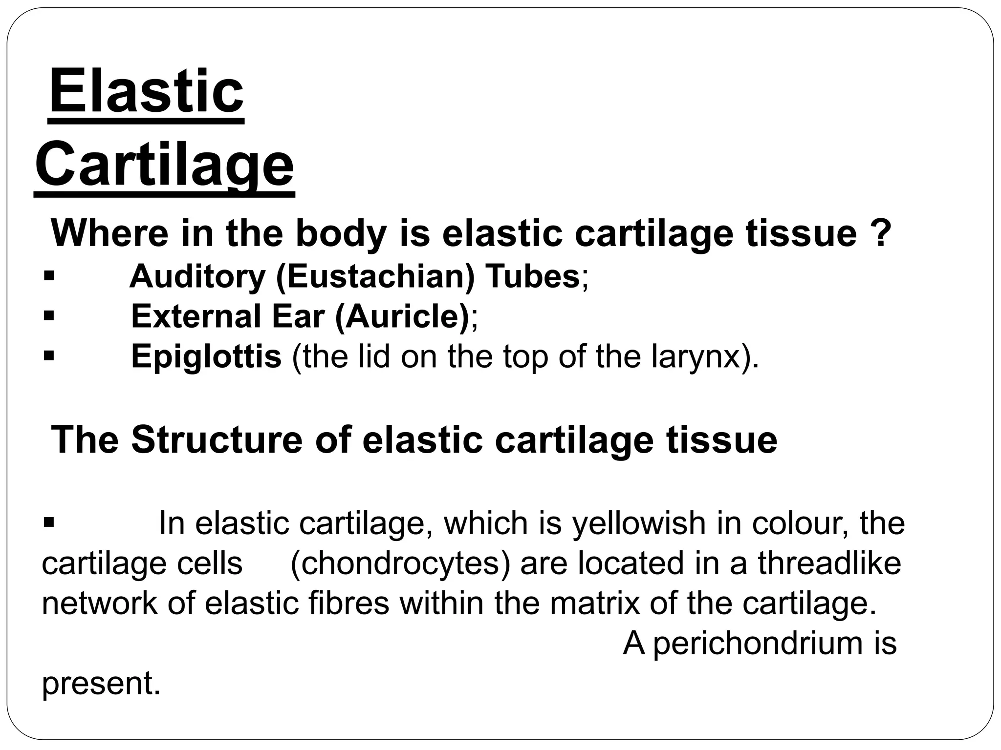
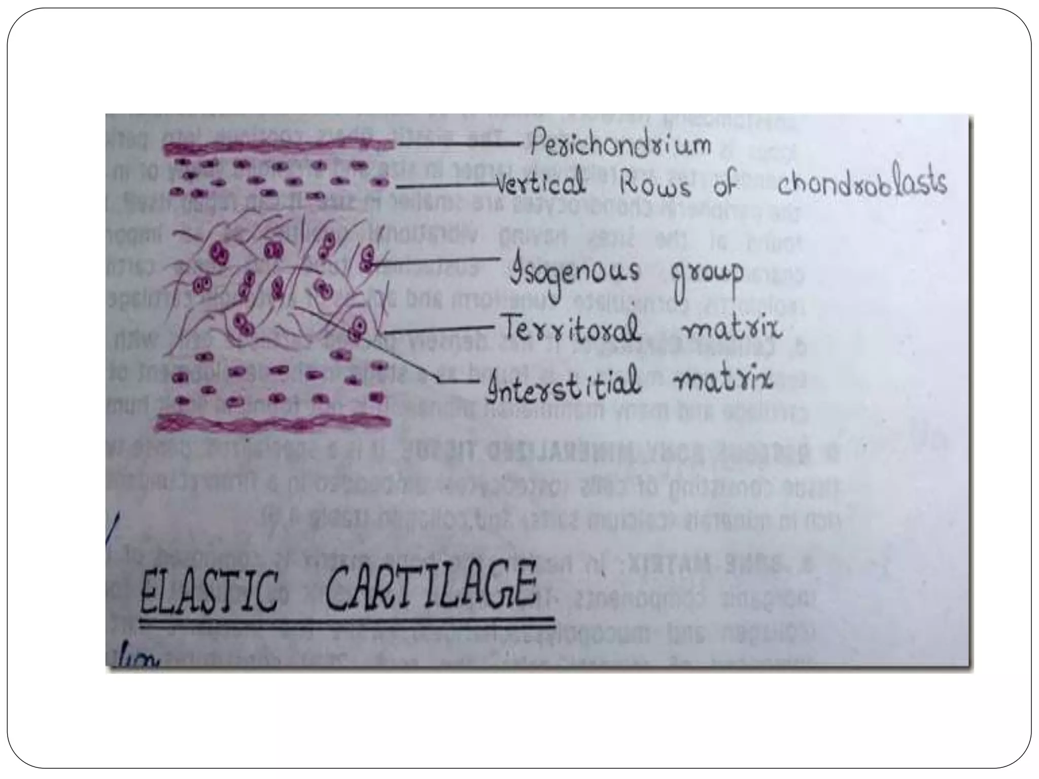
V.Nivash – 117012305040
M.Tech (INT)_Nanotechnology
TECHNICAL COMMUNICATION
CARTILAGE STRUCTURE AND
FUNCTION
CONTENT:
Define Cartilage
Types Cartilage
Hyaline cartilage structure &
function
Fibrocartilage structure &
function
Elastic cartilage structure &
function
Cartilage is an important structural component of
the body. It is a firm tissue but is softer and much
more flexible than bone.
Cartilage is a connective tissue found in many
areas of the body including:
 Joints between bones e.g. the elbows, knees
and ankles
 Ends of the ribs
 Between the vertebrae in the spine
 Ears and nose
 Bronchial tubes or airways
CARTILAGE
Cartilage is made up of specialized cells
called chondrocytes.
 These chondrocytes produce large amount
of extracellular matrix composed of collagen
fibres, proteoglycan, and elastin fibers. There
are no blood vessels in cartilage to supply the
chondrocytes with nutrients.
Instead, nutrients diffuse through a dense
connective tissue surrounding the cartilage
(called the perichondrium) and into the core of
the cartilage. Due to the lack of blood vessels,
cartilage grows and repairs more slowly than
<CON>
TYPES
CARTILAGE:There are three types of cartilage
Hyaline Cartilage
FibroCartilage
ElasticCartilage
Hyaline Cartilage
Where in the body is hyaline cartilage tissue
Hyaline cartilage is the most abundant of the three types of cartilage.
It is found in many locations in the body, including:
•Bronchi, bronchial tubes, costal cartilages, larynx(voice-
box), nose, trachea
•Covering the surface of bones at joints - especially in areas where
damage due to wear may lead to osteoarthritis incl. e.g. the ends of the
long bones, and also the anterior ends of the ribs.
•Embryonic skeleton (i.e. in the fetus).
The Structure of hyaline cartilage
tissue
 Hyaline cartilage consists of a bluish-
white, shiny ground elastic material with a
matrix of chondroitin sulphate into which many
fine collagen fibrils are embedded.
 It contains numerous chondrocytes.
The Functions of hyaline cartilage tissue
 Hyaline cartilage tissue
provides smooth surfaces, enabling tissues to
move/slide easily over each other, e.g. facilitating
smooth movements at joint.
 It is also provides flexibility and
support.
Fibrocartilage
Where in the body is fibrocartilage tissue
Examples include:
Calli (sing. callus), which is the tissue formed between the
ends of the bone at the site of a healing fracture (blood clot
-> granulation tissue -> cartilage -> bone);
Intevertebral discs (i.e. the discs between the vertebrae of
the spine);
Menisci (cartilage pads) of the knee joint.
Pubic symphysis, which is the position at which the hip
bones join at the front of the body.
Also in the portions of the tendons that insert into the
cartilage tissue, especially at joints.
The Structure of fibrocartilage tissue
 Is a tough form of cartilage that
consists of chondrocytes scattered among
clearly visible dense bundles of collagen fibres
within the matrix.
 Fibrocartilage lacks a perichondrium.
The Functions of fibrocartilage
tissue
• Fibrocartilage tissue provides
support and rigidity to
attached/surrounding structures and is
the strongestof the three types of
cartilage.
Elastic
Cartilage
Where in the body is elastic cartilage tissue ?
 Auditory (Eustachian) Tubes;
 External Ear (Auricle);
 Epiglottis (the lid on the top of the larynx).
The Structure of elastic cartilage tissue
 In elastic cartilage, which is yellowish in colour, the
cartilage cells (chondrocytes) are located in a threadlike
network of elastic fibres within the matrix of the cartilage.
A perichondrium is
present.
The Functions of elastic cartilage
tissue
 Elastic cartilages provides support to
surrounding structures and helps the define and
maintain the shape of the area in which it is
present,
e.g. the
external ear.
cartilage structure and function

cartilage structure and function

  • 1.
    V.Nivash – 117012305040 M.Tech(INT)_Nanotechnology TECHNICAL COMMUNICATION CARTILAGE STRUCTURE AND FUNCTION
  • 2.
    CONTENT: Define Cartilage Types Cartilage Hyalinecartilage structure & function Fibrocartilage structure & function Elastic cartilage structure & function
  • 3.
    Cartilage is animportant structural component of the body. It is a firm tissue but is softer and much more flexible than bone. Cartilage is a connective tissue found in many areas of the body including:  Joints between bones e.g. the elbows, knees and ankles  Ends of the ribs  Between the vertebrae in the spine  Ears and nose  Bronchial tubes or airways CARTILAGE
  • 4.
    Cartilage is madeup of specialized cells called chondrocytes.  These chondrocytes produce large amount of extracellular matrix composed of collagen fibres, proteoglycan, and elastin fibers. There are no blood vessels in cartilage to supply the chondrocytes with nutrients. Instead, nutrients diffuse through a dense connective tissue surrounding the cartilage (called the perichondrium) and into the core of the cartilage. Due to the lack of blood vessels, cartilage grows and repairs more slowly than <CON>
  • 5.
    TYPES CARTILAGE:There are threetypes of cartilage Hyaline Cartilage FibroCartilage ElasticCartilage
  • 6.
    Hyaline Cartilage Where inthe body is hyaline cartilage tissue Hyaline cartilage is the most abundant of the three types of cartilage. It is found in many locations in the body, including: •Bronchi, bronchial tubes, costal cartilages, larynx(voice- box), nose, trachea •Covering the surface of bones at joints - especially in areas where damage due to wear may lead to osteoarthritis incl. e.g. the ends of the long bones, and also the anterior ends of the ribs. •Embryonic skeleton (i.e. in the fetus).
  • 8.
    The Structure ofhyaline cartilage tissue  Hyaline cartilage consists of a bluish- white, shiny ground elastic material with a matrix of chondroitin sulphate into which many fine collagen fibrils are embedded.  It contains numerous chondrocytes.
  • 9.
    The Functions ofhyaline cartilage tissue  Hyaline cartilage tissue provides smooth surfaces, enabling tissues to move/slide easily over each other, e.g. facilitating smooth movements at joint.  It is also provides flexibility and support.
  • 10.
    Fibrocartilage Where in thebody is fibrocartilage tissue Examples include: Calli (sing. callus), which is the tissue formed between the ends of the bone at the site of a healing fracture (blood clot -> granulation tissue -> cartilage -> bone); Intevertebral discs (i.e. the discs between the vertebrae of the spine); Menisci (cartilage pads) of the knee joint. Pubic symphysis, which is the position at which the hip bones join at the front of the body. Also in the portions of the tendons that insert into the cartilage tissue, especially at joints.
  • 11.
    The Structure offibrocartilage tissue  Is a tough form of cartilage that consists of chondrocytes scattered among clearly visible dense bundles of collagen fibres within the matrix.  Fibrocartilage lacks a perichondrium.
  • 13.
    The Functions offibrocartilage tissue • Fibrocartilage tissue provides support and rigidity to attached/surrounding structures and is the strongestof the three types of cartilage.
  • 14.
    Elastic Cartilage Where in thebody is elastic cartilage tissue ?  Auditory (Eustachian) Tubes;  External Ear (Auricle);  Epiglottis (the lid on the top of the larynx). The Structure of elastic cartilage tissue  In elastic cartilage, which is yellowish in colour, the cartilage cells (chondrocytes) are located in a threadlike network of elastic fibres within the matrix of the cartilage. A perichondrium is present.
  • 16.
    The Functions ofelastic cartilage tissue  Elastic cartilages provides support to surrounding structures and helps the define and maintain the shape of the area in which it is present, e.g. the external ear.