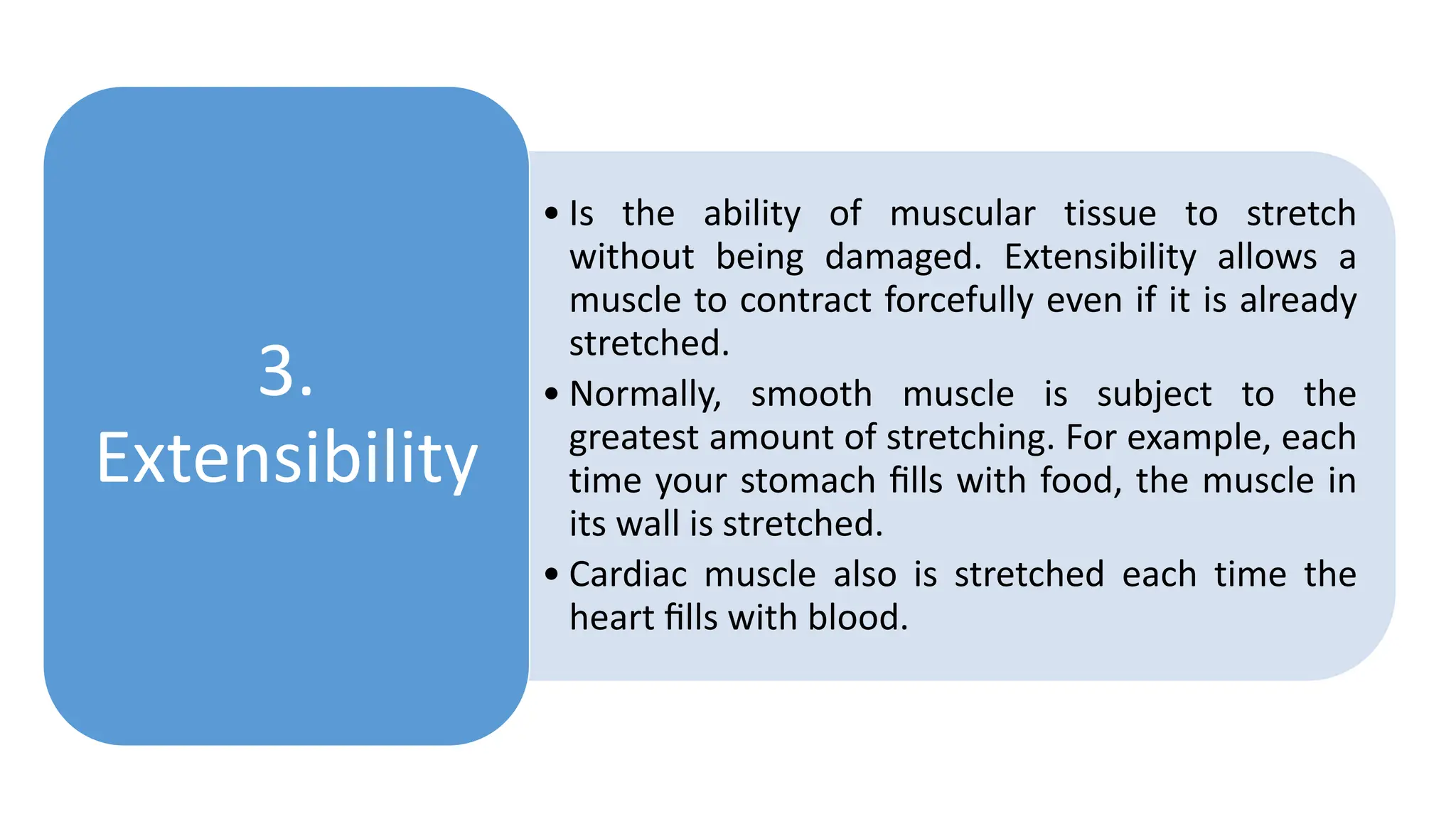
M U S C U L A R T I S S U E
• Skeletal muscle tissue is so named because
most skeletal muscles move bones of the
skeleton. (A few skeletal muscles attach to
and move the skin or other skeletal muscles.)
• Skeletal muscle tissue is striated: Alternating
light and dark bands (striations) are seen
when the tissue is examined with a
microscope.
• Skeletal muscle tissue works mainly in a
voluntary manner. Its activity can be
consciously controlled by neurons (nerve
cells) that are part of the somatic (voluntary)
division of the nervous system.
Skeletal
Muscle
• Only the heart contains cardiac muscle tissue,
which forms most of the heart wall.
• Cardiac muscle is also striated, but its action is
involuntary. The alternating contraction and
relaxation of the heart is not consciously
controlled.
• Rather, the heart beats because it has a
pacemaker that initiates each contraction. This
built-in rhythm is termed autorhythmicity.
• Several hormones and neurotransmitters can
adjust heart rate by speeding or slowing the
pacemaker
Cardiac
Muscle
• Smooth muscle tissue is located in the walls of hollow
internal structures, such as blood vessels, airways, and most
organs in the abdominopelvic cavity. It is also found in the
skin, attached to hair follicles.
• Under a microscope, this tissue lacks the striations of skeletal
and cardiac muscle tissue. For this reason, it looks
nonstriated, which is why it is referred to as smooth.
• The action of smooth muscle is usually involuntary, and some
smooth muscle tissue, such as the muscles that propel food
through your gastrointestinal tract, has autorhythmicity.
• Both cardiac muscle and smooth muscle are regulated by
neurons that are part of the autonomic (involuntary) division
of the nervous system and by hormones released by
endocrine glands.
Smooth
Muscle
•Producing body movements
•Stabilizing body positions
•Storing and moving
substances within the body.
•Generating heat
Functions
of
Muscular
Tissue
•Muscular tissue has four
special properties that
enable it to function and
contribute to homeostasis.
Properties
of
Muscular
Tissue
• A property of both muscle and nerve cells is the ability
to respond to certain stimuli by producing electrical
signals called action potentials. Action potentials can
travel along a cell’s plasma membrane due to the
presence of specific voltage-gated channels. For
muscle cells, two main types of stimuli trigger action
potentials.
• One is autorhythmic electrical signals arising in the
muscular tissue itself, as in the heart’s pacemaker.
• The other is chemical stimuli, such as
neurotransmitters released by neurons, hormones
distributed by the blood, or even local changes in pH.
1.
Electrical
excitability
• Is the ability of muscular tissue to contract
forcefully when stimulated by an action
potential.
• When a muscle contracts, it generates
tension (force of contraction) while pulling
on its attachment points.
• If the tension generated is great enough to
overcome the resistance of the object to be
moved, the muscle shortens and
movement occurs.
2.
Contractility
• Is the ability of muscular tissue to stretch
without being damaged. Extensibility allows a
muscle to contract forcefully even if it is already
stretched.
• Normally, smooth muscle is subject to the
greatest amount of stretching. For example, each
time your stomach fills with food, the muscle in
its wall is stretched.
• Cardiac muscle also is stretched each time the
heart fills with blood.
3.
Extensibility
•Is the ability of muscular
tissue to return to its
original length and
shape after contraction
or extension.
4.
Elasticity
•Each skeletal muscle is a separate organ
composed of hundreds to thousands of cells,
which are called muscle fibers because of their
elongated shapes.
•Thus, muscle cell and muscle fiber are two
terms for the same structure.
•Skeletal muscle also contains connective tissues
surrounding muscle fibers and whole muscles,
and blood vessels and nerves.
CONNECTIVE TISSUE COMPONENTS
• Connective tissue surrounds and protects muscular tissue.
• The subcutaneous layer or hypodermis, which separates muscle
from skin, is composed of areolar connective tissue and adipose
tissue.
• It provides a pathway for nerves, blood vessels, and lymphatic
vessels to enter and exit muscles.
• The adipose tissue of subcutaneous layer stores most of the body’s
triglycerides, serves as an insulating layer that reduces heat loss,
and protects muscles from physical trauma.
 Fascia (bandage) is a dense sheet or broad band of irregular connective
tissue that lines the body wall and limbs and supports and surrounds
muscles and other organs of the body.
 Fascia holds muscles with similar functions together.
 Fascia allows free movement of muscles, carries nerves, blood vessels,
and lymphatic vessels, and fills spaces between muscles.
Fascia is a thin casing of connective tissue that surrounds and holds every organ,
blood vessel, bone, nerve fiber and muscle in place. The tissue does more than
provide internal structure; fascia has nerves that make it almost as sensitive as skin
 Three layers of connective tissue extend from the fascia to protect and strengthen
skeletal muscle.
 The outermost layer, encircling the entire muscle, is the epimysium.
 Perimysium surrounds groups of 10 to 100 or more muscle fibers, separating them
into bundles called fascicles.
 Both epimysium and perimysium are dense irregular connective tissue. Penetrating
the interior of each fascicle and separating individual muscle fibers from one another
is endomysium, a thin sheath of areolar connective tissue
Areolar connective tissue holds organs in place and attaches
epithelial tissue to other underlying tissues. It also serves as a reservoir of
water and salts for surrounding tissues. Almost all cells obtain their nutrients
from and release their wastes into areolar connective tissue
 The epimysium, perimysium, and endomysium all are continuous with the connective
tissue that attaches skeletal muscle to other structures, such as bone or another
muscle.
 For example, all three connective tissue layers may extend beyond the muscle fibers
to form a tendon—a cord of dense regular connective tissue composed of parallel
bundles of collagen fibers that attach a muscle to the periosteum of a bone
A muscle belly is
basically the sum of
all the muscle fibers
in any given muscle
NERVE AND BLOOD SUPPLY
• Skeletal muscles are well supplied with nerves and blood
vessels. Generally, an artery and one or two veins accompany
each nerve that penetrates a skeletal muscle.
• The neurons that stimulate skeletal muscle to contract are
somatic motor neurons.
• Each somatic motor neuron has a threadlike axon that extends
from the brain or spinal cord to a group of skeletal muscle
fibers.
• The axon of a somatic motor neuron typically branches many
times, each branch extending to a different skeletal muscle fiber.
Terminal bouton is the specialized presynaptic terminal at the end of an
axon. Terminal boutons contain necessary organelles, proteins and molecules
needed to transmit chemical/electrical information to the postsynaptic cell.
 Microscopic blood vessels called capillaries are plentiful in muscular
tissue; each muscle fiber is in close contact with one or more capillaries .
 The blood capillaries bring in oxygen and nutrients and remove heat and
the waste products of muscle metabolism.
 Especially during contraction, a muscle fiber synthesizes and uses
considerable ATP (adenosine triphosphate).
Microscopic Anatomy of a Skeletal Muscle Fiber
• The most important components of a skeletal muscle are the muscle
fibers themselves. The diameter of a mature skeletal muscle fiber
ranges from 10 to 100 um.
• The typical length of a mature skeletal muscle fiber is about 10 cm
(4 in.), although some are as long as 30 cm (12 in.).
• Because each skeletal muscle fiber arises during embryonic
development from the fusion of a hundred or more small
mesodermal cells called myoblasts, each mature skeletal muscle
fiber has a hundred or more nuclei.
• Once fusion has occurred, the muscle fiber loses its ability to
undergo cell division.
 The dramatic muscle growth that occurs after birth occurs by hypertrophy,
an enlargement of existing muscle fibers, rather than by hyperplasia, an
increase in the number of fibers.
 During childhood, human growth hormone and other hormones stimulate
an increase in the size of skeletal muscle fibers.
 The hormone testosterone (from the testes in males and in small amounts
from other tissues, such as the ovaries, in females) promotes further
enlargement of muscle fibers.
 A few myoblasts do persist in mature skeletal muscle as satellite cells.
 These cells retain the capacity to fuse with one another or with damaged
muscle fibers to regenerate functional muscle fibers.
 However, the number of new skeletal muscle fibers formed is not enough
to compensate for significant skeletal muscle damage or degeneration.
 In such cases, skeletal muscle tissue undergoes fibrosis, the replacement of
muscle fibers by fibrous scar tissue
• The multiple nuclei of a skeletal muscle fiber are
located just beneath the sarcolemma the plasma
membrane of a muscle cell.
• Thousands of tiny invaginations of the
sarcolemma, called transverse (T) tubules, tunnel
in from the surface toward the center of each
muscle fiber.
• T tubules are open to the outside of the fiber and
thus are filled with interstitial fluid.
• Muscle action potentials travel along the
sarcolemma and through the T tubules, quickly
spreading throughout the muscle fiber
Sarcolemma,
Transverse
Tubules, and
Sarcoplasm
 Within the sarcolemma is the sarcoplasm, the cytoplasm of a muscle
fiber.
 Sarcoplasm includes a substantial amount of glycogen, which is a large
molecule composed of many glucose molecules.
 Glycogen can be used for synthesis of ATP. In addition, the sarcoplasm
contains a red-colored protein called myoglobin.
 This protein, found only in muscle, binds oxygen molecules that diffuse
into muscle fibers from interstitial fluid.
 Myoglobin releases oxygen when it is needed by the mitochondria for
ATP production
• At high magnification, the sarcoplasm
appears stuffed with little threads.
• These small structures are the myofibrils,
the contractile organelles of skeletal
muscle.
• Myofibrils are about 2 um in diameter and
extend the entire length of a muscle fiber.
Their prominent striations make the entire
skeletal muscle fiber appear striated.
Myofibrils
and
Sarcoplasmic
Reticulum
 A fluid-filled system of membranous sacs called the sarcoplasmic
reticulum or SR encircles each myofibril.
 This elaborate system is similar to smooth endoplasmic reticulum in
nonmuscular cells.
 Dilated end sacs of the sarcoplasmic reticulum called terminal cisterns
butt against the T tubule from both sides.
 A transverse tubule and the two terminal cisterns on either side of it form
a triad (tri- three).
 In a relaxed muscle fiber, the sarcoplasmic reticulum stores calcium ions
(Ca2+ ). Release of Ca2+ from the terminal cistern
• Within myofibrils are smaller structures
called filaments .
• Thin filaments are 8 nm in diameter and
1–2 um long, while thick filaments are 16
nm in diameter and 1–2 um long.
• Both thin and thick filaments are directly
involved in the contractile process.
• Overall, there are two thin filaments for
every thick filament in the regions of
filament overlap.
Filaments
and the
Sarcomere
 The filaments inside a myofibril do not extend the entire
length of a muscle fiber.
 Instead, they are arranged in compartments called
sarcomeres, which are the basic functional units of a
myofibril
 Narrow, plate-shaped regions of dense protein material
called Z discs separate one sarcomere from the next.
 Thus, a sarcomere extends from one Z disc to the next Z
disc.
 The thick and thin filaments overlap one another to a greater or lesser extent,
depending on whether the muscle is contracted, relaxed, or stretched.
 The pattern of their overlap, consisting of a variety of zones and bands, creates
the striations that can be seen both in single myofibrils and in whole muscle
fibers.
 The darker middle part of the sarcomere is the A band, which extends the
entire length of the thick filaments.
 Toward each end of the A band is a zone of overlap, where the thick and thin
filaments lie side by side. The I band is a lighter, less dense area that contains
the rest of the thin filaments but no thick filaments.
 A Z disc passes through the center of each I band. A narrow H zone in the center
of each A band contains thick but not thin filaments.
 Supporting proteins that hold the thick filaments together at the center of the H
zone form the M line, so named because it is at the middle of the sarcomere.
Muscle Proteins
• Myofibrils are built from three kinds of proteins:
• contractile proteins, which generate force during
contraction;
• regulatory proteins, which help switch the contraction
process on and off; and
• structural proteins, which keep the thick and thin filaments
in the proper alignment, give the myofibril elasticity and
extensibility, and link the myofibrils to the sarcolemma and
extracellular matrix
 The two contractile proteins in muscle are myosin and actin,
which are the main components of thick and thin filaments,
respectively.
 Myosin functions as a motor protein in all three types of
muscle tissue.
 Motor proteins push or pull various cellular structures to
achieve movement by converting the chemical energy in ATP to
the mechanical energy of motion or the production of force
 In skeletal muscle, about 300 molecules of myosin form a single thick
filament.
 Each myosin molecule is shaped like two golf clubs twisted together.
 The myosin tail (twisted golf club handles) points toward the M line in the
center of the sarcomere.
 Tails of neighboring myosin molecules lie parallel to one another, forming
the shaft of the thick filament.
 The two projections of each myosin molecule (golf club heads) are called
myosin heads.
 The heads project outward from the shaft in a spiraling fashion, each
extending toward one of the six thin filaments that surround each thick
filament.
 Thin filaments are anchored to Z disc.
 Their main component is the protein actin.
 Individual actin molecules join to form an actin filament that is twisted
into a helix.
 On each actin molecule is a myosin-binding site, where a myosin head
can attach.
 Smaller amounts of two regulatory proteins—tropomyosin and
troponin—are also part of the thin filament.
 In relaxed muscle, myosin is blocked from binding to actin because
strands of tropomyosin cover the myosin-binding sites on actin.
 The tropomyosin strands in turn are held in place by troponin molecules
• Titin is the third most plentiful protein in skeletal muscle
(after actin and myosin).
• This molecule’s name reflects its huge size. With a molecular
weight of about 3 million daltons, titin is 50 times larger than
an average sized protein. Each titin molecule spans half a
sarcomere, from a Z disc to an M line, a distance of 1 to 1.2
um in relaxed muscle.
• Each titin molecule connects a Z disc to the M line of the
sarcomere, thereby helping stabilize the position of the thick
filament.
• The part of the titin molecule that extends from the Z disc is
very elastic. Because it can stretch to at least four times its
resting length and then spring back unharmed, titin accounts
for much of the elasticity and extensibility of myofibrils.
• Titin probably helps the sarcomere return to its resting length
after a muscle has contracted or been stretched, may help
prevent overextension of sarcomeres, and maintains the
central location of the A bands.
TITIN
PROTEIN
CONSTRATION AND
RELAXATION OF
SKELETAL MUSCLE
FIBERS
• Muscle contraction occurs because myosin heads attach
to and “walk” along the thin filaments at both ends of a
sarcomere, progressively pulling the thin filaments
toward the M line .
• As a result, the thin filaments slide inward and meet at
the center of a sarcomere. They may even move so far
inward that their ends overlap.
• As the thin filaments slide inward, the Z discs come
closer together, and the sarcomere shortens.
• However, the lengths of the individual thick and thin
filaments do not change.
• Shortening of the sarcomeres causes shortening of the
whole muscle fiber, which in turn leads to shortening of
the entire muscle.
The Sliding
Filament
Mechanism.
• At the onset of contraction, the sarcoplasmic
reticulum releases calcium ions (Ca2+) into the
cytosol.
• There, they bind to troponin.
• Troponin then moves tropomyosin away from
the myosin binding sites on actin.
• Once the binding sites are “free,” the contraction
cycle—the repeating sequence of events that
causes the filaments to slide—begins.
• The contraction cycle consists of four steps
The
Contraction
Cycle
• The myosin head includes an ATP-binding site and an ATPase, an
enzyme that hydrolyzes ATP into ADP (adenosine diphosphate) and
a phosphate group.
• This hydrolysis reaction reorients and energizes the myosin head.
• Notice that the products of ATP hydrolysis—ADP and a phosphate
group—are still attached to the myosin head.
1. ATP
hydrolysis.
• The energized myosin head attaches to the myosin-binding site on
actin and releases the previously hydrolyzed phosphate group.
• When the myosin heads attach to actin during contraction, they are
referred to as crossbridges
2. Attachment
of myosin to
actin to form
crossbridges.
• After the crossbridges form, the power stroke occurs.
• During the power stroke, the site on the crossbridge where ADP
is still bound opens.
• As a result, the crossbridge rotates and releases the ADP.
• The crossbridge generates force as it rotates toward the center
of the sarcomere, sliding the thin filament past the thick
filament toward the M line.
3. Power
stroke.
• At the end of the power stroke, the crossbridge remains firmly
attached to actin until it binds another molecule of ATP.
• As ATP binds to the ATPbinding site on the myosin head, the
myosin head detaches from actin.
4.
Detachment
of myosin
from actin.
• As a muscle action potential propagates along the sarcolemma and
into the T tubules, it causes Ca2+ release channels in the SR
membrane to open.
• When these channels open, Ca2+ flows out of the SR into the
cytosol around the thick and thin filaments.
• As a result, the Ca2+ concentration in the cytosol rises tenfold or
more.
• The released calcium ions combine with troponin, causing it to
change shape.
• This conformational change moves tropomyosin away from the
myosin-binding sites on actin.
• An increase in Ca2+ concentration in the
cytosol starts muscle contraction, and a
decrease stops it.
• When a muscle fiber is relaxed, the
concentration of Ca2+ in its cytosol is
very low, only about 0.1 micromole per
liter (0.1 um/L).
• However, a huge amount of Ca2+ is
stored inside the sarcoplasmic reticulum
Excitation–
Contraction
Coupling
• The contraction cycle repeats as the myosin ATPase hydrolyzes the
newly bound molecule of ATP, and continues as long as ATP is
available and the Ca2+ level near the thin filament is sufficiently
high.
• The crossbridges keep rotating back and forth with each power
stroke, pulling the thin filaments toward the M line.
• Each of the 600 crossbridges in one thick filament attaches and
detaches about five times per second.
• At any one instant, some of the myosin heads are attached to
actin, forming crossbridges and generating force, and other myosin
heads are detached from actin, getting ready to bind again.
•Once these binding sites are free, myosin heads
bind to them to form crossbridges, and the
contraction cycle begins.
•The events just described constitute excitation–
contraction coupling, the steps that connect
excitation (a muscle action potential propagating
along the sarcolemma and into the T tubules) to
contraction (sliding of the filaments).
• The sarcoplasmic reticulum membrane also contains Ca2+
active transport pumps that use ATP to move Ca2+ constantly
from the cytosol into the SR.
• While muscle action potentials continue to propagate through
the T tubules, the Ca2+ release channels are open.
• Calcium ions flow into the cytosol more rapidly than they are
transported back by the pumps.
• After the last action potential has propagated throughout the T
tubules, the Ca2+ release channels close.
• As the pumps move Ca2+ back into the SR, the concentration of
calcium ions in the cytosol quickly decreases.
• Inside the SR, molecules of a calciumbinding protein,
appropriately called calsequestrin, bind to the Ca2+,
enabling even more Ca2+ to be sequestered or stored
within the SR.
• As a result, the concentration of Ca2+ is 10,000 times
higher in the SR than in the cytosol in a relaxed muscle
fiber.
• As the Ca2+ level in the cytosol drops, tropomyosin covers
the myosin-binding sites, and the muscle fiber relaxes
The Neuromuscular Junction
• The neurons that stimulate skeletal muscle fibers to contract are called
somatic motor neurons.
• Each somatic motor neuron has a threadlike axon that extends from the
brain or spinal cord to a group of skeletal muscle fibers.
• A muscle fiber contracts in response to one or more action potentials
propagating along its sarcolemma and through its system of T tubules.
• Muscle action potentials arise at the neuromuscular junction (NMJ), the
synapse between a somatic motor neuron and a skeletal muscle fiber.
• A synapse is a region where communication occurs between two
neurons, or between a neuron and a target cell—in this case, between a
somatic motor neuron and a muscle fiber
 A synapse is a region where communication occurs between
two neurons, or between a neuron and a target cell—in this
case, between a somatic motor neuron and a muscle fiber. At
most synapses a small gap, called the synaptic cleft, separates
the two cells.
 Because the cells do not physically touch, the action potential
cannot “jump the gap” from one cell to another.
 Instead, the first cell communicates with the second by
releasing a chemical called a neurotransmitter.
 At the NMJ, the end of the motor neuron, called the axon terminal,
divides into a cluster of synaptic end bulbs.
 Suspended in the cytosol within each synaptic end bulb are
hundreds of membrane-enclosed sacs called synaptic vesicles.
 Inside each synaptic vesicle are thousands of molecules of
acetylcholine, abbreviated ACh, the neurotransmitter released at the
NMJ
 The region of the sarcolemma opposite the synaptic end bulbs,
called the motor end plate, is the muscle fiber part of the NMJ.
 Within each motor end plate are 30 to 40 million acetylcholine
receptors, integral transmembrane proteins that bind
specifically to ACh.
 These receptors are abundant in junctional folds, deep grooves
in the motor end plate that provide a large surface area for
ACh
• Arrival of the nerve impulse at the synaptic end bulbs causes
many synaptic vesicles to undergo exocytosis.
• During exocytosis, the synaptic vesicles fuse with the motor
neuron’s plasma membrane, liberating ACh into the synaptic
cleft.
• The ACh then diffuses across the synaptic cleft between the
motor neuron and the motor end plate.
1. Release of
acetylcholine.
• Binding of two molecules of ACh to the receptor on the motor
end plate opens an ion channel in the ACh receptor.
• Once the channel is open, small cations, most importantly Na+,
can flow across the membrane.
2. Activation
of ACh
receptors.
• The inflow of Na+(down its electrochemical gradient) makes the inside of
the muscle fiber more positively charged.
• This change in the membrane potential triggers a muscle action potential.
• Each nerve impulse normally elicits one muscle action potential.
• The muscle action potential then propagates along the sarcolemma into the
T tubule system.
• This causes the sarcoplasmic reticulum to release its stored Ca2+ into the
sarcoplasm and the muscle fiber subsequently contracts.
3. Production
of muscle
action
potential.
• The effect of ACh binding lasts only briefly because ACh is rapidly broken
down by an enzyme called acetylcholinesterase (AChE).
• This enzyme is attached to collagen fibers in the extracellular matrix of the
synaptic cleft.
• AChE breaks down ACh into acetyl and choline, products that cannot
activate the ACh receptor.
4 .Termination
of ACh
activity.
 Several plant products and drugs selectively block certain events
at the NMJ.
 Botulinum toxin, produced by the bacterium Clostridium
botulinum, blocks exocytosis of synaptic vesicles at the NMJ.
 As a result, ACh is not released, and muscle contraction does not
occur.
 The bacteria proliferate in improperly canned foods, and their
toxin is one of the most lethal chemicals known.
 A tiny amount can cause death by paralyzing skeletal muscles.
Breathing stops due to paralysis of respiratory muscles, including
the diaphragm.
 Yet it is also the first bacterial toxin to be used as a medicine (Botox®).
 Injections of Botox into the affected muscles can help patients who have strabismus
(crossed eyes), blepharospasm (uncontrollable blinking), or spasms of the vocal cords that
interfere with speech.
 It is also used as a cosmetic treatment to relax muscles that cause facial wrinkles and to
alleviate chronic back pain due to muscle spasms in the lumbar region.
 The plant derivative curare, a poison used by South American Indians on
arrows and blowgun darts, causes muscle paralysis by binding to and
blocking ACh receptors.
 In the presence of curare, the ion channels do not open.
 Curare-like drugs are often used during surgery to relax skeletal muscles
 A family of chemicals called anticholinesterase agents have
the property of slowing the enzymatic activity of
acetylcholinesterase, thus slowing removal of ACh from the
synaptic cleft.
 At low doses, these agents can strengthen weak muscle
contractions.
 One example is neostigmine, which is used to treat patients
with myasthenia gravis
MUSCLE METABOLISM
Muscle fibers have three ways to produce ATP:
 from creatine phosphate
 by anaerobic cellular respiration, and
 by aerobic cellular respiration.
The use of creatine phosphate for ATP production is unique to muscle fibers, but all
body cells make ATP by the reactions of anaerobic and aerobic cellular respiration.
 While muscle fibers are relaxed, they produce more ATP than they
need for resting metabolism.
 The excess ATP is used to synthesize creatine phosphate, an energy-
rich molecule that is found only in muscle fibers.
 The enzyme creatine kinase (CK) catalyzes the transfer of one of the
high-energy phosphate groups from ATP to creatine, forming creatine
phosphate and ADP.
 Creatine is a small, amino acid–like molecule that is synthesized in the
liver, kidneys, and pancreas and then transported to muscle fibers.
Creatine Phosphate
Creatine phosphate is three to six times more plentiful than
ATP in the sarcoplasm of a relaxed muscle fiber.
When contraction begins and the ADP level starts to rise, CK
catalyzes the transfer of a high-energy phosphate group from
creatine phosphate back to ADP.
This direct phosphorylation reaction quickly regenerates new
ATP molecules.
 Together, creatine phosphate and ATP provide enough
energy for muscles to contract maximally for about 15
seconds.
 Anaerobic cellular respiration is a series of ATP-producing reactions that do not
require oxygen.
 When muscle activity continues and the supply of creatine phosphate within the
muscle fiber is depleted, glucose is catabolized to generate ATP.
 Glucose easily passes from the blood into contracting muscle fibers via facilitated
diffusion, and it is also produced by the breakdown of glycogen within muscle
fibers.
 Then, a series of 10 reactions known as glycolysis quickly breaks down each
glucose molecule into two molecules of pyruvic acid.
 These reactions use two molecules of ATP but produce four, for a net gain of two
molecules of ATP.
Anaerobic Cellular Respiration
 Ordinarily, the pyruvic acid formed by glycolysis in the cytosol enters
mitochondria, where it undergoes a series of oxygen-requiring
reactions called aerobic cellular respiration (described next) that
produce a large amount of ATP.
 During some activities, however, not enough oxygen is available. In
such cases, anaerobic reactions convert most of the pyruvic acid to
lactic acid in the cytosol.
 About 80% of the lactic acid produced in this way diffuses out of the
skeletal muscle fibers into the blood. Liver cells can convert some of
the lactic acid back to glucose.
 Muscular activity that lasts longer than half a minute depends increasingly on aerobic
cellular respiration, a series of oxygen-requiring reactions that produce ATP in
mitochondria.
 If sufficient oxygen is present, pyruvic acid enters the mitochondria, where it is
completely oxidized in reactions that generate ATP, carbon dioxide, water, and heat
(Figure 10.12c).
 Although aerobic cellular respiration is slower than glycolysis, it yields much more ATP.
 Each molecule of glucose yields about 36 molecules of ATP; a typical fatty acid
molecule yields more than 100 molecules of ATP via aerobic cellular respiration.
Aerobic Cellular Respiration
• The inability of a muscle to maintain force of contraction
after prolonged activity is called muscle fatigue.
• Fatigue results mainly from changes within muscle fibers.
• Even before actual muscle fatigue occurs, a person may
have feelings of tiredness and the desire to cease
activity; this response, called central fatigue, is caused by
changes in the central nervous system (brain and spinal
cord).
• Although its exact mechanism is unknown, it may be a
protective mechanism to stop a person from exercising
before muscles become damaged. As you will see,
certain types of skeletal muscle fibers fatigue more
quickly than others
Muscle
Fatigue
 Although the precise mechanisms that cause muscle fatigue are still not clear, several
factors are thought to contribute.
 One is inadequate release of calcium ions from the SR, resulting in a decline of Ca2
concentration in the sarcoplasm.
 Depletion of creatine phosphate also is associated with fatigue, but surprisingly,
the ATP levels in fatigued muscle often are not much lower than those in resting
muscle.
 Other factors that contribute to muscle fatigue include insufficient oxygen,
depletion of glycogen and other nutrients, buildup of lactic acid and ADP, and
failure of action potentials in the motor neuron to release enough acetylcholine.
 The principal tissue in the heart wall is cardiac muscle tissue.
 Between the layers of cardiac muscle fibers, the contractile cells
of the heart, are sheets of connective tissue that contain blood
vessels, nerves, and the conduction system of the heart.
 Cardiac muscle fibers have the same arrangement of actin and
myosin and the same bands, zones, and Z discs as skeletal muscle
fibers
Cardiac Muscle Tissue
 Intercalated discs are unique to cardiac muscle fibers.
 These microscopic structures are irregular transverse thickenings of
the sarcolemma that connect the ends of cardiac muscle fibers to
one another.
 The discs contain desmosomes, which hold the fibers together, and
gap junctions, which allow muscle action potentials to spread from
one cardiac muscle fiber to another.
 Cardiac muscle tissue has an endomysium and perimysium, but lacks
an epimysium.
 In response to a single action potential, cardiac muscle tissue
remains contracted 10 to 15 times longer than skeletal muscle
tissue.
 The long contraction is due to prolonged delivery of Ca2+into the
sarcoplasm.
 In cardiac muscle fibers, Ca2+ enters the sarcoplasm both from the
sarcoplasmic reticulum and from the interstitial fluid that bathes
the fibers.
 Because the channels that allow inflow of Ca2+ from interstitial
fluid stay open for a relatively long time, a cardiac muscle
contraction lasts much longer than a skeletal muscle twitch.
 We have seen that skeletal muscle tissue contracts only when stimulated by
acetylcholine released by a nerve impulse in a motor neuron.
 In contrast, cardiac muscle tissue contracts when stimulated by its own
autorhythmic muscle fibers.
 Under normal resting conditions, cardiac muscle tissue contracts and relaxes about
75 times a minute.
 This continuous, rhythmic activity is a major physiological difference between
cardiac and skeletal muscle tissue.
 The mitochondria in cardiac muscle fibers are larger and more numerous than in
skeletal muscle fibers.
 This structural feature correctly suggests that cardiac muscle depends largely on
aerobic cellular respiration to generate ATP, and thus requires a constant supply of
oxygen.
 Cardiac muscle fibers can also use lactic acid produced by skeletal muscle fibers to
make ATP, a benefit during exercise
 Like cardiac muscle tissue, smooth muscle tissue is usually activated involuntarily.
 Of the two types of smooth muscle tissue, the more common type is visceral
(single-unit) smooth muscle tissue.
 It is found in tubular arrangements that form part of the walls of small arteries and
veins and of hollow organs such as the stomach, intestines, uterus, and urinary
bladder.
 Like cardiac muscle, visceral smooth muscle is autorhythmic.
 The fibers connect to one another by gap junctions, forming a network through
which muscle action potentials can spread.
 When a neurotransmitter, hormone, or autorhythmic signal stimulates one fiber,
the muscle action potential is transmitted to neighboring fibers, which then
contract in unison, as a single unit.
Smooth Muscle Tissue
 The second type of smooth muscle tissue, multiunit smooth muscle
tissue, consists of individual fibers, each with its own motor neuron
terminals and with few gap junctions between neighboring fibers.
 Stimulation of one visceral muscle fiber causes contraction of many
adjacent fibers, but stimulation of one multiunit fiber causes contraction
of that fiber only.
 Multiunit smooth muscle tissue is found in the walls of large arteries, in
airways to the lungs, in the arrector pili muscles that attach to hair
follicles, in the muscles of the iris that adjust pupil diameter, and in the
ciliary body that adjusts focus of the lens in the eye.
 A single relaxed smooth muscle fiber is 30–200 um long.
 It is thickest in the middle (3–8 um) and tapers at each end.
 Within each fiber is a single, oval, centrally located nucleus.
 The sarcoplasm of smooth muscle fibers contains both thick filaments
and thin filaments, in ratios between 1:10 and 1:15, but they are not
arranged in orderly sarcomeres as in striated muscle.
 Smooth muscle fibers also contain intermediate filaments. Because the
various filaments have no regular pattern of overlap, smooth muscle
fibers do not exhibit striations, causing a smooth appearance
Microscopic Anatomy of Smooth Muscle
 Smooth muscle fibers also lack transverse tubules and have only a small
amount of sarcoplasmic reticulum for storage of Ca2+.
 Although there are no transverse tubules in smooth muscle tissue,
there are small pouchlike invaginations of the plasma membrane called
caveolae that contain extracellular Ca2+ that can be used for muscular
contraction.
 In smooth muscle fibers, the thin filaments attach to structures called dense
bodies, which are functionally similar to Z discs in striated muscle fibers. Some
dense bodies are dispersed throughout the sarcoplasm; others are attached to the
sarcolemma. Bundles of intermediate filaments also attach to dense bodies and
stretch from one dense body to another.
 During contraction, the sliding filament mechanism involving thick and thin
filaments generates tension that is transmitted to intermediate filaments. These in
turn pull on the dense bodies attached to the sarcolemma, causing a lengthwise
shortening of the muscle fiber. As a smooth muscle fiber contracts, it rotates as a
corkscrew turns. The fiber twists in a helix as it contracts, and rotates in the
opposite direction as it relaxes.
 Although the principles of contraction are similar, smooth muscle
tissue exhibits some important physiological differences from cardiac
and skeletal muscle tissue.
 Contraction in a smooth muscle fiber starts more slowly and lasts
much longer than skeletal muscle fiber contraction.
 Another difference is that smooth muscle can both shorten and
stretch to a greater extent than the other muscle types.
Physiology of Smooth Muscle
An increase in the concentration of Ca2+ in the cytosol of a smooth
muscle fiber initiates contraction, just as in striated muscle.
Sarcoplasmic reticulum (the reservoir for Ca2+ in striated muscle) is
found in small amounts in smooth muscle.
Calcium ions flow into smooth muscle cytosol from both the
interstitial fluid and sarcoplasmic reticulum.
Because there are no transverse tubules in smooth muscle fibers
(there are caveolae instead), it takes longer for Ca2+ to reach the
filaments in the center of the fiber and trigger the contractile
process.
This accounts, in part, for the slow onset of contraction of smooth
muscle.
 Several mechanisms regulate contraction and relaxation of smooth muscle cells.
 In one such mechanism, a regulatory protein called calmodulin binds to Ca2+ in the
cytosol. (Recall that troponin takes this role in striated muscle fibers.)
 After binding to Ca2+, calmodulin activates an enzyme called myosin light chain
kinase.
 This enzyme uses ATP to add a phosphate group to a portion of the myosin head.
 Once the phosphate group is attached, the myosin head can bind to actin, and
contraction can occur.
 Because myosin light chain kinase works rather slowly, it contributes to the
slowness of smooth muscle contraction.
 Not only do calcium ions enter smooth muscle fibers slowly, they also move slowly
out of the muscle fiber, which delays relaxation.
 The prolonged presence of Ca2+ in the cytosol provides for smooth muscle tone, a
state of continued partial contraction.
 Smooth muscle tissue can thus sustain long-term tone, which is important in the
gastrointestinal tract, where the walls maintain a steady pressure on the contents of
the tract, and in the walls of blood vessels called arterioles, which maintain a steady
pressure on blood

luctur muscular tissue physiology .pptx

  • 1.
    M U SC U L A R T I S S U E
  • 2.
    • Skeletal muscletissue is so named because most skeletal muscles move bones of the skeleton. (A few skeletal muscles attach to and move the skin or other skeletal muscles.) • Skeletal muscle tissue is striated: Alternating light and dark bands (striations) are seen when the tissue is examined with a microscope. • Skeletal muscle tissue works mainly in a voluntary manner. Its activity can be consciously controlled by neurons (nerve cells) that are part of the somatic (voluntary) division of the nervous system. Skeletal Muscle
  • 3.
    • Only theheart contains cardiac muscle tissue, which forms most of the heart wall. • Cardiac muscle is also striated, but its action is involuntary. The alternating contraction and relaxation of the heart is not consciously controlled. • Rather, the heart beats because it has a pacemaker that initiates each contraction. This built-in rhythm is termed autorhythmicity. • Several hormones and neurotransmitters can adjust heart rate by speeding or slowing the pacemaker Cardiac Muscle
  • 4.
    • Smooth muscletissue is located in the walls of hollow internal structures, such as blood vessels, airways, and most organs in the abdominopelvic cavity. It is also found in the skin, attached to hair follicles. • Under a microscope, this tissue lacks the striations of skeletal and cardiac muscle tissue. For this reason, it looks nonstriated, which is why it is referred to as smooth. • The action of smooth muscle is usually involuntary, and some smooth muscle tissue, such as the muscles that propel food through your gastrointestinal tract, has autorhythmicity. • Both cardiac muscle and smooth muscle are regulated by neurons that are part of the autonomic (involuntary) division of the nervous system and by hormones released by endocrine glands. Smooth Muscle
  • 6.
    •Producing body movements •Stabilizingbody positions •Storing and moving substances within the body. •Generating heat Functions of Muscular Tissue
  • 7.
    •Muscular tissue hasfour special properties that enable it to function and contribute to homeostasis. Properties of Muscular Tissue
  • 8.
    • A propertyof both muscle and nerve cells is the ability to respond to certain stimuli by producing electrical signals called action potentials. Action potentials can travel along a cell’s plasma membrane due to the presence of specific voltage-gated channels. For muscle cells, two main types of stimuli trigger action potentials. • One is autorhythmic electrical signals arising in the muscular tissue itself, as in the heart’s pacemaker. • The other is chemical stimuli, such as neurotransmitters released by neurons, hormones distributed by the blood, or even local changes in pH. 1. Electrical excitability
  • 9.
    • Is theability of muscular tissue to contract forcefully when stimulated by an action potential. • When a muscle contracts, it generates tension (force of contraction) while pulling on its attachment points. • If the tension generated is great enough to overcome the resistance of the object to be moved, the muscle shortens and movement occurs. 2. Contractility
  • 10.
    • Is theability of muscular tissue to stretch without being damaged. Extensibility allows a muscle to contract forcefully even if it is already stretched. • Normally, smooth muscle is subject to the greatest amount of stretching. For example, each time your stomach fills with food, the muscle in its wall is stretched. • Cardiac muscle also is stretched each time the heart fills with blood. 3. Extensibility
  • 11.
    •Is the abilityof muscular tissue to return to its original length and shape after contraction or extension. 4. Elasticity
  • 14.
    •Each skeletal muscleis a separate organ composed of hundreds to thousands of cells, which are called muscle fibers because of their elongated shapes. •Thus, muscle cell and muscle fiber are two terms for the same structure. •Skeletal muscle also contains connective tissues surrounding muscle fibers and whole muscles, and blood vessels and nerves.
  • 15.
    CONNECTIVE TISSUE COMPONENTS •Connective tissue surrounds and protects muscular tissue. • The subcutaneous layer or hypodermis, which separates muscle from skin, is composed of areolar connective tissue and adipose tissue. • It provides a pathway for nerves, blood vessels, and lymphatic vessels to enter and exit muscles. • The adipose tissue of subcutaneous layer stores most of the body’s triglycerides, serves as an insulating layer that reduces heat loss, and protects muscles from physical trauma.
  • 17.
     Fascia (bandage)is a dense sheet or broad band of irregular connective tissue that lines the body wall and limbs and supports and surrounds muscles and other organs of the body.  Fascia holds muscles with similar functions together.  Fascia allows free movement of muscles, carries nerves, blood vessels, and lymphatic vessels, and fills spaces between muscles.
  • 18.
    Fascia is athin casing of connective tissue that surrounds and holds every organ, blood vessel, bone, nerve fiber and muscle in place. The tissue does more than provide internal structure; fascia has nerves that make it almost as sensitive as skin
  • 19.
     Three layersof connective tissue extend from the fascia to protect and strengthen skeletal muscle.  The outermost layer, encircling the entire muscle, is the epimysium.  Perimysium surrounds groups of 10 to 100 or more muscle fibers, separating them into bundles called fascicles.  Both epimysium and perimysium are dense irregular connective tissue. Penetrating the interior of each fascicle and separating individual muscle fibers from one another is endomysium, a thin sheath of areolar connective tissue Areolar connective tissue holds organs in place and attaches epithelial tissue to other underlying tissues. It also serves as a reservoir of water and salts for surrounding tissues. Almost all cells obtain their nutrients from and release their wastes into areolar connective tissue
  • 21.
     The epimysium,perimysium, and endomysium all are continuous with the connective tissue that attaches skeletal muscle to other structures, such as bone or another muscle.  For example, all three connective tissue layers may extend beyond the muscle fibers to form a tendon—a cord of dense regular connective tissue composed of parallel bundles of collagen fibers that attach a muscle to the periosteum of a bone
  • 22.
    A muscle bellyis basically the sum of all the muscle fibers in any given muscle
  • 23.
    NERVE AND BLOODSUPPLY • Skeletal muscles are well supplied with nerves and blood vessels. Generally, an artery and one or two veins accompany each nerve that penetrates a skeletal muscle. • The neurons that stimulate skeletal muscle to contract are somatic motor neurons. • Each somatic motor neuron has a threadlike axon that extends from the brain or spinal cord to a group of skeletal muscle fibers. • The axon of a somatic motor neuron typically branches many times, each branch extending to a different skeletal muscle fiber.
  • 24.
    Terminal bouton isthe specialized presynaptic terminal at the end of an axon. Terminal boutons contain necessary organelles, proteins and molecules needed to transmit chemical/electrical information to the postsynaptic cell.
  • 25.
     Microscopic bloodvessels called capillaries are plentiful in muscular tissue; each muscle fiber is in close contact with one or more capillaries .  The blood capillaries bring in oxygen and nutrients and remove heat and the waste products of muscle metabolism.  Especially during contraction, a muscle fiber synthesizes and uses considerable ATP (adenosine triphosphate).
  • 26.
    Microscopic Anatomy ofa Skeletal Muscle Fiber • The most important components of a skeletal muscle are the muscle fibers themselves. The diameter of a mature skeletal muscle fiber ranges from 10 to 100 um. • The typical length of a mature skeletal muscle fiber is about 10 cm (4 in.), although some are as long as 30 cm (12 in.). • Because each skeletal muscle fiber arises during embryonic development from the fusion of a hundred or more small mesodermal cells called myoblasts, each mature skeletal muscle fiber has a hundred or more nuclei. • Once fusion has occurred, the muscle fiber loses its ability to undergo cell division.
  • 27.
     The dramaticmuscle growth that occurs after birth occurs by hypertrophy, an enlargement of existing muscle fibers, rather than by hyperplasia, an increase in the number of fibers.  During childhood, human growth hormone and other hormones stimulate an increase in the size of skeletal muscle fibers.  The hormone testosterone (from the testes in males and in small amounts from other tissues, such as the ovaries, in females) promotes further enlargement of muscle fibers.  A few myoblasts do persist in mature skeletal muscle as satellite cells.  These cells retain the capacity to fuse with one another or with damaged muscle fibers to regenerate functional muscle fibers.  However, the number of new skeletal muscle fibers formed is not enough to compensate for significant skeletal muscle damage or degeneration.  In such cases, skeletal muscle tissue undergoes fibrosis, the replacement of muscle fibers by fibrous scar tissue
  • 30.
    • The multiplenuclei of a skeletal muscle fiber are located just beneath the sarcolemma the plasma membrane of a muscle cell. • Thousands of tiny invaginations of the sarcolemma, called transverse (T) tubules, tunnel in from the surface toward the center of each muscle fiber. • T tubules are open to the outside of the fiber and thus are filled with interstitial fluid. • Muscle action potentials travel along the sarcolemma and through the T tubules, quickly spreading throughout the muscle fiber Sarcolemma, Transverse Tubules, and Sarcoplasm
  • 33.
     Within thesarcolemma is the sarcoplasm, the cytoplasm of a muscle fiber.  Sarcoplasm includes a substantial amount of glycogen, which is a large molecule composed of many glucose molecules.  Glycogen can be used for synthesis of ATP. In addition, the sarcoplasm contains a red-colored protein called myoglobin.  This protein, found only in muscle, binds oxygen molecules that diffuse into muscle fibers from interstitial fluid.  Myoglobin releases oxygen when it is needed by the mitochondria for ATP production
  • 34.
    • At highmagnification, the sarcoplasm appears stuffed with little threads. • These small structures are the myofibrils, the contractile organelles of skeletal muscle. • Myofibrils are about 2 um in diameter and extend the entire length of a muscle fiber. Their prominent striations make the entire skeletal muscle fiber appear striated. Myofibrils and Sarcoplasmic Reticulum
  • 35.
     A fluid-filledsystem of membranous sacs called the sarcoplasmic reticulum or SR encircles each myofibril.  This elaborate system is similar to smooth endoplasmic reticulum in nonmuscular cells.  Dilated end sacs of the sarcoplasmic reticulum called terminal cisterns butt against the T tubule from both sides.  A transverse tubule and the two terminal cisterns on either side of it form a triad (tri- three).  In a relaxed muscle fiber, the sarcoplasmic reticulum stores calcium ions (Ca2+ ). Release of Ca2+ from the terminal cistern
  • 38.
    • Within myofibrilsare smaller structures called filaments . • Thin filaments are 8 nm in diameter and 1–2 um long, while thick filaments are 16 nm in diameter and 1–2 um long. • Both thin and thick filaments are directly involved in the contractile process. • Overall, there are two thin filaments for every thick filament in the regions of filament overlap. Filaments and the Sarcomere
  • 39.
     The filamentsinside a myofibril do not extend the entire length of a muscle fiber.  Instead, they are arranged in compartments called sarcomeres, which are the basic functional units of a myofibril  Narrow, plate-shaped regions of dense protein material called Z discs separate one sarcomere from the next.  Thus, a sarcomere extends from one Z disc to the next Z disc.
  • 41.
     The thickand thin filaments overlap one another to a greater or lesser extent, depending on whether the muscle is contracted, relaxed, or stretched.  The pattern of their overlap, consisting of a variety of zones and bands, creates the striations that can be seen both in single myofibrils and in whole muscle fibers.  The darker middle part of the sarcomere is the A band, which extends the entire length of the thick filaments.  Toward each end of the A band is a zone of overlap, where the thick and thin filaments lie side by side. The I band is a lighter, less dense area that contains the rest of the thin filaments but no thick filaments.  A Z disc passes through the center of each I band. A narrow H zone in the center of each A band contains thick but not thin filaments.  Supporting proteins that hold the thick filaments together at the center of the H zone form the M line, so named because it is at the middle of the sarcomere.
  • 43.
    Muscle Proteins • Myofibrilsare built from three kinds of proteins: • contractile proteins, which generate force during contraction; • regulatory proteins, which help switch the contraction process on and off; and • structural proteins, which keep the thick and thin filaments in the proper alignment, give the myofibril elasticity and extensibility, and link the myofibrils to the sarcolemma and extracellular matrix
  • 44.
     The twocontractile proteins in muscle are myosin and actin, which are the main components of thick and thin filaments, respectively.  Myosin functions as a motor protein in all three types of muscle tissue.  Motor proteins push or pull various cellular structures to achieve movement by converting the chemical energy in ATP to the mechanical energy of motion or the production of force
  • 45.
     In skeletalmuscle, about 300 molecules of myosin form a single thick filament.  Each myosin molecule is shaped like two golf clubs twisted together.  The myosin tail (twisted golf club handles) points toward the M line in the center of the sarcomere.  Tails of neighboring myosin molecules lie parallel to one another, forming the shaft of the thick filament.  The two projections of each myosin molecule (golf club heads) are called myosin heads.  The heads project outward from the shaft in a spiraling fashion, each extending toward one of the six thin filaments that surround each thick filament.
  • 48.
     Thin filamentsare anchored to Z disc.  Their main component is the protein actin.  Individual actin molecules join to form an actin filament that is twisted into a helix.  On each actin molecule is a myosin-binding site, where a myosin head can attach.  Smaller amounts of two regulatory proteins—tropomyosin and troponin—are also part of the thin filament.  In relaxed muscle, myosin is blocked from binding to actin because strands of tropomyosin cover the myosin-binding sites on actin.  The tropomyosin strands in turn are held in place by troponin molecules
  • 50.
    • Titin isthe third most plentiful protein in skeletal muscle (after actin and myosin). • This molecule’s name reflects its huge size. With a molecular weight of about 3 million daltons, titin is 50 times larger than an average sized protein. Each titin molecule spans half a sarcomere, from a Z disc to an M line, a distance of 1 to 1.2 um in relaxed muscle. • Each titin molecule connects a Z disc to the M line of the sarcomere, thereby helping stabilize the position of the thick filament. • The part of the titin molecule that extends from the Z disc is very elastic. Because it can stretch to at least four times its resting length and then spring back unharmed, titin accounts for much of the elasticity and extensibility of myofibrils. • Titin probably helps the sarcomere return to its resting length after a muscle has contracted or been stretched, may help prevent overextension of sarcomeres, and maintains the central location of the A bands. TITIN PROTEIN
  • 54.
  • 55.
    • Muscle contractionoccurs because myosin heads attach to and “walk” along the thin filaments at both ends of a sarcomere, progressively pulling the thin filaments toward the M line . • As a result, the thin filaments slide inward and meet at the center of a sarcomere. They may even move so far inward that their ends overlap. • As the thin filaments slide inward, the Z discs come closer together, and the sarcomere shortens. • However, the lengths of the individual thick and thin filaments do not change. • Shortening of the sarcomeres causes shortening of the whole muscle fiber, which in turn leads to shortening of the entire muscle. The Sliding Filament Mechanism.
  • 56.
    • At theonset of contraction, the sarcoplasmic reticulum releases calcium ions (Ca2+) into the cytosol. • There, they bind to troponin. • Troponin then moves tropomyosin away from the myosin binding sites on actin. • Once the binding sites are “free,” the contraction cycle—the repeating sequence of events that causes the filaments to slide—begins. • The contraction cycle consists of four steps The Contraction Cycle
  • 57.
    • The myosinhead includes an ATP-binding site and an ATPase, an enzyme that hydrolyzes ATP into ADP (adenosine diphosphate) and a phosphate group. • This hydrolysis reaction reorients and energizes the myosin head. • Notice that the products of ATP hydrolysis—ADP and a phosphate group—are still attached to the myosin head. 1. ATP hydrolysis. • The energized myosin head attaches to the myosin-binding site on actin and releases the previously hydrolyzed phosphate group. • When the myosin heads attach to actin during contraction, they are referred to as crossbridges 2. Attachment of myosin to actin to form crossbridges.
  • 58.
    • After thecrossbridges form, the power stroke occurs. • During the power stroke, the site on the crossbridge where ADP is still bound opens. • As a result, the crossbridge rotates and releases the ADP. • The crossbridge generates force as it rotates toward the center of the sarcomere, sliding the thin filament past the thick filament toward the M line. 3. Power stroke. • At the end of the power stroke, the crossbridge remains firmly attached to actin until it binds another molecule of ATP. • As ATP binds to the ATPbinding site on the myosin head, the myosin head detaches from actin. 4. Detachment of myosin from actin.
  • 59.
    • As amuscle action potential propagates along the sarcolemma and into the T tubules, it causes Ca2+ release channels in the SR membrane to open. • When these channels open, Ca2+ flows out of the SR into the cytosol around the thick and thin filaments. • As a result, the Ca2+ concentration in the cytosol rises tenfold or more. • The released calcium ions combine with troponin, causing it to change shape. • This conformational change moves tropomyosin away from the myosin-binding sites on actin.
  • 62.
    • An increasein Ca2+ concentration in the cytosol starts muscle contraction, and a decrease stops it. • When a muscle fiber is relaxed, the concentration of Ca2+ in its cytosol is very low, only about 0.1 micromole per liter (0.1 um/L). • However, a huge amount of Ca2+ is stored inside the sarcoplasmic reticulum Excitation– Contraction Coupling
  • 63.
    • The contractioncycle repeats as the myosin ATPase hydrolyzes the newly bound molecule of ATP, and continues as long as ATP is available and the Ca2+ level near the thin filament is sufficiently high. • The crossbridges keep rotating back and forth with each power stroke, pulling the thin filaments toward the M line. • Each of the 600 crossbridges in one thick filament attaches and detaches about five times per second. • At any one instant, some of the myosin heads are attached to actin, forming crossbridges and generating force, and other myosin heads are detached from actin, getting ready to bind again.
  • 64.
    •Once these bindingsites are free, myosin heads bind to them to form crossbridges, and the contraction cycle begins. •The events just described constitute excitation– contraction coupling, the steps that connect excitation (a muscle action potential propagating along the sarcolemma and into the T tubules) to contraction (sliding of the filaments).
  • 65.
    • The sarcoplasmicreticulum membrane also contains Ca2+ active transport pumps that use ATP to move Ca2+ constantly from the cytosol into the SR. • While muscle action potentials continue to propagate through the T tubules, the Ca2+ release channels are open. • Calcium ions flow into the cytosol more rapidly than they are transported back by the pumps. • After the last action potential has propagated throughout the T tubules, the Ca2+ release channels close. • As the pumps move Ca2+ back into the SR, the concentration of calcium ions in the cytosol quickly decreases.
  • 66.
    • Inside theSR, molecules of a calciumbinding protein, appropriately called calsequestrin, bind to the Ca2+, enabling even more Ca2+ to be sequestered or stored within the SR. • As a result, the concentration of Ca2+ is 10,000 times higher in the SR than in the cytosol in a relaxed muscle fiber. • As the Ca2+ level in the cytosol drops, tropomyosin covers the myosin-binding sites, and the muscle fiber relaxes
  • 69.
    The Neuromuscular Junction •The neurons that stimulate skeletal muscle fibers to contract are called somatic motor neurons. • Each somatic motor neuron has a threadlike axon that extends from the brain or spinal cord to a group of skeletal muscle fibers. • A muscle fiber contracts in response to one or more action potentials propagating along its sarcolemma and through its system of T tubules. • Muscle action potentials arise at the neuromuscular junction (NMJ), the synapse between a somatic motor neuron and a skeletal muscle fiber. • A synapse is a region where communication occurs between two neurons, or between a neuron and a target cell—in this case, between a somatic motor neuron and a muscle fiber
  • 70.
     A synapseis a region where communication occurs between two neurons, or between a neuron and a target cell—in this case, between a somatic motor neuron and a muscle fiber. At most synapses a small gap, called the synaptic cleft, separates the two cells.  Because the cells do not physically touch, the action potential cannot “jump the gap” from one cell to another.  Instead, the first cell communicates with the second by releasing a chemical called a neurotransmitter.
  • 72.
     At theNMJ, the end of the motor neuron, called the axon terminal, divides into a cluster of synaptic end bulbs.  Suspended in the cytosol within each synaptic end bulb are hundreds of membrane-enclosed sacs called synaptic vesicles.  Inside each synaptic vesicle are thousands of molecules of acetylcholine, abbreviated ACh, the neurotransmitter released at the NMJ
  • 73.
     The regionof the sarcolemma opposite the synaptic end bulbs, called the motor end plate, is the muscle fiber part of the NMJ.  Within each motor end plate are 30 to 40 million acetylcholine receptors, integral transmembrane proteins that bind specifically to ACh.  These receptors are abundant in junctional folds, deep grooves in the motor end plate that provide a large surface area for ACh
  • 74.
    • Arrival ofthe nerve impulse at the synaptic end bulbs causes many synaptic vesicles to undergo exocytosis. • During exocytosis, the synaptic vesicles fuse with the motor neuron’s plasma membrane, liberating ACh into the synaptic cleft. • The ACh then diffuses across the synaptic cleft between the motor neuron and the motor end plate. 1. Release of acetylcholine. • Binding of two molecules of ACh to the receptor on the motor end plate opens an ion channel in the ACh receptor. • Once the channel is open, small cations, most importantly Na+, can flow across the membrane. 2. Activation of ACh receptors.
  • 75.
    • The inflowof Na+(down its electrochemical gradient) makes the inside of the muscle fiber more positively charged. • This change in the membrane potential triggers a muscle action potential. • Each nerve impulse normally elicits one muscle action potential. • The muscle action potential then propagates along the sarcolemma into the T tubule system. • This causes the sarcoplasmic reticulum to release its stored Ca2+ into the sarcoplasm and the muscle fiber subsequently contracts. 3. Production of muscle action potential. • The effect of ACh binding lasts only briefly because ACh is rapidly broken down by an enzyme called acetylcholinesterase (AChE). • This enzyme is attached to collagen fibers in the extracellular matrix of the synaptic cleft. • AChE breaks down ACh into acetyl and choline, products that cannot activate the ACh receptor. 4 .Termination of ACh activity.
  • 78.
     Several plantproducts and drugs selectively block certain events at the NMJ.  Botulinum toxin, produced by the bacterium Clostridium botulinum, blocks exocytosis of synaptic vesicles at the NMJ.  As a result, ACh is not released, and muscle contraction does not occur.  The bacteria proliferate in improperly canned foods, and their toxin is one of the most lethal chemicals known.  A tiny amount can cause death by paralyzing skeletal muscles. Breathing stops due to paralysis of respiratory muscles, including the diaphragm.
  • 79.
     Yet itis also the first bacterial toxin to be used as a medicine (Botox®).  Injections of Botox into the affected muscles can help patients who have strabismus (crossed eyes), blepharospasm (uncontrollable blinking), or spasms of the vocal cords that interfere with speech.  It is also used as a cosmetic treatment to relax muscles that cause facial wrinkles and to alleviate chronic back pain due to muscle spasms in the lumbar region.
  • 80.
     The plantderivative curare, a poison used by South American Indians on arrows and blowgun darts, causes muscle paralysis by binding to and blocking ACh receptors.  In the presence of curare, the ion channels do not open.  Curare-like drugs are often used during surgery to relax skeletal muscles
  • 81.
     A familyof chemicals called anticholinesterase agents have the property of slowing the enzymatic activity of acetylcholinesterase, thus slowing removal of ACh from the synaptic cleft.  At low doses, these agents can strengthen weak muscle contractions.  One example is neostigmine, which is used to treat patients with myasthenia gravis
  • 82.
  • 83.
    Muscle fibers havethree ways to produce ATP:  from creatine phosphate  by anaerobic cellular respiration, and  by aerobic cellular respiration. The use of creatine phosphate for ATP production is unique to muscle fibers, but all body cells make ATP by the reactions of anaerobic and aerobic cellular respiration.
  • 84.
     While musclefibers are relaxed, they produce more ATP than they need for resting metabolism.  The excess ATP is used to synthesize creatine phosphate, an energy- rich molecule that is found only in muscle fibers.  The enzyme creatine kinase (CK) catalyzes the transfer of one of the high-energy phosphate groups from ATP to creatine, forming creatine phosphate and ADP.  Creatine is a small, amino acid–like molecule that is synthesized in the liver, kidneys, and pancreas and then transported to muscle fibers. Creatine Phosphate
  • 85.
    Creatine phosphate isthree to six times more plentiful than ATP in the sarcoplasm of a relaxed muscle fiber. When contraction begins and the ADP level starts to rise, CK catalyzes the transfer of a high-energy phosphate group from creatine phosphate back to ADP. This direct phosphorylation reaction quickly regenerates new ATP molecules.  Together, creatine phosphate and ATP provide enough energy for muscles to contract maximally for about 15 seconds.
  • 89.
     Anaerobic cellularrespiration is a series of ATP-producing reactions that do not require oxygen.  When muscle activity continues and the supply of creatine phosphate within the muscle fiber is depleted, glucose is catabolized to generate ATP.  Glucose easily passes from the blood into contracting muscle fibers via facilitated diffusion, and it is also produced by the breakdown of glycogen within muscle fibers.  Then, a series of 10 reactions known as glycolysis quickly breaks down each glucose molecule into two molecules of pyruvic acid.  These reactions use two molecules of ATP but produce four, for a net gain of two molecules of ATP. Anaerobic Cellular Respiration
  • 90.
     Ordinarily, thepyruvic acid formed by glycolysis in the cytosol enters mitochondria, where it undergoes a series of oxygen-requiring reactions called aerobic cellular respiration (described next) that produce a large amount of ATP.  During some activities, however, not enough oxygen is available. In such cases, anaerobic reactions convert most of the pyruvic acid to lactic acid in the cytosol.  About 80% of the lactic acid produced in this way diffuses out of the skeletal muscle fibers into the blood. Liver cells can convert some of the lactic acid back to glucose.
  • 92.
     Muscular activitythat lasts longer than half a minute depends increasingly on aerobic cellular respiration, a series of oxygen-requiring reactions that produce ATP in mitochondria.  If sufficient oxygen is present, pyruvic acid enters the mitochondria, where it is completely oxidized in reactions that generate ATP, carbon dioxide, water, and heat (Figure 10.12c).  Although aerobic cellular respiration is slower than glycolysis, it yields much more ATP.  Each molecule of glucose yields about 36 molecules of ATP; a typical fatty acid molecule yields more than 100 molecules of ATP via aerobic cellular respiration. Aerobic Cellular Respiration
  • 94.
    • The inabilityof a muscle to maintain force of contraction after prolonged activity is called muscle fatigue. • Fatigue results mainly from changes within muscle fibers. • Even before actual muscle fatigue occurs, a person may have feelings of tiredness and the desire to cease activity; this response, called central fatigue, is caused by changes in the central nervous system (brain and spinal cord). • Although its exact mechanism is unknown, it may be a protective mechanism to stop a person from exercising before muscles become damaged. As you will see, certain types of skeletal muscle fibers fatigue more quickly than others Muscle Fatigue
  • 95.
     Although theprecise mechanisms that cause muscle fatigue are still not clear, several factors are thought to contribute.  One is inadequate release of calcium ions from the SR, resulting in a decline of Ca2 concentration in the sarcoplasm.  Depletion of creatine phosphate also is associated with fatigue, but surprisingly, the ATP levels in fatigued muscle often are not much lower than those in resting muscle.  Other factors that contribute to muscle fatigue include insufficient oxygen, depletion of glycogen and other nutrients, buildup of lactic acid and ADP, and failure of action potentials in the motor neuron to release enough acetylcholine.
  • 96.
     The principaltissue in the heart wall is cardiac muscle tissue.  Between the layers of cardiac muscle fibers, the contractile cells of the heart, are sheets of connective tissue that contain blood vessels, nerves, and the conduction system of the heart.  Cardiac muscle fibers have the same arrangement of actin and myosin and the same bands, zones, and Z discs as skeletal muscle fibers Cardiac Muscle Tissue
  • 97.
     Intercalated discsare unique to cardiac muscle fibers.  These microscopic structures are irregular transverse thickenings of the sarcolemma that connect the ends of cardiac muscle fibers to one another.  The discs contain desmosomes, which hold the fibers together, and gap junctions, which allow muscle action potentials to spread from one cardiac muscle fiber to another.  Cardiac muscle tissue has an endomysium and perimysium, but lacks an epimysium.
  • 101.
     In responseto a single action potential, cardiac muscle tissue remains contracted 10 to 15 times longer than skeletal muscle tissue.  The long contraction is due to prolonged delivery of Ca2+into the sarcoplasm.  In cardiac muscle fibers, Ca2+ enters the sarcoplasm both from the sarcoplasmic reticulum and from the interstitial fluid that bathes the fibers.  Because the channels that allow inflow of Ca2+ from interstitial fluid stay open for a relatively long time, a cardiac muscle contraction lasts much longer than a skeletal muscle twitch.
  • 102.
     We haveseen that skeletal muscle tissue contracts only when stimulated by acetylcholine released by a nerve impulse in a motor neuron.  In contrast, cardiac muscle tissue contracts when stimulated by its own autorhythmic muscle fibers.  Under normal resting conditions, cardiac muscle tissue contracts and relaxes about 75 times a minute.  This continuous, rhythmic activity is a major physiological difference between cardiac and skeletal muscle tissue.  The mitochondria in cardiac muscle fibers are larger and more numerous than in skeletal muscle fibers.  This structural feature correctly suggests that cardiac muscle depends largely on aerobic cellular respiration to generate ATP, and thus requires a constant supply of oxygen.  Cardiac muscle fibers can also use lactic acid produced by skeletal muscle fibers to make ATP, a benefit during exercise
  • 103.
     Like cardiacmuscle tissue, smooth muscle tissue is usually activated involuntarily.  Of the two types of smooth muscle tissue, the more common type is visceral (single-unit) smooth muscle tissue.  It is found in tubular arrangements that form part of the walls of small arteries and veins and of hollow organs such as the stomach, intestines, uterus, and urinary bladder.  Like cardiac muscle, visceral smooth muscle is autorhythmic.  The fibers connect to one another by gap junctions, forming a network through which muscle action potentials can spread.  When a neurotransmitter, hormone, or autorhythmic signal stimulates one fiber, the muscle action potential is transmitted to neighboring fibers, which then contract in unison, as a single unit. Smooth Muscle Tissue
  • 104.
     The secondtype of smooth muscle tissue, multiunit smooth muscle tissue, consists of individual fibers, each with its own motor neuron terminals and with few gap junctions between neighboring fibers.  Stimulation of one visceral muscle fiber causes contraction of many adjacent fibers, but stimulation of one multiunit fiber causes contraction of that fiber only.  Multiunit smooth muscle tissue is found in the walls of large arteries, in airways to the lungs, in the arrector pili muscles that attach to hair follicles, in the muscles of the iris that adjust pupil diameter, and in the ciliary body that adjusts focus of the lens in the eye.
  • 107.
     A singlerelaxed smooth muscle fiber is 30–200 um long.  It is thickest in the middle (3–8 um) and tapers at each end.  Within each fiber is a single, oval, centrally located nucleus.  The sarcoplasm of smooth muscle fibers contains both thick filaments and thin filaments, in ratios between 1:10 and 1:15, but they are not arranged in orderly sarcomeres as in striated muscle.  Smooth muscle fibers also contain intermediate filaments. Because the various filaments have no regular pattern of overlap, smooth muscle fibers do not exhibit striations, causing a smooth appearance Microscopic Anatomy of Smooth Muscle
  • 108.
     Smooth musclefibers also lack transverse tubules and have only a small amount of sarcoplasmic reticulum for storage of Ca2+.  Although there are no transverse tubules in smooth muscle tissue, there are small pouchlike invaginations of the plasma membrane called caveolae that contain extracellular Ca2+ that can be used for muscular contraction.
  • 109.
     In smoothmuscle fibers, the thin filaments attach to structures called dense bodies, which are functionally similar to Z discs in striated muscle fibers. Some dense bodies are dispersed throughout the sarcoplasm; others are attached to the sarcolemma. Bundles of intermediate filaments also attach to dense bodies and stretch from one dense body to another.  During contraction, the sliding filament mechanism involving thick and thin filaments generates tension that is transmitted to intermediate filaments. These in turn pull on the dense bodies attached to the sarcolemma, causing a lengthwise shortening of the muscle fiber. As a smooth muscle fiber contracts, it rotates as a corkscrew turns. The fiber twists in a helix as it contracts, and rotates in the opposite direction as it relaxes.
  • 111.
     Although theprinciples of contraction are similar, smooth muscle tissue exhibits some important physiological differences from cardiac and skeletal muscle tissue.  Contraction in a smooth muscle fiber starts more slowly and lasts much longer than skeletal muscle fiber contraction.  Another difference is that smooth muscle can both shorten and stretch to a greater extent than the other muscle types. Physiology of Smooth Muscle
  • 112.
    An increase inthe concentration of Ca2+ in the cytosol of a smooth muscle fiber initiates contraction, just as in striated muscle. Sarcoplasmic reticulum (the reservoir for Ca2+ in striated muscle) is found in small amounts in smooth muscle. Calcium ions flow into smooth muscle cytosol from both the interstitial fluid and sarcoplasmic reticulum. Because there are no transverse tubules in smooth muscle fibers (there are caveolae instead), it takes longer for Ca2+ to reach the filaments in the center of the fiber and trigger the contractile process. This accounts, in part, for the slow onset of contraction of smooth muscle.
  • 113.
     Several mechanismsregulate contraction and relaxation of smooth muscle cells.  In one such mechanism, a regulatory protein called calmodulin binds to Ca2+ in the cytosol. (Recall that troponin takes this role in striated muscle fibers.)  After binding to Ca2+, calmodulin activates an enzyme called myosin light chain kinase.  This enzyme uses ATP to add a phosphate group to a portion of the myosin head.  Once the phosphate group is attached, the myosin head can bind to actin, and contraction can occur.  Because myosin light chain kinase works rather slowly, it contributes to the slowness of smooth muscle contraction.
  • 116.
     Not onlydo calcium ions enter smooth muscle fibers slowly, they also move slowly out of the muscle fiber, which delays relaxation.  The prolonged presence of Ca2+ in the cytosol provides for smooth muscle tone, a state of continued partial contraction.  Smooth muscle tissue can thus sustain long-term tone, which is important in the gastrointestinal tract, where the walls maintain a steady pressure on the contents of the tract, and in the walls of blood vessels called arterioles, which maintain a steady pressure on blood