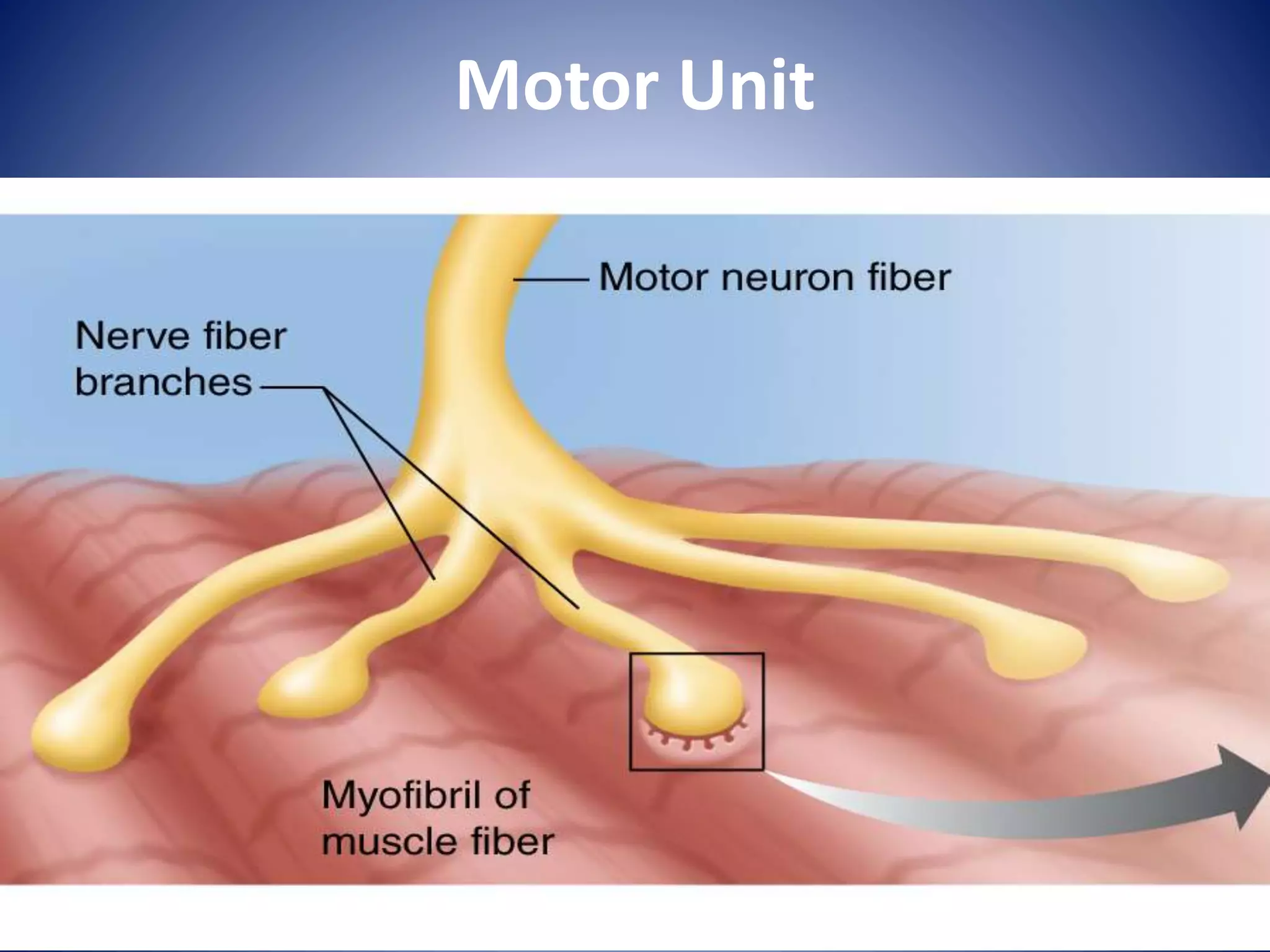
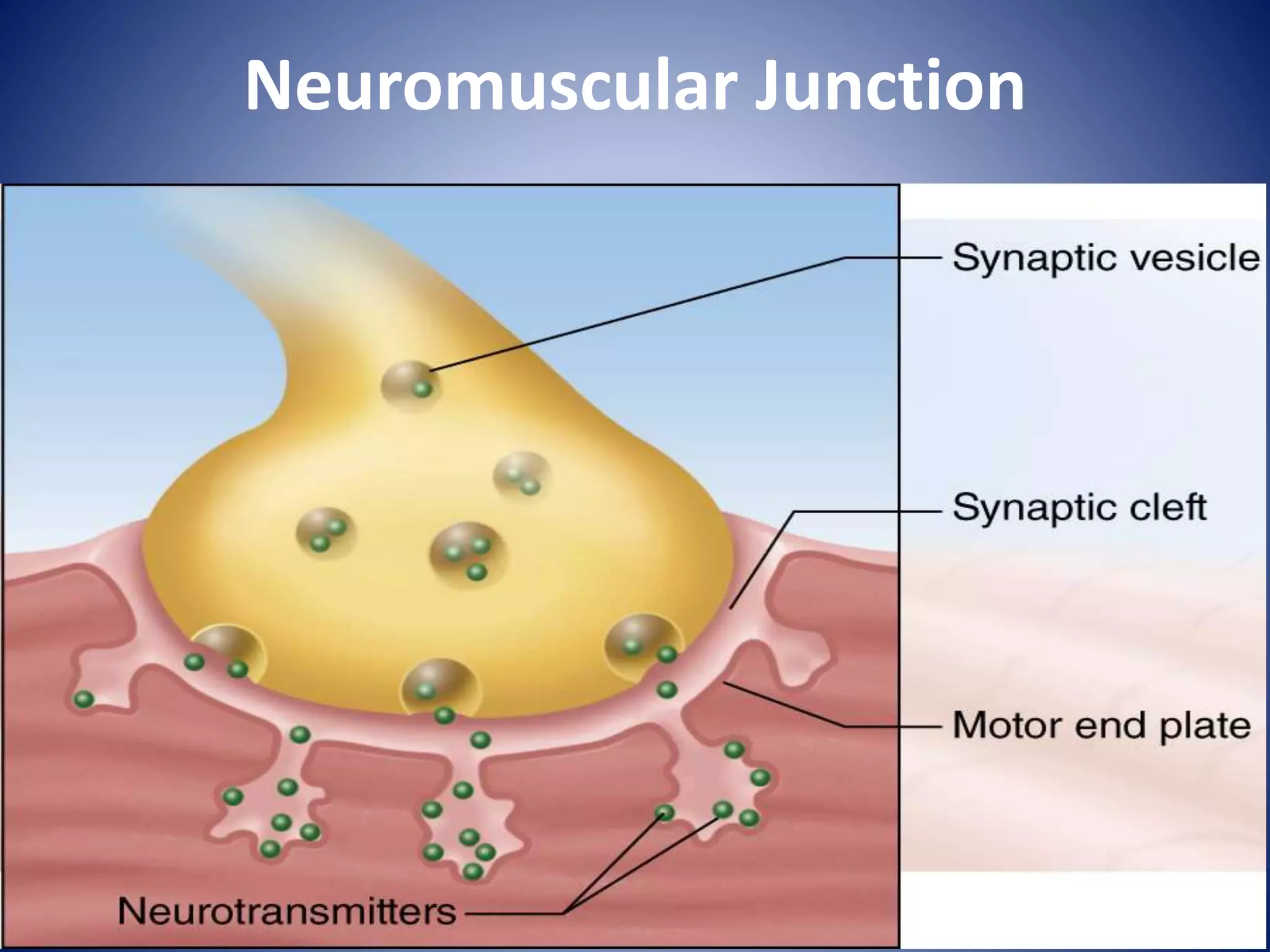
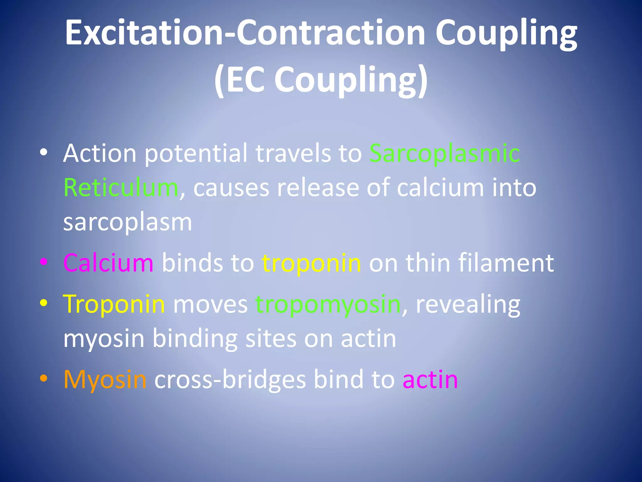
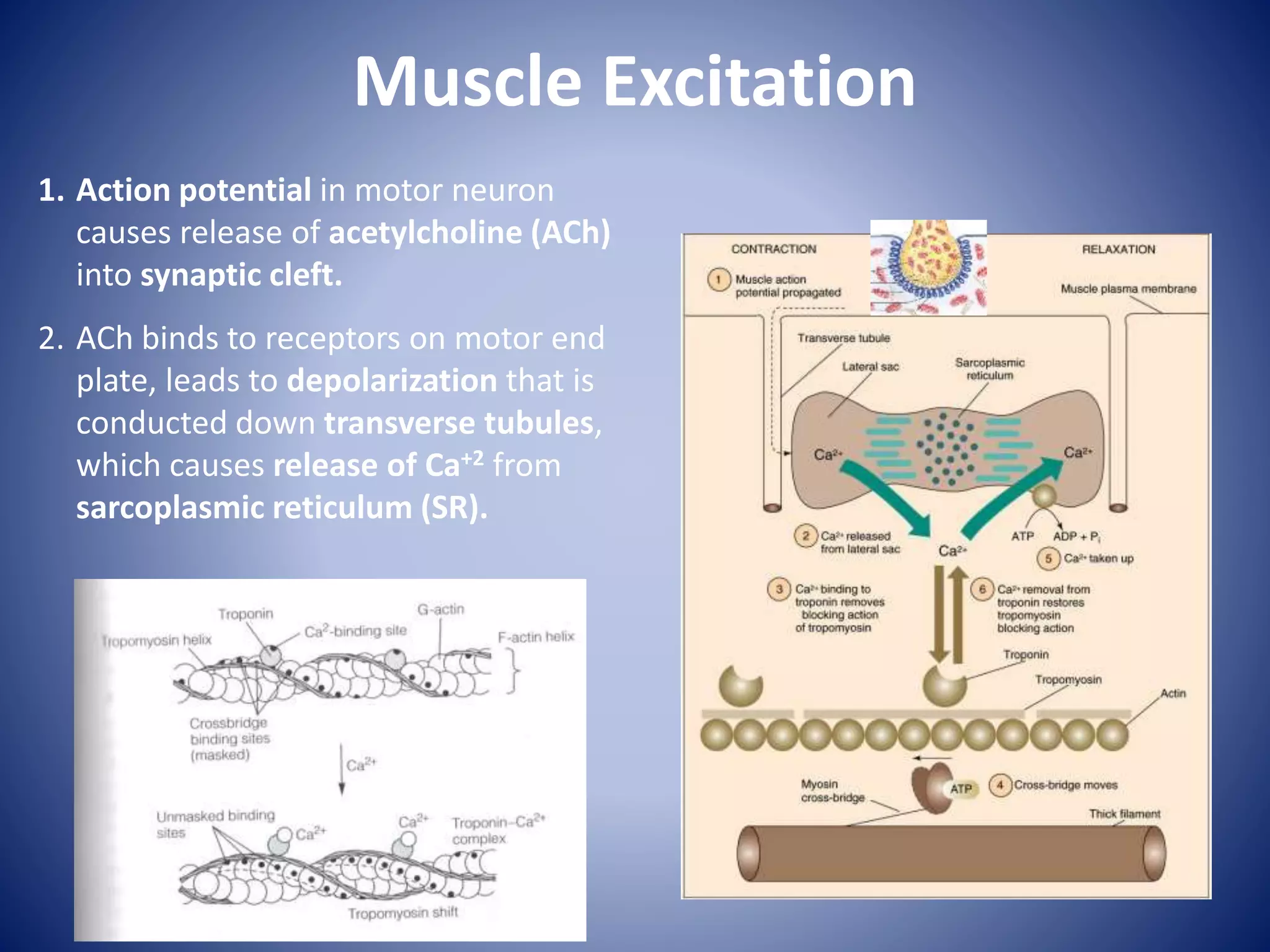
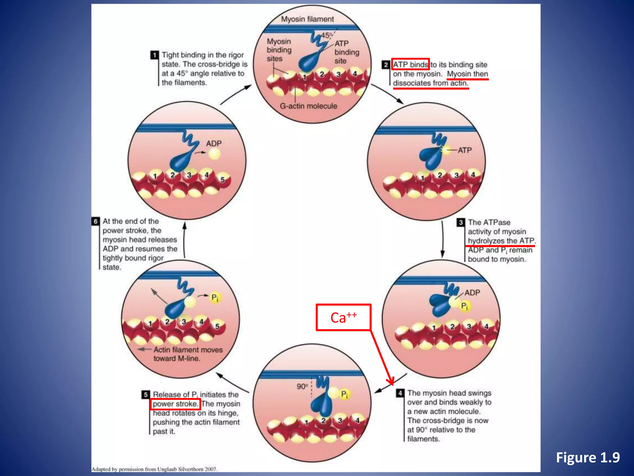
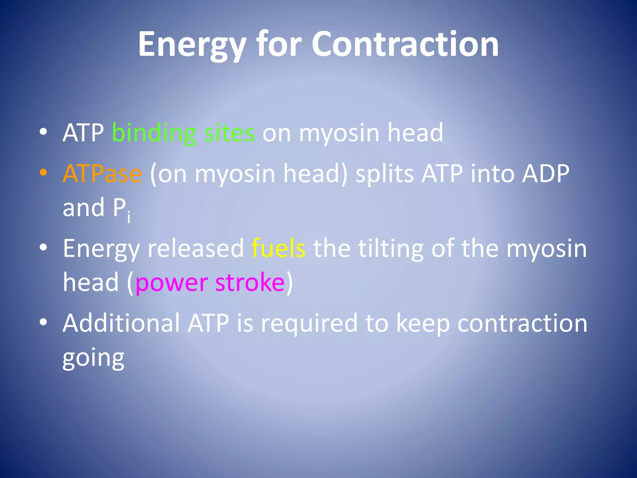
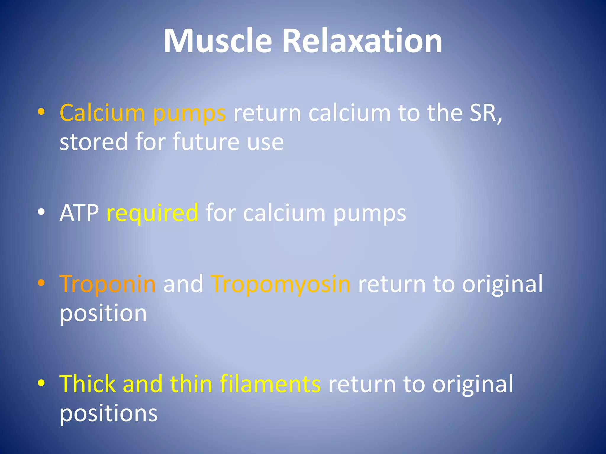
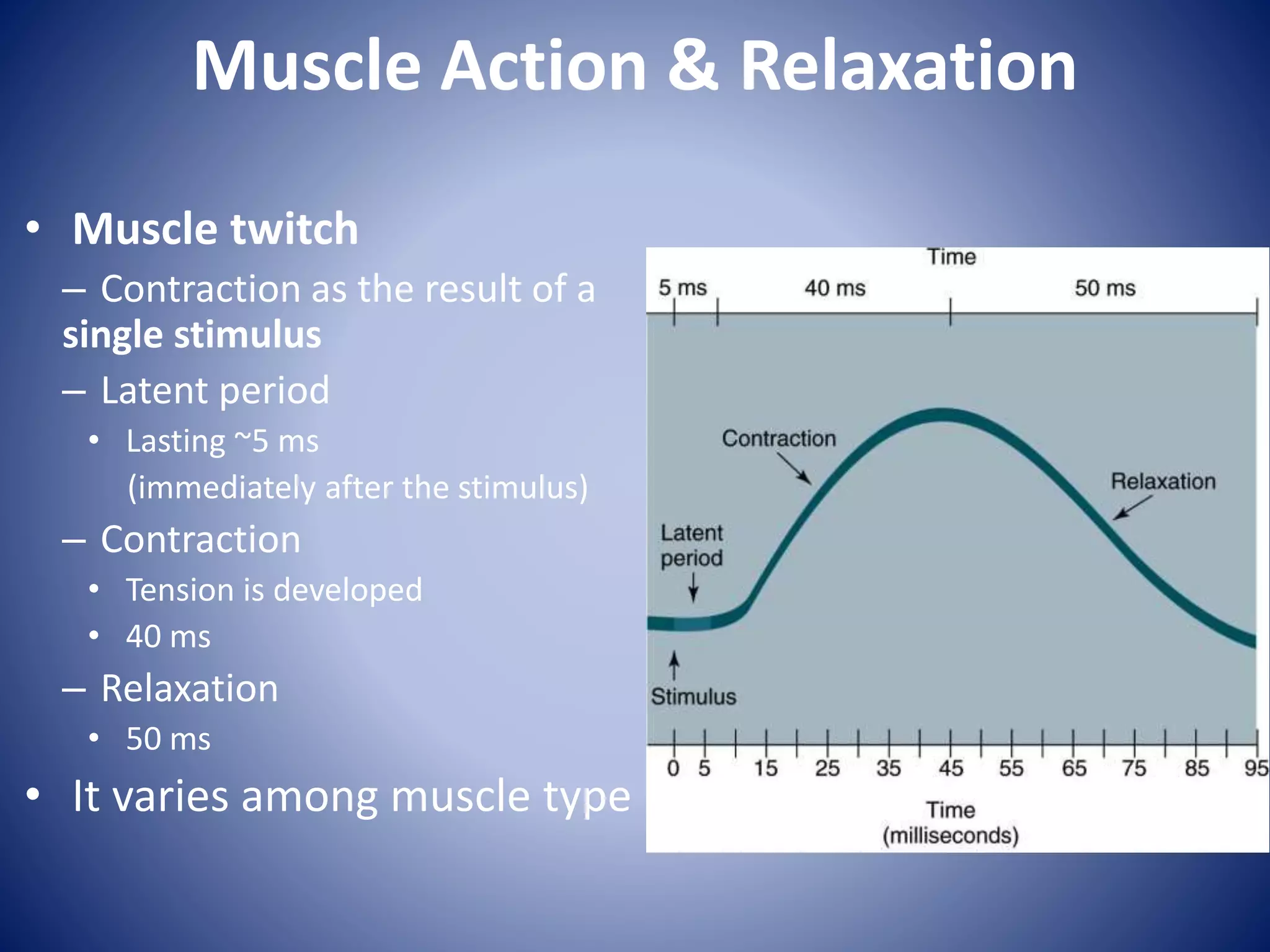
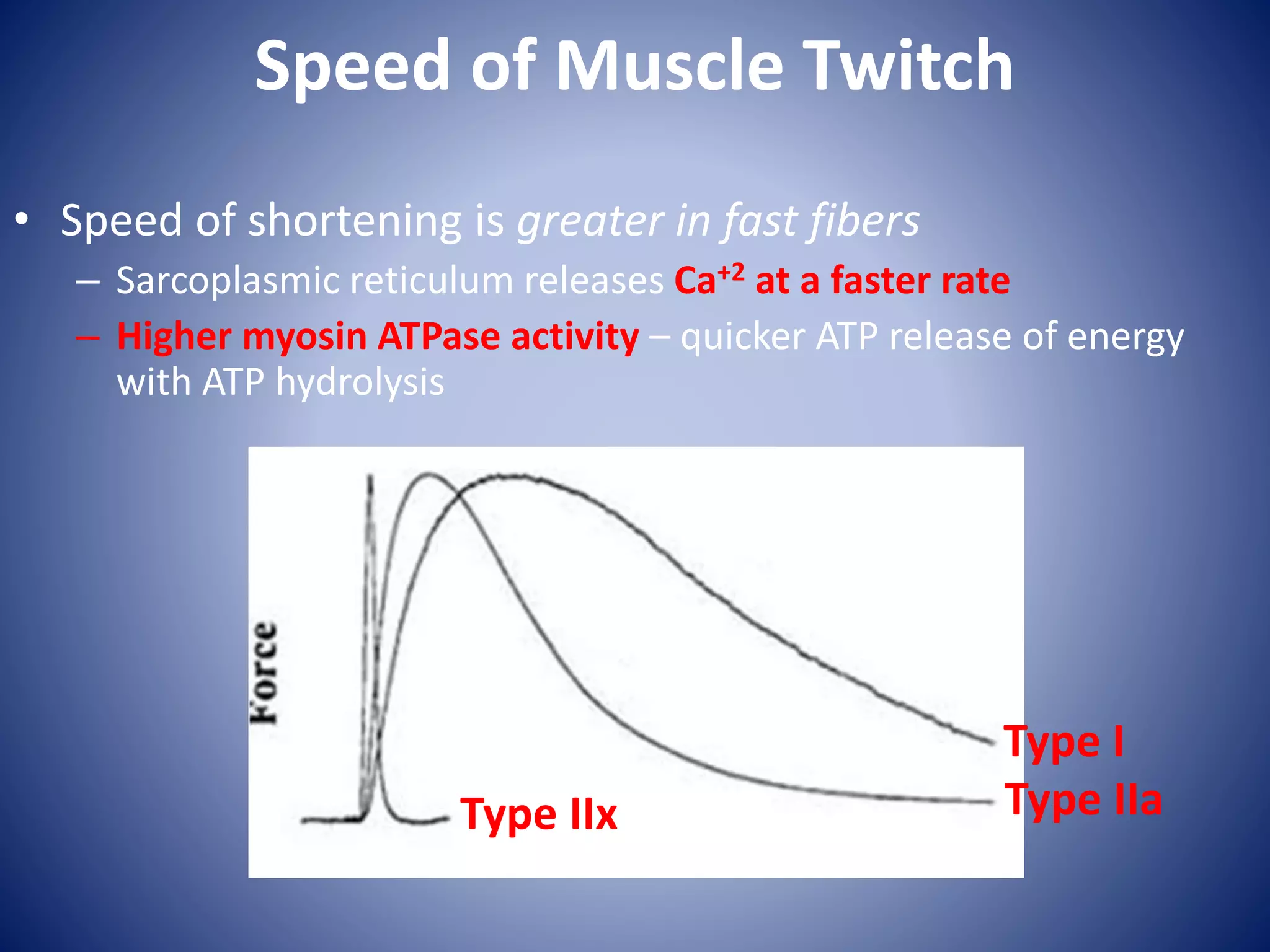
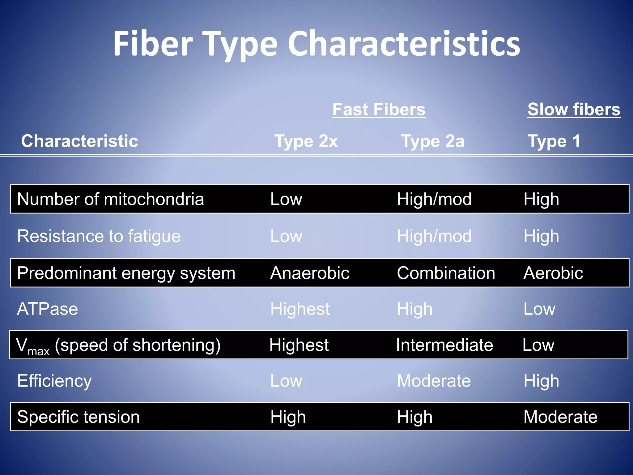
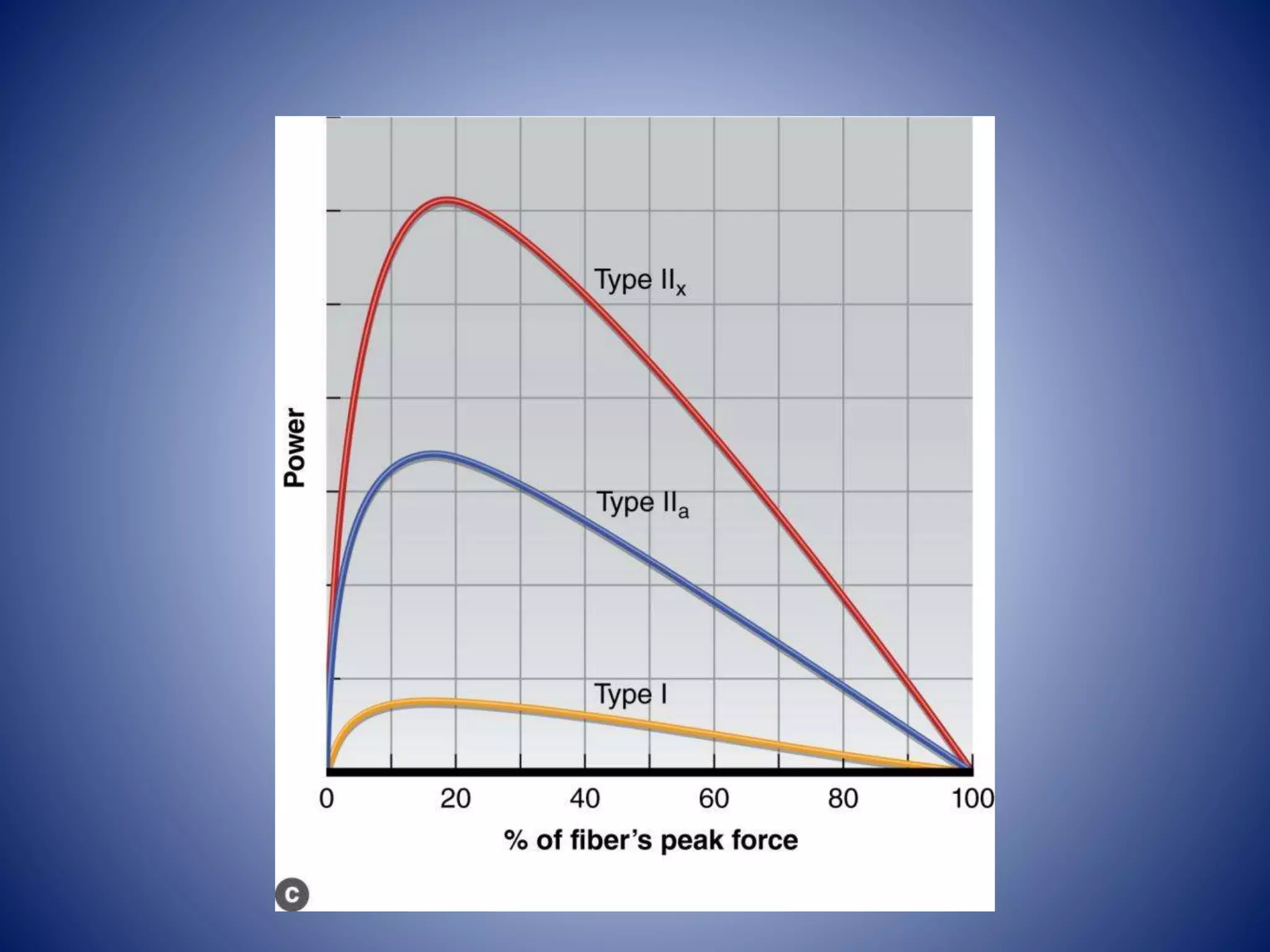
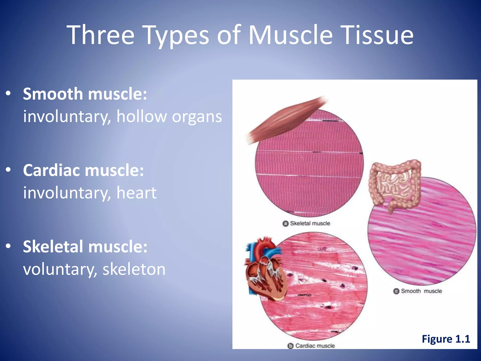
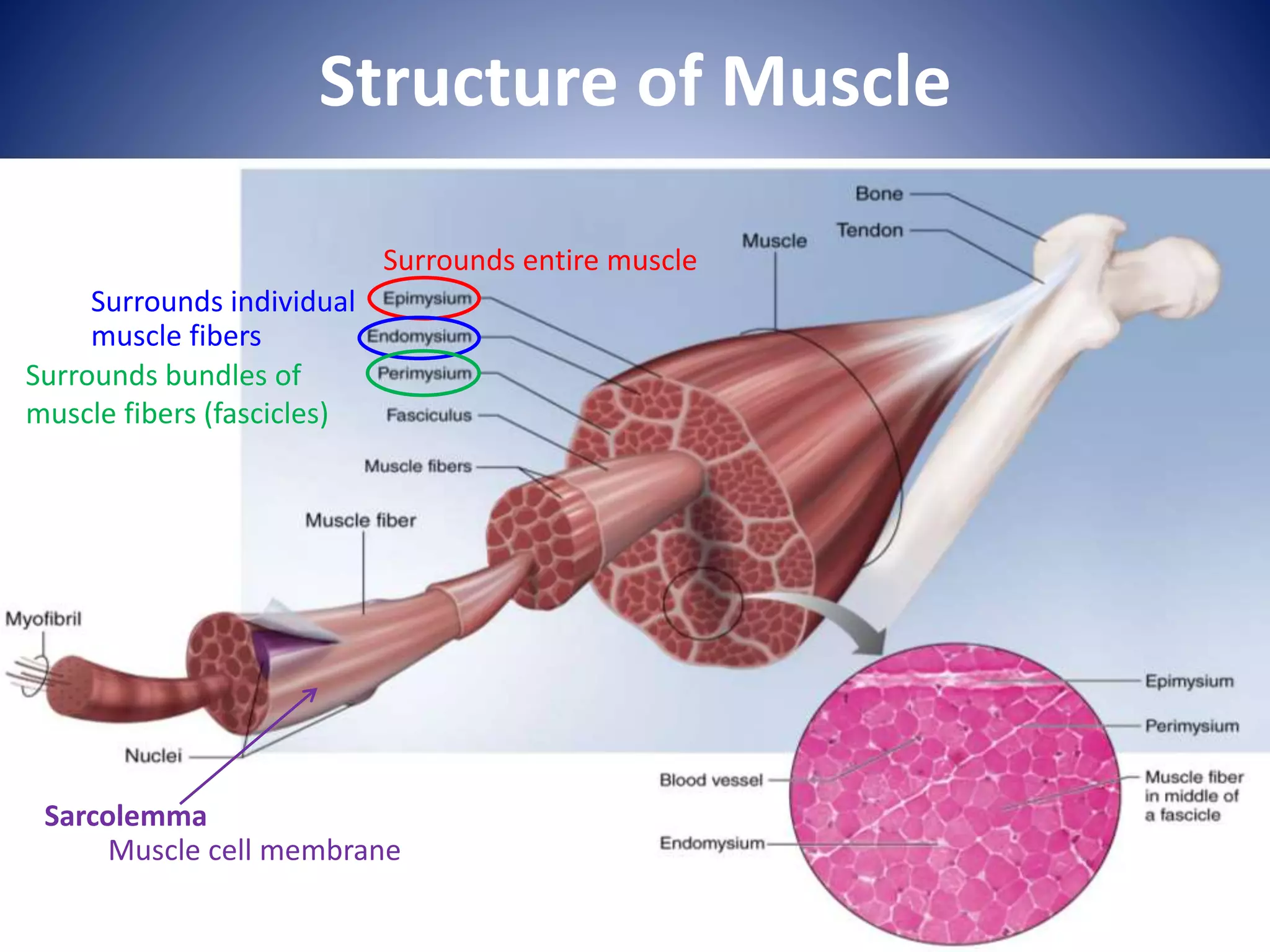
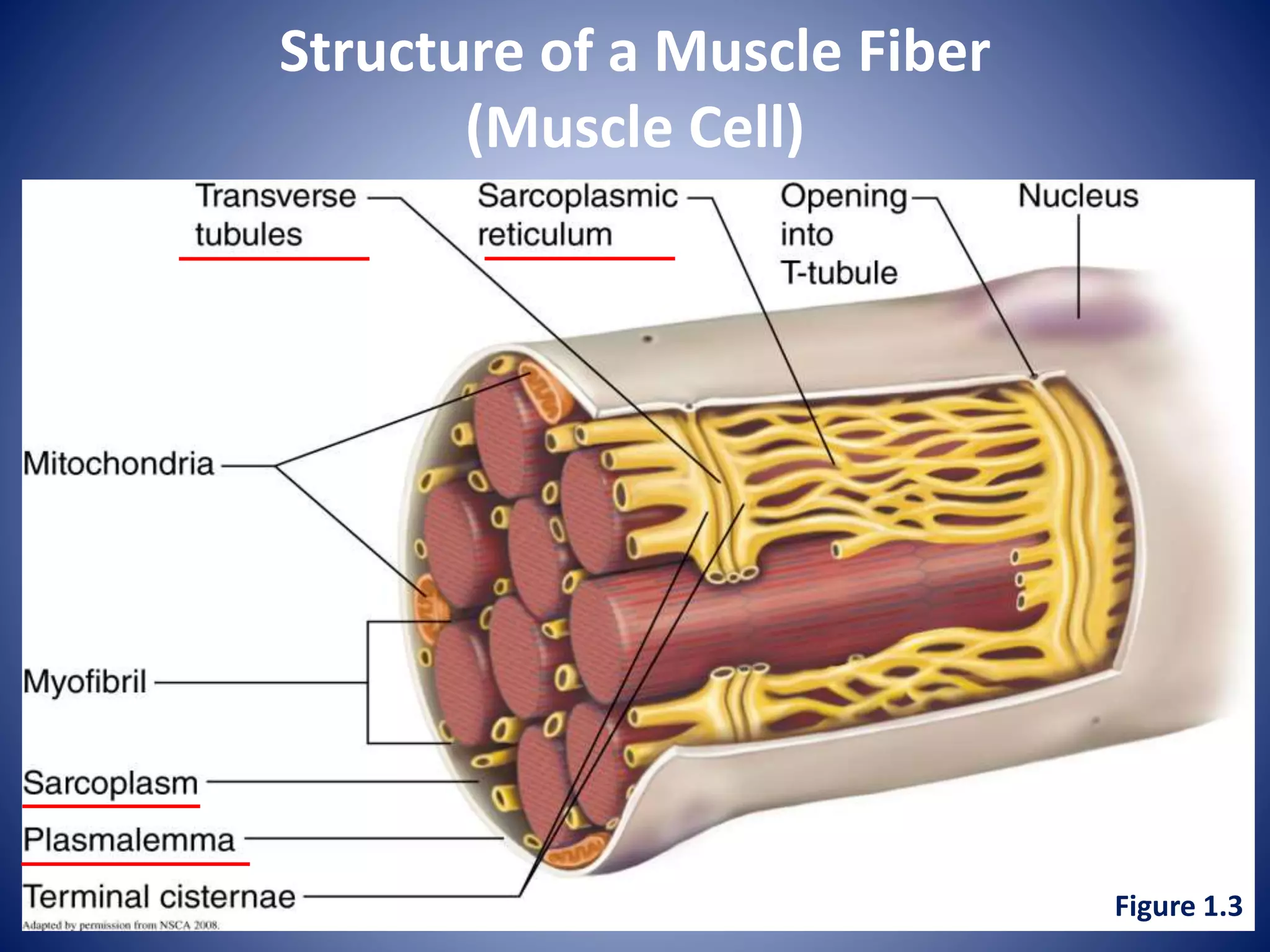
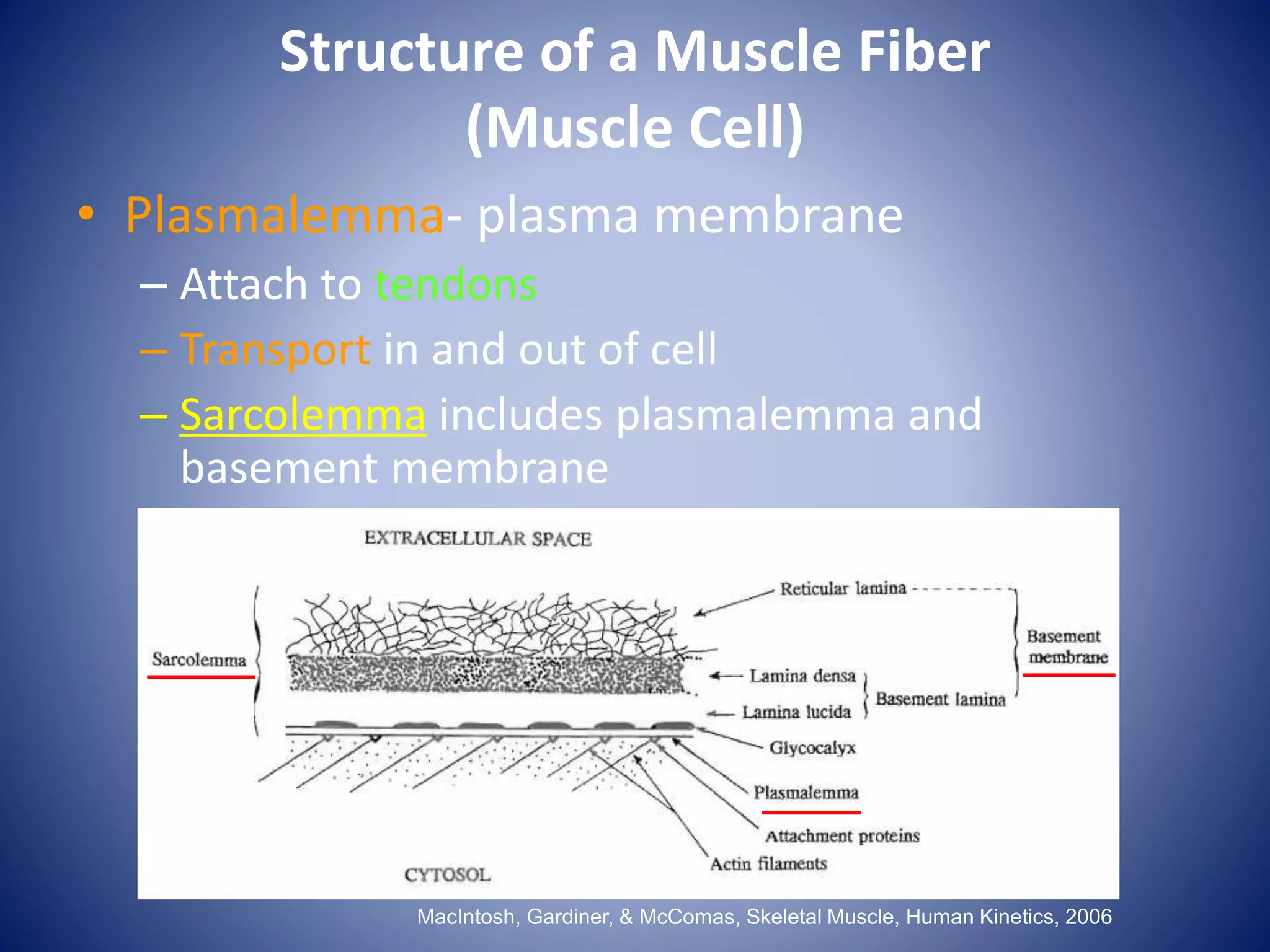
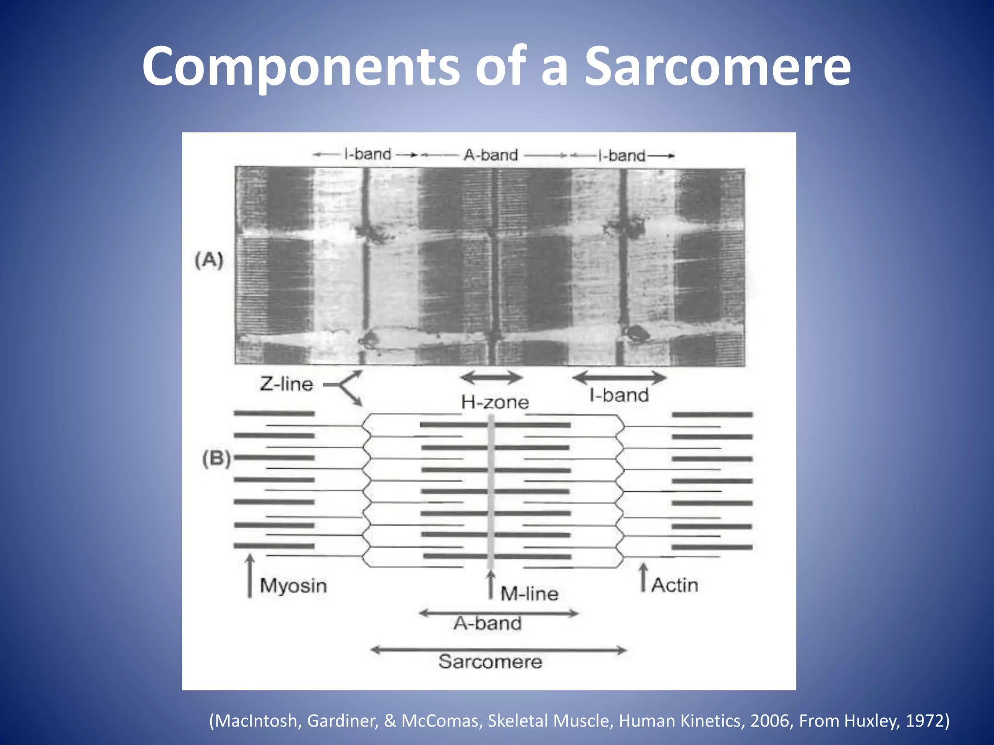
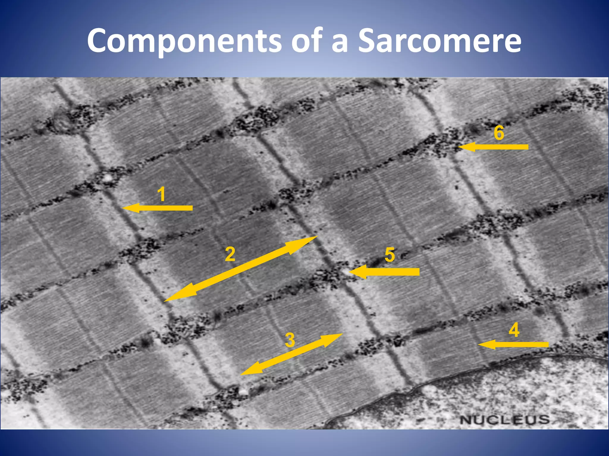
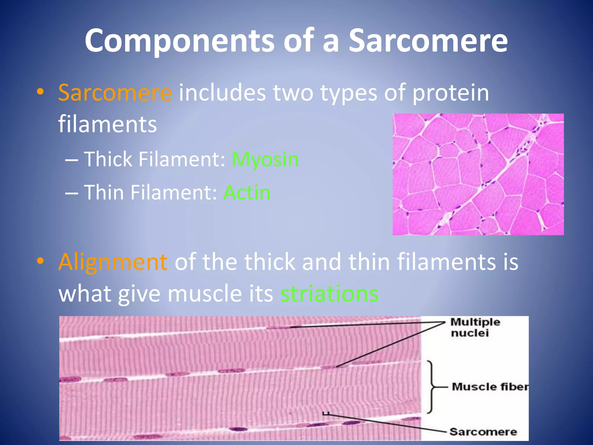
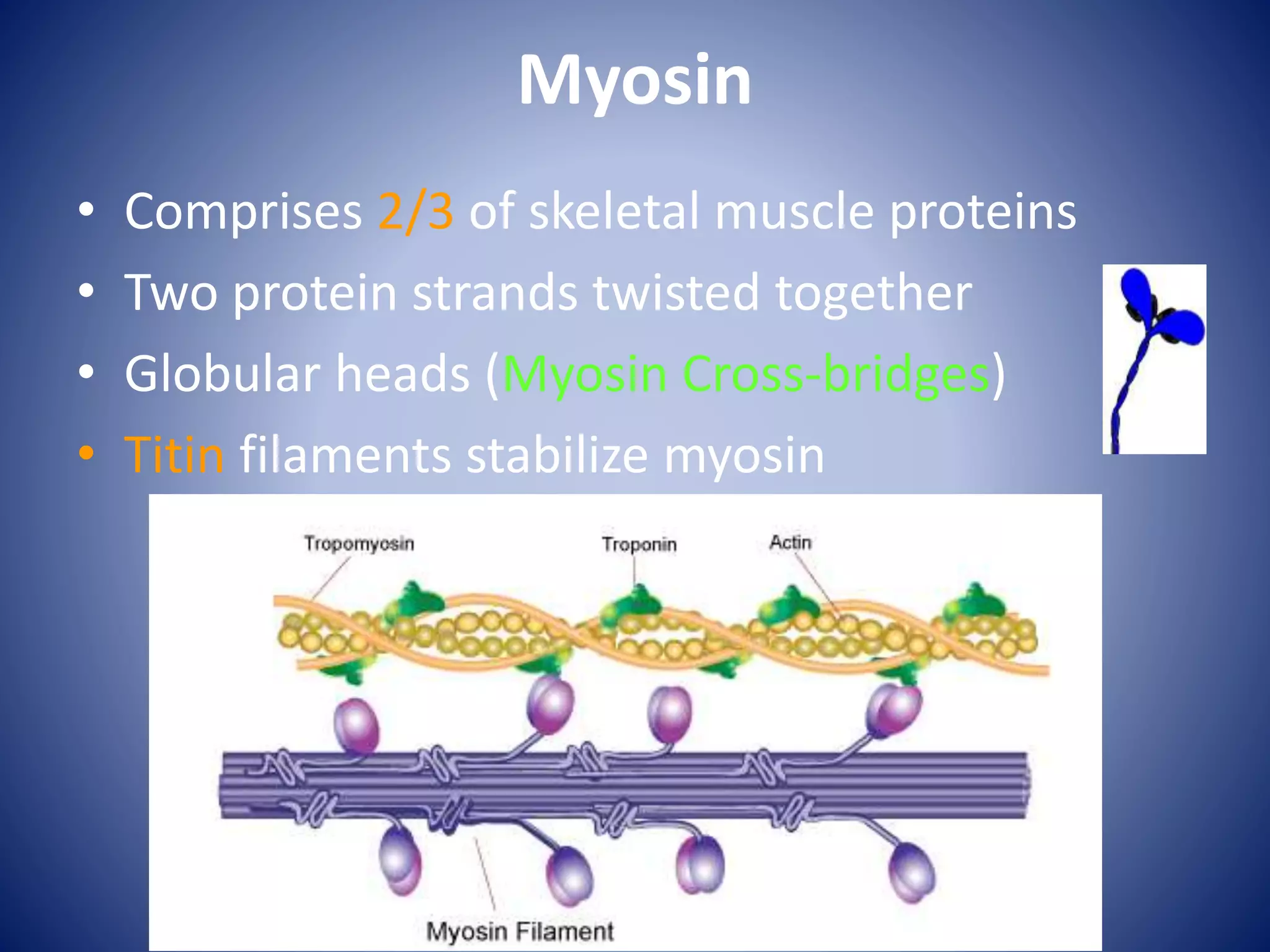
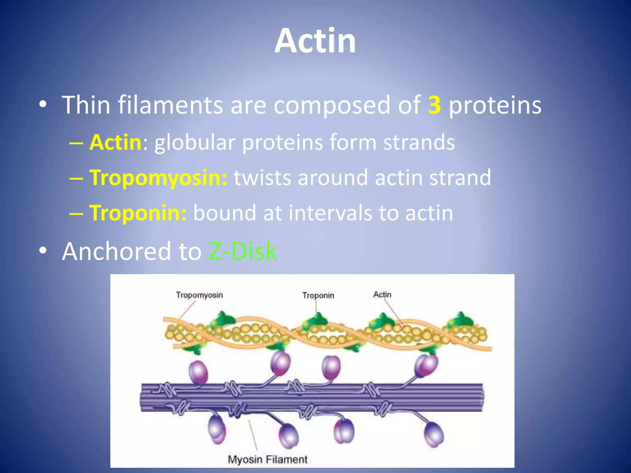
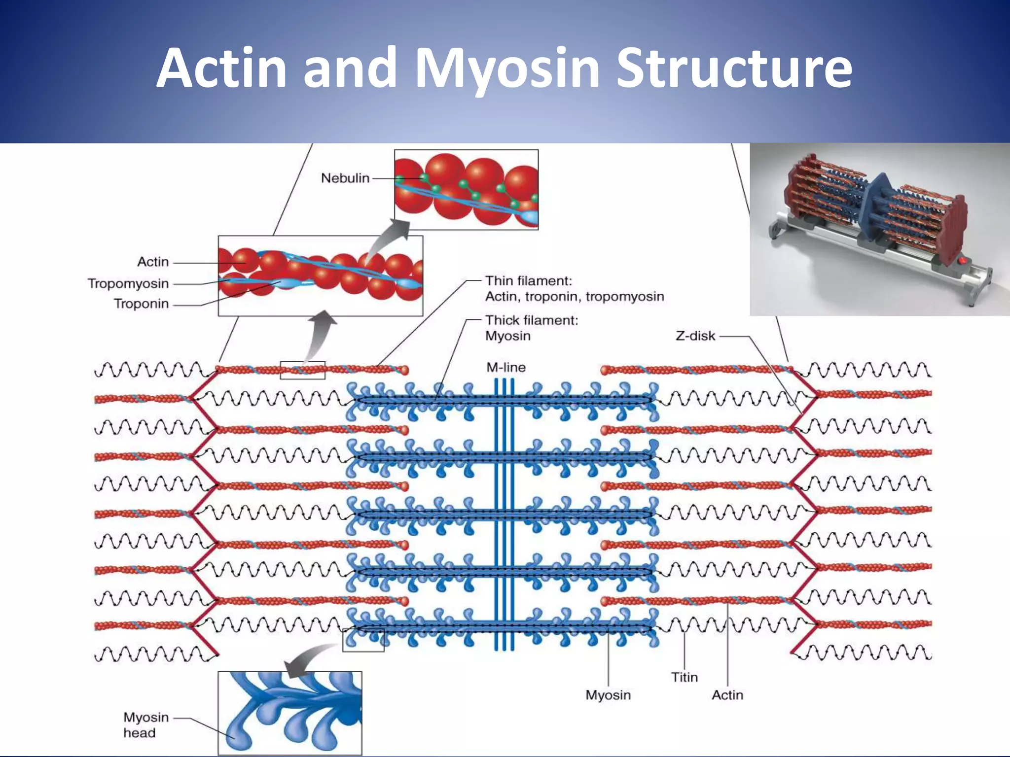
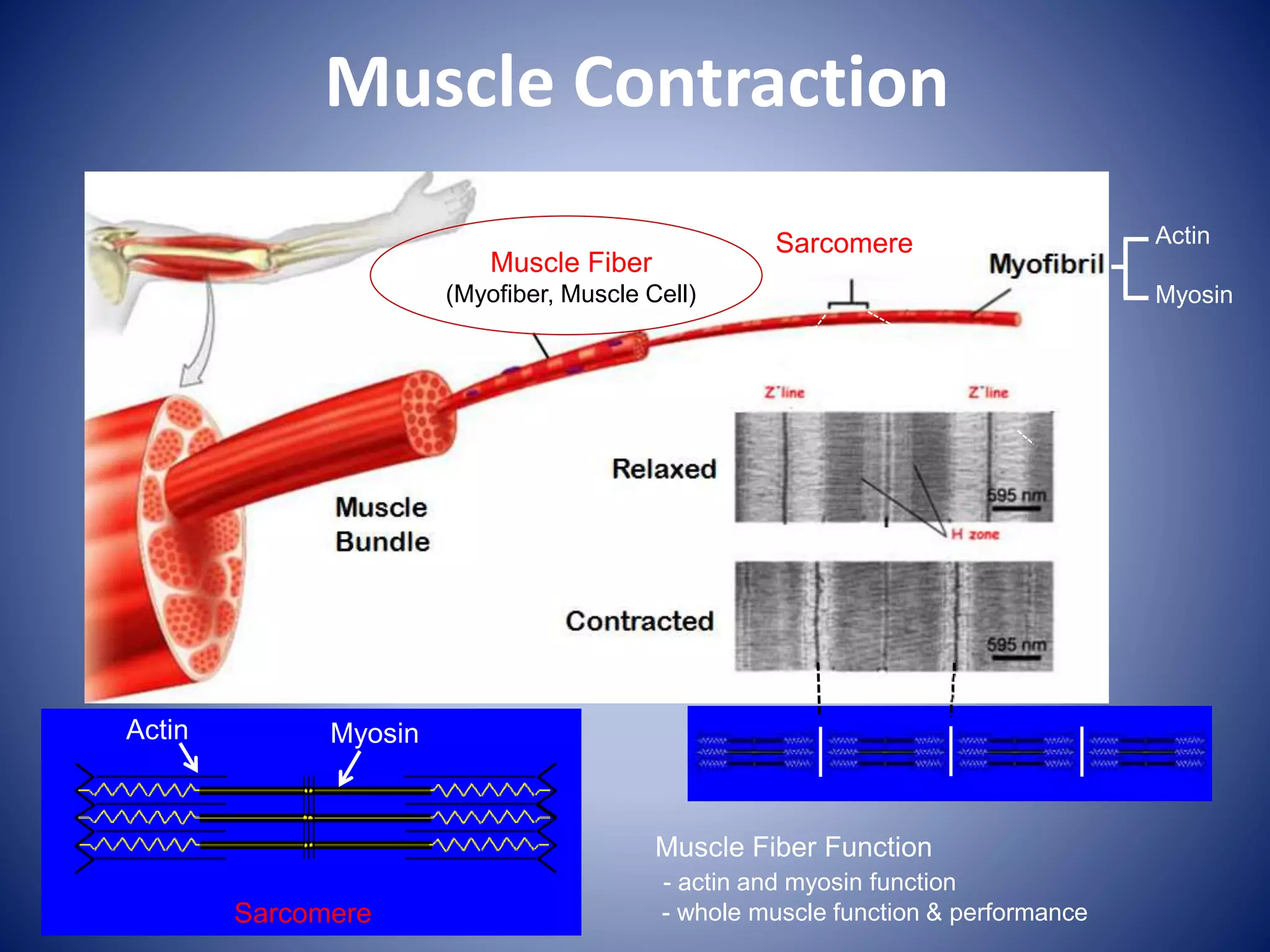
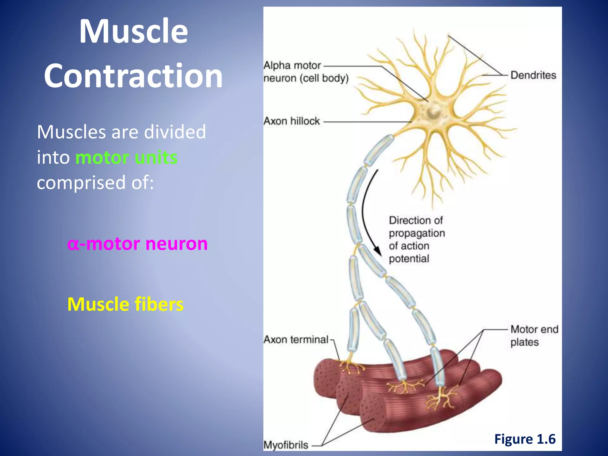
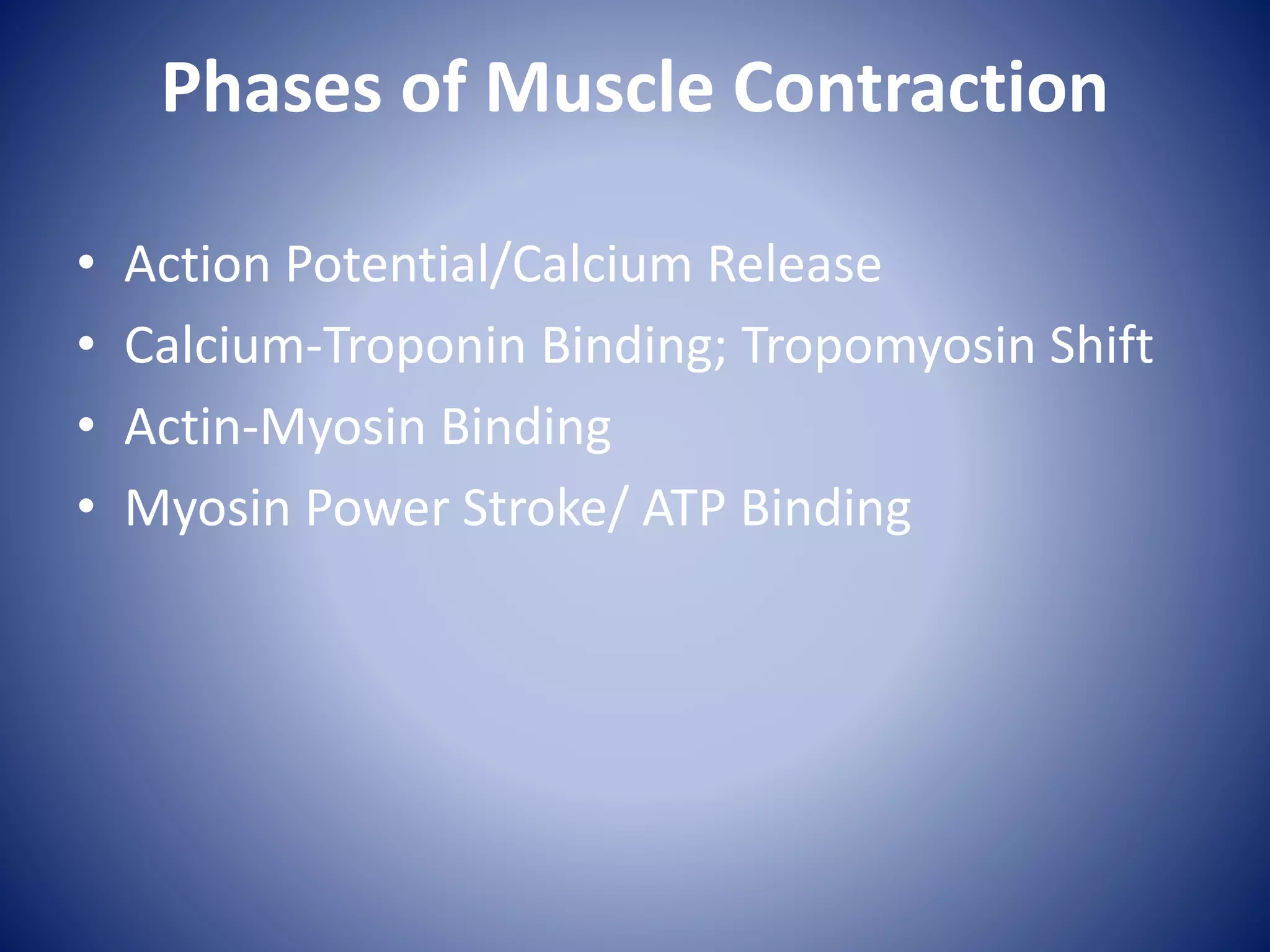
The document discusses the structure and function of skeletal muscle. It describes the hierarchy of muscle organization from the whole muscle down to the sarcomere level. Key components of muscle fibers and sarcomeres such as myofibrils, actin, and myosin are defined. The process of muscle contraction is summarized in three steps: 1) an action potential causes calcium release and binding to troponin, 2) myosin binds to actin and generates force through its power stroke, and 3) relaxation occurs when calcium is reabsorbed and the sarcomere returns to its resting state. Muscle fiber types and their roles in athletic performance are also overviewed.
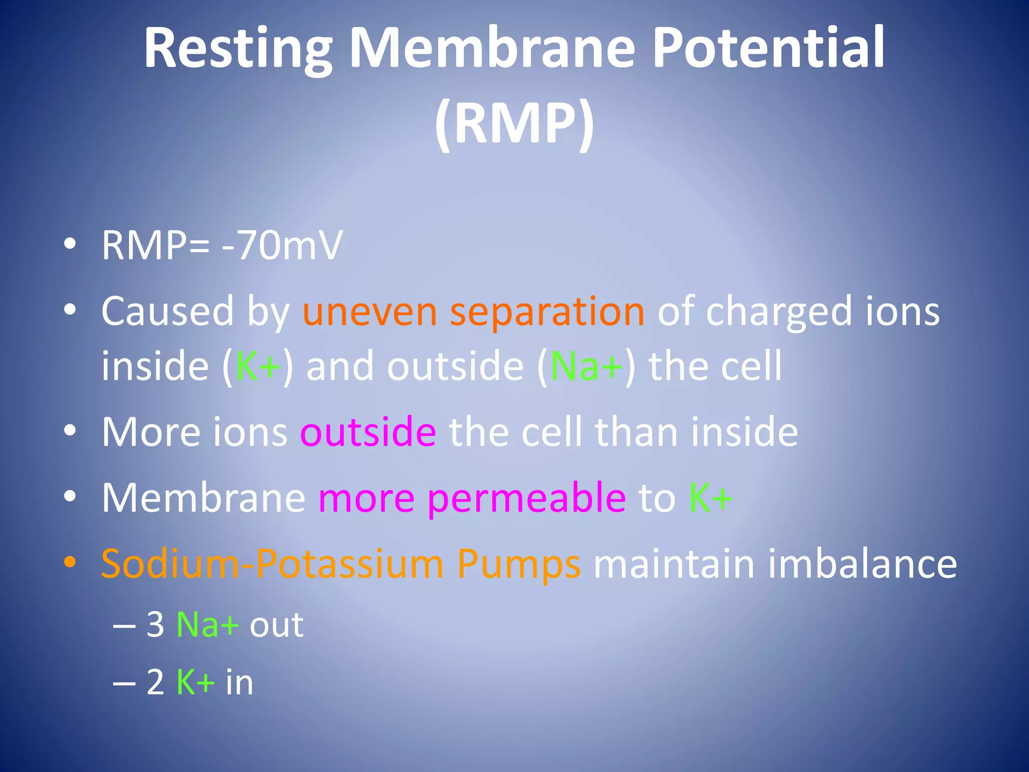
![Ions Channels
• At rest, almost all the
Na+ channels are
closed.
• At rest, few K+
channels are open.
– Leaking due to [ ]
gradient](https://image.slidesharecdn.com/lecture4-musclephysiology1-150726203813-lva1-app6892/75/Lecture-4-muscle-physiology-1-21-2048.jpg)