
Recommended
More Related Content
What's hot
What's hot (20)
Similar to Sex Differences in Adolescent Brain Activity and Depression
Similar to Sex Differences in Adolescent Brain Activity and Depression (20)
Recently uploaded
Recently uploaded (20)
Sex Differences in Adolescent Brain Activity and Depression
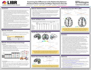
- 1. Introduction This project is part of a Centers of Biomedical Research Excellence grant (P20 GM199097, Hays-Grudo) funded by the National Institutes of Health. Examining Sex Differences in the Relationship Between Adolescent Brain Activity and Major Depressive Disorder Chase A. Antonacci • Kara L. Kerr • Kelly T. Cosgrove • Erin L. Ratliff • Jerzy Bodurka • Amanda Sheffield Morris Methods: Participants Methods: Participants & Experimental Design • AFNI’s 3dttest++ was used to examine adolescents’ BOLD activation during feigned error conditions in comparison to trials where both members answered correctly. • Depressive symptoms were assessed using the Mood and Feelings Questionnaire (MFQ) while ER skill/competency was assessed using the Difficulties in Emotion Regulation Scale (DERS). • Within a priori anatomical masks of the dorsolateral prefrontal cortex and posterior cingulate cortex, a voxelwise threshold of p < 0.005 was set with a clusterwise correction for multiple comparisons at p < 0.05 (corrected). Results: Self-Report Scores • No group-level differences emerged between boys and girls in self-report measures of depressive symptoms (MFQ) or difficulty in emotion regulation (DERS). 0 2 4 6 8 10 12 14 16 -0.4 -0.3 -0.2 -0.1 0 0.1 0.2 0.3 0.4 0.5 dlPFC Right: Activation by MFQ Score Gender Differences in the Relationship Between Adolescent Brain Activity and Depressive Symptoms Figure 2 Dorsolateral Prefrontal Cortex Right Z = 28 +4.59-1.59 t-Statistic 0 2 4 6 8 10 12 14 16 -0.8 -0.6 -0.4 -0.2 0 0.2 0.4 0.6 0.8 Beta dlPFC Left: Activation by MFQ Score +4.08-1.94 t-Statistic Left Z = 35 Fig. 2 Boys and girls differed in the relationship between MFQ scores and both right and left dlPFC activation. Correlation plots illustrate a positive slope for boys and a negative slope for girls in both the right and left dlPFC. Beta MFQScore MFQScore Results: Boys versus Girls: MFQ in PCC • In most countries around the world, women face nearly 2x the risk for depression in comparison to men1 though it remains unclear why this disparity exists. • Adolescence appears to be a key period in the developmental timeline for understanding this differential in depressive diagnoses; it’s during this critical period of vulnerability between the ages of 13 and 15 that girls begin to outpace boys in rates of depression2. • While sex differences in depressive disorders have been explored in adults3, little research has focused on understanding sex differences at their key starting point during adolescence and, in particular, how variation in developmental neurobiology might to help explain why women remain at greater risk. • Because depression is often characterized as a disorder of impaired emotion regulation (ER)4, understanding how ER strategies emerge and develop in adolescence remains an important component for understanding the progression of depressive psychopathology. • The current study aims to examine neurobiological differences between boys and girls that underlie depressive symptomatology in a socially-situated emotion regulation task. • Though there are a number of competing theories explaining why women remain at greater risk for depression5,6 the current study examines what brain regions associated with ER may be useful in helping to understand, and in the long term, potentially predict which adolescents are at increased risk for developing internalizing symptoms and ultimately clinical depression. • Parent-adolescent dyads underwent fMRI hyperscanning (concurrent scanning to simultaneously measure neural responses of two interacting individuals in separate scanners). • All participants were psychiatrically healthy as per the MINI Neuropsychiatric Interview (MINI 7.0). Task Design: • Dyads completed the Testing Emotional Attachment and Mutuality (TEAM) Task during simultaneous fMRI hyperscanning. • The TEAM task is a collaborative game designed to assess brain activity when either member of the dyad makes a costly mistake. • The task includes fixed trials in which each member of the dyad believes the other has made an error, costing them both five dollars. Scan Parameters: • Two identical GE MR750 3.0T MRI scanners using NOVA 32-channel phased array head coils • Scan time = 7min 50 sec; 231 EPI volumes; TR/TE = 2000/25 ms, 41 axial slices with 2.9 mm thickness; image matrix = 96x96 reconstructed into a 128x128 resulting in 1.9x1.9x2.9 mm3 voxel volume; voxel size = 1.9x1.875x2.90mm3; 6mm FWHM spatial smoothing • All functional imaging data were analyzed using AFNI. 0 2 4 6 8 10 12 14 16 -0.6 -0.4 -0.2 0 0.2 0.4 0.6 PCC Left: Activation by MFQ Score Figure 1 Posterior Cingulate Cortex (PCC) Left X = 3 Right X = -15 Y = 65 +4.84-1.50 t-Statistic 0 2 4 6 8 10 12 14 16 -0.5 -0.3 -0.1 0.1 0.3 0.5 0.7 PCC Right: Activation by MFQ Score MFQScore BetaBeta Gender Differences in the Relationship Between Adolescent Brain Activity and Depressive Symptoms MFQScore Fig. 1 Boys and girls differed in the relationship between MFQ scores and PCC activation. Correlation plot between MFQ score and PCC beta value, illustrating a positive slope for boys and a negative slope for girls in both the right and left PCC. Methods: Statistical Analyses Boys and girls differed in the relationship between depressive symptoms and bilateral PCC activation. Correlation plot between MFQ scores and PCC beta values illustrates a positive slope for boys and a negative slope for girls Results: Boys versus Girls: DERS in rPCC 0 20 40 60 80 100 120 -1.25 -0.75 -0.25 0.25 0.75 DERSScore Beta PCC Right: Activation by DERS Score +4.69-0.66 t-Statistic X = -4.4 Boys and girls differed in the relationship between ER difficulty and rPCC activation. Correlation plot illustrates a positive slope for boys and a negative slope for girls. Results: Boys versus Girls: MFQ in dlPFC Boys and girls differed in the relationship between depressive symptoms and bilateral dlPFC activation. Correlation plot between MFQ scores and dlPFC beta values illustrates a positive slope for boys and a negative slope for girls Table 1: Self-Report Measures Descriptives Inferential Boys (n = 12) Girls (n = 17) Mean Median SD Mean Median SD t df p Cohen's d Age 14.86 15.00 0.86 14.83 14.50 0.92 MFQ 4.71 3.50 4.95 5.17 4.50 3.67 -0.286 23.218 0.777 0.106 DERS Composite 71.14 74.50 17.30 68.17 65.50 16.01 0.499 26.949 0.622 0.179 DERS Non-Acceptance 9.71 8.50 3.31 10.39 9.00 4.13 -0.512 29.954 0.612 0.178 DERS Goals 12.71 12.50 4.58 10.89 11.50 3.77 1.206 25.003 0.239 0.441 DERS Impulse 10.00 7.50 5.64 8.28 8.00 2.67 1.054 17.530 0.306 0.408 DERS Awareness 16.00 14.00 7.39 15.83 16.00 5.29 0.071 22.686 0.944 0.027 DERS Strategies 12.57 11.50 4.54 12.17 11.00 4.27 0.257 27.226 0.799 0.092 DERS Clarity 10.14 9.00 3.68 10.61 9.00 3.88 -0.349 28.777 0.730 0.123 References 1Hankin, B. L., Abramson, L. Y., Moffitt, T. E., Silva, P. A., McGee, R., & Angell, K. E. (1998). Development of depression from preadolescence to young adulthood: emerging gender differences in a 10-year longitudinal study. Journal of Abnormal Psychology, 107(1), 128–140. 2NIMH » Major Depression. (n.d.). Retrieved January 11, 2019, from https://www.nimh.nih.gov/health/statistics/major-depression.shtml 3McRae, K., Ochsner, K. N., Mauss, I. B., Gabrieli, J. J. D., & Gross, J. J. (2008). Gender Differences in Emotion Regulation: An fMRI Study of Cognitive Reappraisal. Group Processes & Intergroup Relations : GPIR, 11(2), 143–162. https://doi.org/10.1177/1368430207088035 4Joormann, J., & Gotlib, I. H. (2010). Emotion Regulation in Depression: Relation to Cognitive Inhibition. Cognition & Emotion, 24(2), 281–298. https://doi.org/10.1080/02699930903407948 5Nolen-Hoeksema, S. (2001). Gender Differences in Depression. Current Directions in Psychological Science, 10(5), 173–176. Retrieved from JSTOR. 6Bangasser, D. A. (2013). Sex differences in stress-related receptors: “micro” differences with “macro” implications for mood and anxiety disorders. Biology of Sex Differences, 4(1), 2. https://doi.org/10.1186/2042-6410-4-2 Conclusion • When faced with an emotionally-vexing stimulus – a parent’s costly error – boys experiencing greater depressive symptoms exhibited increased activation in these emotion regulation regions while girls exhibited decreased activation. • When experiencing depressive symptoms, boys may be upregulating key ER regions, helping them to engage more adaptive/effective emotion regulation strategies while girls, for whatever reason, appear to be downregulating these regions and conceivably not invoking adaptive emotional responses, potentially allowing for more maladaptive strategies to take hold. • Though the results of this study do not suggest an obvious neurobiological mechanism by which we can account for the vast differences in MDD rates between men and women, our findings do highlight a sharp dissimilarity in ER-related functional brain activity between adolescent boys and girls. • Given that during this ‘critical period of vulnerability’ for MDD, boys and girls exhibited not only diverging but indeed opposite trends in activation in relation to depressive symptoms and ER skill, it’s reasonable to presume that this difference may be central in accounting for differential rates of MDD diagnoses, even if we have yet to substantiate a causal mechanism. • Future research should address the longitudinal implications of this study. These data may enable us to examine whether sex differences in adolescent brain activity related to emotion regulation do in fact mediate and predict the relationship between internalizing symptoms and clinical depression.