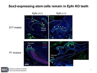
1. The document presents data on the expression of Epfn and Tbx1 during mouse molar and incisor development. Epfn and Tbx1 are co-expressed in the proliferating inner dental epithelium.
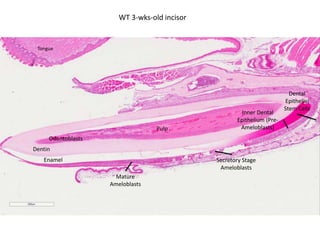
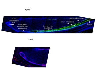
2. Tbx1 expression is downregulated at the preameloblast stage, while Epfn expression continues into ameloblast development. Tbx1 expression is also reduced in Epfn KO molars, indicating Epfn acts upstream of Tbx1.
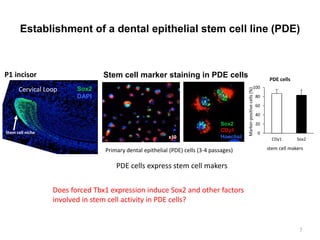
3. Experiments using dental epithelial stem cells show Epfn and Tbx1 promote proliferation and Epfn binds to the Tbx1 promoter region, suggesting a physical interaction between the two proteins.


![DEVELOPMENT_OF_PERIODONTIUM[1] anoushka.pptx](https://cdn.slidesharecdn.com/ss_thumbnails/developmentofperiodontium1anoushka-251216170022-7613e1d2-thumbnail.jpg?width=640&height=640&fit=bounds)


