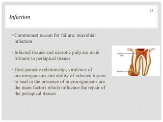
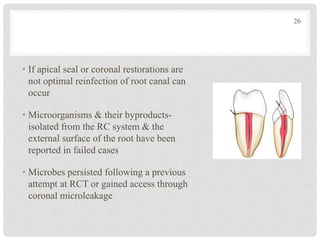
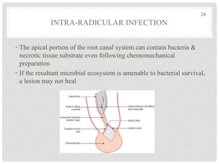
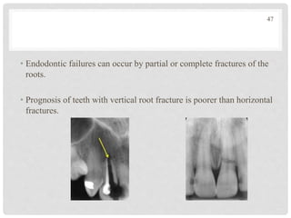
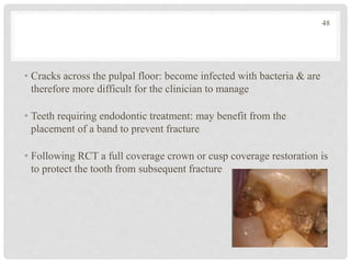
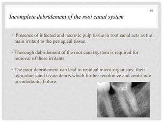
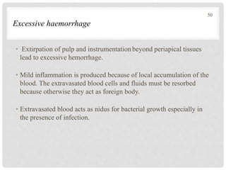
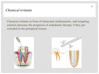
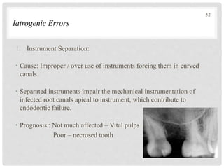
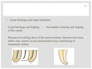
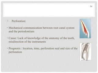
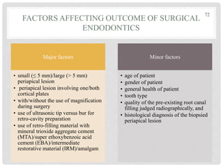
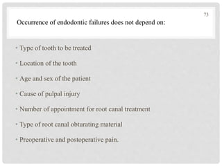
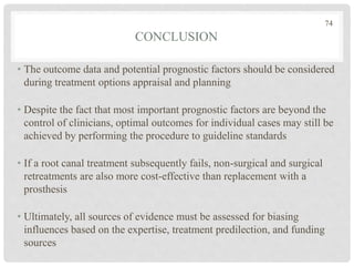
Call Girls Secunderabad 7001305949 all area service COD available Any Time
Success and failures in Endodontics
1. GUIDED BY:
DR HEMANT VAGARALI
SUCCESS AND FAILURES
IN ENDODONTICS
PRESENTED BY:
DR SAHANA UMESH
2. CONTENTS
• Introduction
• Endodontic treatment outcome
• Evaluation of success of RCT
• Clinical
• Radiographic
• Histological
• Causes of endodontic failures
• Local factors affecting success or failure
• Infection
• Excessive haemorrhage
• Incomplete debridement
2
3. • Iatrogenic errors
• Chemical irritants
• Systemic factors affecting success or failure
• Factors to be considered before retreatment
• Factors affecting prognosis of endodontic treatment
• Contraindications for endodontic retreatment
• Problems of endodontic retreatment
• Treatment options
• Conclusion
• References
3
4. INTRODUCTION
• Success is defined by goals established to be achieved, so the goal of
endodontic treatment is to heal or prevent the disease.
• The definition of success of RCT is ambiguous :
• Stringent: Radiographic and clinical normalcy
• Lenient: Clinical normalcy
• In different studies success rate ranges from 54-95% for RCT. Non
surgical retreatment showed a higher rate of success(83%) compared
with endodontic surgery (71.8%)
4
5. • Communication with patients can be improved by replacing the
value-laden terms ‘‘success’’ and ‘‘failure’’ with neutral expressions:
• ‘‘chance of healing’’
• ‘‘risk of inflammation’’
• Avoid the terms ‘‘success’’ and ‘‘failure’’ in defining the outcome of
endodontic treatment
5
6. ENDODONTIC TREATMENT OUTCOME
• Aim of endodontic treatment is to
prevent or cure apical periodontitis
• Healed: Both clinical and
radiographic presentations are
normal
• Healing: It is a dynamic process,
reduced radiolucency combined
with normal clinical presentation
• Not healed: No change or increase
in radiolucency, clinical signs may
or may not be present or vice
versa.
4 year
follow
up
Immediate
post op
3 years 6 years
6
7. • Dynamics of healing: The potential of teeth to remain free of apical
periodontitis after nonsurgical endodontic treatment is 93–98%.
• The signs of healing are evident within the first year after treatment
in nearly 90% of the cases
• Rud et al. introduced a classification for outcome assessment after
apical surgery that referred to healing:
• Complete
• Incomplete
• Uncertain
• Unsatisfactory
7
8. SUGGESTED CLINICALAPPROACHES
• Healed: Advised to monitor coronal restorations periodically.
• Healing: Regular clinical and radiographic check-ups (every 6
months to 1 year). During this period, it is imperative to monitor the
coronal restoration to ensure coronal seal and prevent possible
fractures.
• Not healed: Advised to undergo non-surgical or surgical retreatment
or extraction. The decision is based on local and general factors
8
9. EVALUATION OF SUCCESS OF RCT
CLINICAL
• No tenderness to
percussion or palpation
• Normal tooth mobility
• No evidence of
subjective discomfort
• Tooth having normal
form, function and
aesthetics
• No sign of infection or
swelling
• No sinus tract or
integrated periodontal
disease
• Minimal to no scarring
or discoloration
RADIOGRAPHIC
• Normal or slightly
thickened periodontal
ligament space
• Reduction or
elimination of previous
rarefaction.
• No evidence of
resorption
• Normal lamina dura.
• A dense three
dimensional obturation
of canal space
HISTOLOGICAL
• Absence of
inflammation
• Regeneration of
periodontal ligament
fibers
• Presence of osseous
repair
• Repair of cementum
• Absence of resorption
• Repair of previously
resorbed areas.
9
11. RADIOGRAPHIC CONSIDERATIONS
• Who is reading the radiograph?
• Who performed the treatment?
• Angulations, exposure and processing settings
• Time of recall
• Physical and emotional condition of the operator
• Patient-clinician relationship
11
12. TREATMENT OUTCOME STUDIES
• Ingle & Beveridge undergraduate students at the University of
Washington were capable of obtaining 95% success
• When a carefully followed course of therapy is instituted; little
opportunity to deviate from predetermined patterns of therapy-
results are strongly in favor of success
• Strindberg reported on degree of success, criteria: the point to which
the canal was filled whether past the radiographic apex, exactly to it,
or short of it.
• All types, responded with success more than 90% of the time, teeth
filled slightly short of the apex had the highest ratio of success.
12
13. WASHINGTON STUDY
• To evaluate endodontically treated teeth to determine their rate of
success, rate of failure, causes of failure
• Analysis of the failures in pilot study led to modifications in
technique & treatment
• Improvements in treatment are reflected in the improvement in
success, which increased to 94.45% from a former success rate of
91.10%
• 95% of all endodontically treated teeth were successful
13
14. • Even with the limited number of patients in the pilot study, the
causes of failure became apparent.
• Clinical techniques were then changed in an effort to overcome
failure
• Patients were recalled for follow-up at 6 months, 1 year, 2 years, and
5 years
14
15. • Radiographs were carefully evaluated for improvement or lack of
improvement
• Success group: Decided periradicular improvement & those with
continuing periradicular health
• Failures: Teeth that initially demonstrated periradicular damage and
that had not improved, as well as those that had deteriorated since
treatment
15
16. 2- Year Recall Analysis
• 1229/ 3678: recall rate of 33.41%
• Before improvements instituted: 91.10% success rate— 104 failures
of 1,067 cases
• After these improvements were instituted, the success rate rose to
94.45%—9 failures of 162 cases.
16
17. CRITICISM AGAINST THE WASHINGTON STUDY
• Only a radiographic study.
• Histologic evaluation is a much more accurate method of
determining if inflammation remains at the apex than radiologic
evidence
• But biopsy: impractical in patients
• 26% of the teeth with no radiolucencies showed chronic
inflammation histologically - Walton
• Since histologic evaluation is impractical: comfort and function &
the radiographic findings were considered in the Washington study
17
18. TEMPLE UNIVERSITY STUDY
• 95.2% success rate at the end of 1 year with 458 canals filled by the
gutta-percha
• Vital inflamed pulps: more success (98.2%) than teeth with nonvital
pulps (93.1%)
• Less success with short-filled canals (71.1%) than with flush-filled or
overfilled canals (100%)
18
19. SJOGREN ET AL. FROM SWEDEN
• Remarkable study of 356 endodontic patients, re-examined 8 to 10
years later
• 96% success rate if the teeth had vital pulps prior to treatment
• 86% if the pulps were necrotic & the teeth had periradicular
lesions
• 62% if the teeth had been re-treated
• Direct correlation between success & the point of termination of the
root filling
19
20. OUTCOME DEFINITION AS UNCERTAIN,
QUESTIONABLE, DOUBTFUL OR IMPROVED
• Originally introduced to imply uncertainty of the outcome & also to
define improved outcomes
• Strictly: cases that could not be assessed because of insufficient
radiographic information and thus were not included in either the
successful or unsuccessful outcome categories
• Same terms describe cases with a decrease in size of the
radiolucencies & considered either as a successful or as an uncertain
outcome for nonsurgical treatment & apical surgery
• This modified classification lowered the failure rate in comparison
with the strict classification
20
21. CAUSES OF ENDODONTIC FAILURES
Factors affecting success or failure of endodontic therapy in every
case:
• Diagnosis and the treatment planning
• Radiographic interpretation
• Anatomy of the tooth and root canal system
• Debridement of the root canal space
• Asepsis of treatment regimen
• Quality and extent of apical seal
• Quality of post endodontic restoration
• Systemic health of the patient
• Skill of the operator
21
22. • Pulpal status
• Periodontal status
• Size of periapical radiolucency
• Canal anatomy like degree of canal calcification, presence of
accessory or lateral canals, resorption, degree of curvature of canal
• Crown and root fracture
• Iatrogenic errors
• Occlusal discrepancies
• Extent and quality of the obturation
• Quality of the post endodontic restoration
• Time of post-treatment evaluation
Factors affecting success or failure of a particular case:
22
23. LOCAL FACTORS AFFECTING SUCCESS OR FAILURE
Infection
Incomplete
Debridement
Excessive
Haemorrhage
Chemical
Irritants
Iatrogenic Errors Anatomic Factors
Root Fractures
Periodontal
Considerations
23
24. According to Rhodes JS
Endodontic failure comprises:
• Biological failings (infection)
• Cysts
• Root fracture
• Incorrect diagnosis and primary treatment
• Foreign body reactions
• Healing with scar
• Neuropathic problems
• Economic constraints
24
25. Infection
• Commonest reason for failure: microbial
infection
• Infected tissues and necrotic pulp are main
irritants to periapical tissues
• Host-parasite relationship, virulence of
microorganisms and ability of infected tissues
to heal in the presence of microorganisms are
the main factors which influence the repair of
the periapical tissues
25
26. • If apical seal or coronal restorations are
not optimal reinfection of root canal can
occur
• Microorganisms & their byproducts-
isolated from the RC system & the
external surface of the root have been
reported in failed cases
• Microbes persisted following a previous
attempt at RCT or gained access through
coronal microleakage
26
28. • The apical portion of the root canal system can contain bacteria &
necrotic tissue substrate even following chemomechanical
preparation
• If the resultant microbial ecosystem is amenable to bacterial survival,
a lesion may not heal
28
INTRA-RADICULAR INFECTION
29. • The radiographic appearance of a RC filling does not give an
indication of biological status
• A satisfactory radiographic result could be failing biologically
• Bacterial regulatory systems: survive periods of starvation or nutrient
depletion
• Bacteria may not be completely eliminated after thorough cleaning,
shaping & disinfection
• Moreover, when obturation is postponed, bacteria may be able to
recolonize in the canal
29
30. • No preparation technique can totally eliminate the intracanal irritants,
& a “critical amount” can sustain periradicular inflammation
• Gutta-percha root canal fillings do not resist salivary contamination-
“long term prognosis of treatment seems to correlate directly with the
quality of the coronal seal.”
• Irritants: infected dentin chips, is packed at the apex or pushed
through the apex
• Periapical tissue could become colonized:
• By periodontal contamination
• the virulence of the resistant bacteria
• Extrusion by overaggressive instrument action
30
31. • Organisms survive in periradicular lesions: -
• Actinomyces
• Peptostreptococcus
• Propionibacterium
• Prevotella
• Porphyromonas
• Staphylococcus
• Pseudomonas aeruginosa
• Barnett, stated ‘Pseudomonas refractory periradicular infection could
be “cured” only by heavy doses of metronidazole following the
failure of re-treatment and apicoectomy
31
32. • Bacterial infection: the major cause of persistent periapical
inflammation following RCT
• Technical failings that may predispose RC system to inadequate
disinfection:
1. Poor aseptic technique
2. Inability to prepare the canal to length
3. Missed canals
4. Procedural errors
5. Poor obturation
6. Poor restoration and coronal microleakage
7. Resistant bacteria.
32
33. POOR ASEPTIC TECHNIQUES
• The majority of RCT is carried out without a rubber dam
BENEFITS
• prevention of microbial contamination
• the safe use of sodium hypochlorite
• airway protection
• retraction of the soft tissues
• unimpeded vision, which is useful with magnification
• quicker & more pleasant treatment
• reduction of microbial aerosol
• allows the operative field to be dried.
33
34. • Failure to achieve patency during preparation: inadequate penetration
• Persistent infection & endodontic failure
• Apical 3 mm of a RC- the highest percentage of lateral canals &
deltas
• If mechanical preparation & consequently irrigant penetration: 2–3
mm short of the constriction, the hypothetical length of canal that has
not been disinfected could be as great as 6–7 mm
INABILITY TO PREPARE TO LENGTH
34
35. MISSED CANALS
• Aberrant or unusual anatomy: considered in retreatment cases
• If a root-filled tooth appears satisfactory from a radiographic
perspective but is still symptomatic, a missed canal could be
suspected
• The clinician must be aware of normal root canal anatomy before re-
entering a RC treated tooth and be prepared for added complexity in
retreatment cases
35
36. POOR CORONAL RESTORATION
• Coronal restoration: prevent ingress of bacteria into the internal
environment & assists in providing a total seal
• Good RCT with good coronal restoration achieves the best outcome
• leaking restorations & recurrent caries may compromise the
effectiveness of cleaning and shaping: Microleakage
• Important to achieve an effective seal with a rubber dam to prevent
salivary contamination & reinfection during root canal preparation
36
37. RESISTANT BACTERIA
• The microbiological flora in failing root-treated teeth: different from
that of an untreated canal
• Infected untreated canals: mixed infection in which Gram-negative
anaerobic rods predominate
• Failed root-treated canals may only have 1–2 species of generally
Gram-positive bacteria
37
38. MICROBIAL FLORA OF RC TREATED TEETH
• Predominantly Gram-positive cocci, rods & filaments
• Species belonging to the genera Actinomyces, Enterococcus &
Propionibacterium
• Enterococcus faecalis: it is rarely found in infected but untreated
root canals
• Resistant to most of the intracanal medicaments & can tolerate a pH
up to 11.5
• Grow as mono infection in treated canals in the absence of
synergistic support from other bacteria
• But its presence: not universal
38
39. E.Faecalis
• Sundqvist et al: Enterococcus faecalis- 38% of failing canals
• Increased proportions of E. faecalis in teeth lacking adequate seal
during treatment
• E. faecalis enters the canal during treatment.
• Strains of E. faecalis have shown resistance: Ca(OH)2
• Yeast-like : Candida species- resistant to the most commonly
deployed ICM
39
40. Characteristics of E.faecalis
• Gram positive cocci: singly, in pairs or as short chains
• Facultative anaerobes, possessing the ability to grow in the presence
or absence of oxygen
• Enterococci can grow at 100ºC and 450ºC at pH 9.6 in 6.5% NaCl
broth and survive at 600ºC for 30 minutes (Sherman, 1937)
• Survival in root canal infections, where nutrients are scarce & there
are limited means of escape from root canal medicaments
• 23 enterococci species & they are divided into 5 groups based on
their interaction with mannitol, sorbose & arginine
40
41. Survival of E.faecalis
• E. faecalis is less dependent upon virulence factors
• It has the ability to survive & persist as a pathogen in root canals of
teeth (Rocas et al. 2000)
• Antibiotic resistance genes is from other microbes or by spontaneous
mutation (Mundy et al. 2000)
• Presence of serine protease & collagen binding protein help in the
invasion of E.faecalis into the dentinal tubules (Hubble et al. 2003)
41
42. • Alkaline tolerance due to cell wall associated proton pump: resistant
to the antimicrobial effect of Ca(OH)2 (Fabricus et al.1982;
Tansiverdi et al. 1997)
• Forms biofilm that helps it resist destruction: 1000 times more
resistance to phagocytosis, antibodies & antimicrobials than (Chavez
De Paz Le et al. 2003)
42
43. Eradication of E.faecalis
• Sodium hypochlorite effective against existence as a biofilm (Distel
et al., 2002)
• MTAD ( Abdullah M et al,2005)
• Erythromycin mixed with Ca(OH)2 against monoinfections of
enterococci (Shabahang and Torabinejab, 2003)
• Chlorhexidine better antimicrobial action against E. faecalis (Basrani
et al., 2002)
• Activity of sealers: Roth 811 greatest antimicrobial activity against
E.faecalis
• Nanometric bioactive glass 45s5, the killing efficacy was higher
(Waltimo et al., 2007)
43
44. CYSTIC APICAL PERIODONTITIS
• The recorded incidence of cysts among apical periodontitis lesions
varies from 6% to 55%
• Apical periodontitis cannot be differentially diagnosed into cystic
and non-cystic lesions based on radiographs alone
• Reported incidence of periapical cysts is probably due to the
difference in the interpretation of the sections
• 52% of the lesions were found to be epithelialized but only 15%
were actually periapical cysts
44
45. CYSTS
• D/D: greater than 1 cm in diameter with well-defined margins
• Radicular cysts are categorized as:
45
APICAL TRUE
CYSTS:
lesion is completely
enclosed by the
epithelial lining &
has no
communication
with the RC system
of the tooth
APICAL
POCKET CYSTS:
epithelial lined
sac is in
communication
with the RC
system of the
tooth
46. CRACKED TEETH AND FRACTURES
• Careful assessment of the tooth: operating microscope or loupes, an
indicator dye- evaluate the degree of severity before embarking on
RC retreatment
• Treatment: Severity of the crack
• Exposed to the oral cavity: a crack contains bacteria, reinfection of
the root-filled canal/ inflammation alongside the fracture line in the
PDL
46
47. • Endodontic failures can occur by partial or complete fractures of the
roots.
• Prognosis of teeth with vertical root fracture is poorer than horizontal
fractures.
47
48. • Cracks across the pulpal floor: become infected with bacteria & are
therefore more difficult for the clinician to manage
• Teeth requiring endodontic treatment: may benefit from the
placement of a band to prevent fracture
• Following RCT a full coverage crown or cusp coverage restoration is
to protect the tooth from subsequent fracture
48
49. Incomplete debridement of the root canal system
• Presence of infected and necrotic pulp tissue in root canal acts as the
main irritant to the periapical tissue.
• Thorough debridement of the root canal system is required for
removal of these irritants.
• The poor debridement can lead to residual micro-organisms, their
byproducts and tissue debris which further recolonize and contribute
to endodontic failure.
49
50. Excessive haemorrhage
• Extirpation of pulp and instrumentation beyond periapical tissues
lead to excessive hemorrhage.
• Mild inflammation is produced because of local accumulation of the
blood. The extravasated blood cells and fluids must be resorbed
because otherwise they act as foreign body.
• Extravasated blood acts as nidus for bacterial growth especially in
the presence of infection.
50
51. Chemical irritants
Chemical irritants in form of intracanal medicaments, and irrigating
solution decrease the prognosis of endodontic therapy if they get
extruded in the periapical tissues.
51
52. Iatrogenic Errors
1. Instrument Separation:
• Cause: Improper / over use of instruments forcing them in curved
canals.
• Separated instruments impair the mechanical instrumentation of
infected root canals apical to instrument, which contribute to
endodontic failure.
• Prognosis : Not much affected – Vital pulps
Poor – necrosed tooth
52
53. 2. Canal blockage and ledge formation
• Canal blockage and ledging Incomplete cleaning and shaping
of the canals.
• Because of working short of the canal terminus, bacteria and tissue
debris may remain in non-instrumented area contributing to
endodontic failure.
53
54. 3. Perforation:
• Mechanical communication between root canal system
and the periodontium
• Cause: Lack of knowledge of the anatomy of the tooth,
misdirection of the instruments
• Prognosis : location, time, perforation seal and size of the
perforation
54
55. 4. Incompletely filled canals
• Cause: Incomplete instrumentation or ledge formation, blockage of
canal, and improper measurements of working length
• Several studies have shown that incomplete obturation of more than
2mm short of apex tend to have poor prognosis
55
56. 5. Overfilling of root canals
• Causes:
1. Failure in determining the apical foramen.
2. Absence of apical stop and constriction in mature teeth
3. Incorrect selection of master cone
4. Open apices
• Overfilling of the root canals may cause endodontic failure because
of continuous irritation of the periapical tissues.
56
57. Anatomic factors
• Presence of overly curved canals, calcifications, numerous lateral
and accessory canals, bifurcations, aberrant canal anatomy like C or
S shaped canals may pose problems in adequate cleaning and shaping
57
58. Periodontal considerations
• An endodontic failure may occur because of communication between
the periodontal ligament and the root canal system.
• Recession of attachment apparatus may expose lateral canals to the
oral fluids which can lead to reinfection of the root canal system
because of percolation of fluids
58
59. Healing with scar
• Scar / fibrous healing is not normally failure
• Common following surgical endodontics: buccal & lingual plates
have been perforated by an existing lesion
• Irregular resolution of the previous radiolucent area.
59
60. SYSTEMIC FACTORS AFFECTING SUCCESS OR
FAILURE
• Nutritional deficiencies
• Diabetes mellitus
• Renal failure
• Blood dyscrasias
• Hormonal imbalance
• Autoimmune disorders
• Opportunistic infections
• Aging
• Patients on long term steroid therapy
60
62. FACTORS AFFECTING PROGNOSIS OF ENDODONTIC
TREATMENT
• Presence of any periapical radiolucency
• Quality of the obturation
• Apical extension of the obturation material
• Bacterial status of the canal
• Observation period
• Post endodontic coronal restoration
• Iatrogenic complication
62
63. BEFORE GOING FOR ENDODONTIC RETREATMENT,
FOLLOWING FACTORS SHOULD BE CONSIDERED:
• When should treatment be considered, i.e. if patient is asymptomatic
even if treatment is not proper, the retreatment should be postponed.
• Patient’s needs and expectations.
• Strategic importance of the tooth.
• Periodontal evaluation of the tooth.
• Other interdisciplinary evaluation.
• Chair time and cost.
63
64. CONTRAINDICATIONS OF ENDODONTIC
RETREATMENT
• Unfavorable root anatomy (shape, taper, remaining dentin thickness)
• Presence of untreatable root resorptions or perforations
• Presence of root or bifurcation caries
• Insufficient crown/root ratio
64
65. PROBLEMS COMMONLY ENCOUNTERED DURING
RETREATMENT
• Unpredictable result
• Frustration
• Cost factor
• Time consuming
65
66. WHEN IS ENDODONTICS SUCCESSFUL?
CRITERIA
FOR
SUCCESS
No
evidence
of soft
tissue
destruction
Absence
of pain
Disappear
ance of
sinus tract
Absence
of
swelling
Absence
of
mobility
No loss
of
function
66
67. METHODS TO IMPROVE SUCCESS IN ENDODONTICS
1. Use great care in case selection.
2. Maintain an organized approach. Be certain of instrument
position and procedure before progressing.
3. Establish adequate cavity preparation of both the access cavity,
which can be improved by modifications of the coronal
preparation, and the radicular preparation, which can be
improved by more thorough canal debridement—cleaning and
shaping.
67
68. 4. Determine the exact length of tooth to the foramen and be certain
to operate only to the apical stop, about 0.5 to 1.0 mm from the
external orifice of the foramen.
5. Always use pre-curved, sharp instruments in curved canals
6. Use periradicular surgery only in those cases for which surgery is
definitely indicated
68
69. 7. Always check the apical density of the completed root canal
filling of the patient undergoing periradicular surgical treatment. If
found wanting, the apical foramen is prepared and retro filled.
8. Properly restore each treated pulpless tooth to prevent coronal
fracture and microleakage.
9. Practice endodontic techniques until the procedures become a
routine.
69
71. WHEN TO CONSIDER ENDODONTIC SURGERY?
• Failure of endodontic retreatment
• Non negotiable/presence of lateral canals
• Teeth with long, wide posts – Risk of tooth fracture with
conventional retreatment
• Calcified or obstructed root canal in a symptomatic tooth
• Perforation that can’t be treated non surgically
• Combined endo-perio lesions
71
72. FACTORS AFFECTING OUTCOME OF SURGICAL
ENDODONTICS
Major factors
• small (≤ 5 mm)/large (> 5 mm)
periapical lesion
• periapical lesion involving one/both
cortical plates
• with/without the use of magnification
during surgery
• use of ultrasonic tip versus bur for
retro-cavity preparation
• use of retro-filling material with
mineral trioxide aggregate cement
(MTA)/super ethoxybenzoic acid
cement (EBA)/intermediate
restorative material (IRM)/amalgam
Minor factors
• age of patient
• gender of patient
• general health of patient
• tooth type
• quality of the pre-existing root canal
filling judged radiographically, and
• histological diagnosis of the biopsied
periapical lesion
72
73. • Type of tooth to be treated
• Location of the tooth
• Age and sex of the patient
• Cause of pulpal injury
• Number of appointment for root canal treatment
• Type of root canal obturating material
• Preoperative and postoperative pain.
Occurrence of endodontic failures does not depend on:
73
74. CONCLUSION
• The outcome data and potential prognostic factors should be considered
during treatment options appraisal and planning
• Despite the fact that most important prognostic factors are beyond the
control of clinicians, optimal outcomes for individual cases may still be
achieved by performing the procedure to guideline standards
• If a root canal treatment subsequently fails, non-surgical and surgical
retreatments are also more cost-effective than replacement with a
prosthesis
• Ultimately, all sources of evidence must be assessed for biasing
influences based on the expertise, treatment predilection, and funding
sources
74
75. REFERENCES
1. Ingle’s Endodontics – 7th edition
2. Pathways of the pulp – Cohen – 10th edition
3. Grossman’s endodontic practice – 14th edition
4. Advanced Endodontics. Clinical Retreatment and Surgery. Rhodes
JS
5. Contemporary Endodontic Treatment. Endodontics. Colleagues
for Excellence. Fall/ Winter 2003
6. Nair. P.N.R. On the causes of persistent apical periodontitis: a
review. International Endodontic Journal, 39, 249–281, 2006
7. Orstavik D, Kerekes K, Eriksen HM. The periapical index: A
scoring system for radiographic assessment of apical
periodontitis. Endod Dent Traumatol 1986; 2: 20-34.
75
Editor's Notes
Healed: This category also includes the typical appearance of a scar after apical surgery.
Healing: This denotes a decrease in the size of radiolucency and clinical normalcy after a follow-up period shorter than 4 years.
Disease: refractory/recurrent/emerged apical periodontitis. Exceeding 4 years of observation period
The filling material acts as a foreign body which may generate immunological response. Biofilms are also seen on the extruded material. These biofilms contains the treatment resistant bacteria.
Be wary of the case that will be an obvious failure, but, at the same time, be daring within the limits of capability.