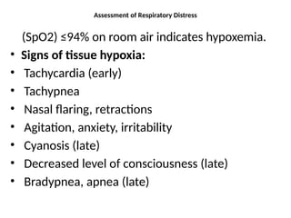
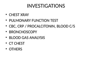
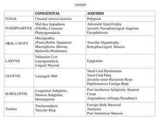
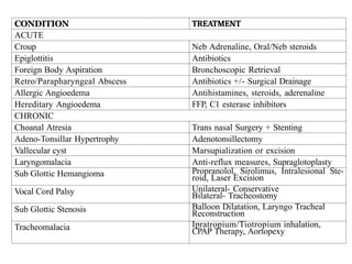
STRIDOR
HOW TO WRITE & REMEMBER
• CAUSES
• CLINICAL FEAUTRES
• DIFFRENTIAL DIAGNOSIS
• INVESTIGATION
• TREATMENT
• COMPLICATIONS
• PROGNOSIS
Assessment of Respiratory Distress
(SpO2) ≤94% on room air indicates hypoxemia.
• Signs of tissue hypoxia:
• Tachycardia (early)
• Tachypnea
• Nasal flaring, retractions
• Agitation, anxiety, irritability
• Cyanosis (late)
• Decreased level of consciousness (late)
• Bradypnea, apnea (late)
INVESTIGATIONS
• CHEST XRAY
• PULMONARY FUNCTION TEST
• CBC, CRP / PROCALCITONIN, BLOOD C/S
• BRONCHOSCOPY
• BLOOD GAS ANALYSIS
• CT CHEST
• OTHERS
• A stridor is a high-pitched sound produced by
airflow turbulence through a partially
obstructed airway involving the supraglottis,
glottis, subglottis, or trachea.
LOCALIZATION
1.Inspiratory stridor- Localizes to the
supraglottis
2.Biphasic stridor- Localizes to the glottis or the
sub-glottis
3.Expiratory stridor- Localizes to the trachea
CAUSES
CONGENITAL ACQUIRED
NASAL Choanal atresia/stenosis Polyposis
NASOPHARYNX
Mid-face hypoplasia
(Pfeiffer, Crouzon)
Pharyngomalacia
Adenoidal hypertrophy
Juvenile Nasopharyngeal Angioma
Encephalocele
ORAL CAVITY
Micrognathia
(Pierre-Robin Sequence)
Macroglossia (Downs,
Beckwith-Wiedmann)
Tonsillar Hypertrophy
Retropharyngeal Abscess
LARYNX
Vallecular Cyst
Laryngomalacia
Lingual Thyroid
Epiglottitis
GLOTTIS Laryngeal Web
Vocal Cord Dysfunction
Vocal Cord Palsy
Juvenile onset Recurrent Resp
Papillomatosis Foreign Body
SUBGLOTTIS
Congenital Subglottic
Stenosis Subglottic
Hemangioma
Post Intubation Subglottic Stenosis
Croup
Angioedema (Allergic/Hereditary)
Trachea
Tracheomalacia
Vascular Ring
Foreign Body Bacterial
Tracheitis
Post Intubation Stenosis
Laryngomalacia
• the most common cause of stridor in an infant
• is inspiratory,
• typically worsens by 4-6 weeks,
• Increases with exertion being relatively silent at
rest and/or in a prone position.
• the first presentation, is often misdiagnosed as
a croup,
• Laryngomalacia is generally a benign condition that
typically gets noticed at 2-4 weeks,
• it progresses over the next 2-4 weeks, and
• stabilizes for 1-2 months after which the intensity of the
sounds starts to decrease
Red flags
• onset at birth, facial dysmorphism,poor weight gain, prior
history of intubation, hypophonia/ Aphonia , recurrent
apneic events, or an acute life threatening event
Inv
• flexible bronchoscopy or a laryngoscopy is the
gold standard investigation for confirmation of
anatomical pathologies of the airway
T/T
• For anatomical defects such as laryngomalacia
proper nutritive care is important so that
adequate weight gain can be achieved.
• GER is a common association observed in as
much as 64%
• Anti-reflux measures
• Supraglotoplasty
Croup
• Acute Laryngo-tracheobronchitis
• typically occurring in children 6months-3years
following a febrile illness
Inv
• Chest X-ray is the most commonly performed
• The steeple sign, considered specific for
croup, indicated edema/ obstruction of the
subglottis and
• can be seen in any subglottic pathologies.
T/T
• Neb Adrenaline,
• Oral/Neb steroids
CONDITION TREATMENT
ACUTE
Croup Neb Adrenaline, Oral/Neb steroids
Epiglottitis Antibiotics
Foreign Body Aspiration Bronchoscopic Retrieval
Retro/Parapharyngeal Abscess Antibiotics +/- Surgical Drainage
Allergic Angioedema Antihistamines, steroids, aderenaline
Hereditary Angioedema FFP, C1 esterase inhibitors
CHRONIC
Choanal Atresia Trans nasal Surgery + Stenting
Adeno-Tonsillar Hypertrophy Adenotonsillectomy
Vallecular cyst Marsupialization or excision
Laryngomalacia Anti-reflux measures, Supraglotoplasty
Sub Glottic Hemangioma Propranolol, Sirolimus, Intralesional Ste-
roid, Laser Excision
Vocal Cord Palsy Unilateral- Conservative
Bilateral- Tracheostomy
Sub Glottic Stenosis Balloon Dilatation, Laryngo Tracheal
Reconstruction
Tracheomalacia Ipratropium/Tiotropium inhalation,
CPAP Therapy, Aortopexy
• T/T-
• 1.ANTIBIOTICS- ampicillin, coamoxiclav, 2nd
or
3rd
gen cephalosporin,Linazolid, vancomycin ,
clindamycin, Azithromycin
• 2. PARACETAMOL
• 3. FLUID INTAKE
• 4. MAINTAINING NUTRITION
THANKYOU

stridor mbbs.pptx upper respiratory track emergency

  • 1.
  • 2.
    HOW TO WRITE& REMEMBER • CAUSES • CLINICAL FEAUTRES • DIFFRENTIAL DIAGNOSIS • INVESTIGATION • TREATMENT • COMPLICATIONS • PROGNOSIS
  • 3.
    Assessment of RespiratoryDistress (SpO2) ≤94% on room air indicates hypoxemia. • Signs of tissue hypoxia: • Tachycardia (early) • Tachypnea • Nasal flaring, retractions • Agitation, anxiety, irritability • Cyanosis (late) • Decreased level of consciousness (late) • Bradypnea, apnea (late)
  • 4.
    INVESTIGATIONS • CHEST XRAY •PULMONARY FUNCTION TEST • CBC, CRP / PROCALCITONIN, BLOOD C/S • BRONCHOSCOPY • BLOOD GAS ANALYSIS • CT CHEST • OTHERS
  • 5.
    • A stridoris a high-pitched sound produced by airflow turbulence through a partially obstructed airway involving the supraglottis, glottis, subglottis, or trachea.
  • 6.
    LOCALIZATION 1.Inspiratory stridor- Localizesto the supraglottis 2.Biphasic stridor- Localizes to the glottis or the sub-glottis 3.Expiratory stridor- Localizes to the trachea
  • 7.
    CAUSES CONGENITAL ACQUIRED NASAL Choanalatresia/stenosis Polyposis NASOPHARYNX Mid-face hypoplasia (Pfeiffer, Crouzon) Pharyngomalacia Adenoidal hypertrophy Juvenile Nasopharyngeal Angioma Encephalocele ORAL CAVITY Micrognathia (Pierre-Robin Sequence) Macroglossia (Downs, Beckwith-Wiedmann) Tonsillar Hypertrophy Retropharyngeal Abscess LARYNX Vallecular Cyst Laryngomalacia Lingual Thyroid Epiglottitis GLOTTIS Laryngeal Web Vocal Cord Dysfunction Vocal Cord Palsy Juvenile onset Recurrent Resp Papillomatosis Foreign Body SUBGLOTTIS Congenital Subglottic Stenosis Subglottic Hemangioma Post Intubation Subglottic Stenosis Croup Angioedema (Allergic/Hereditary) Trachea Tracheomalacia Vascular Ring Foreign Body Bacterial Tracheitis Post Intubation Stenosis
  • 8.
    Laryngomalacia • the mostcommon cause of stridor in an infant • is inspiratory, • typically worsens by 4-6 weeks, • Increases with exertion being relatively silent at rest and/or in a prone position. • the first presentation, is often misdiagnosed as a croup,
  • 9.
    • Laryngomalacia isgenerally a benign condition that typically gets noticed at 2-4 weeks, • it progresses over the next 2-4 weeks, and • stabilizes for 1-2 months after which the intensity of the sounds starts to decrease Red flags • onset at birth, facial dysmorphism,poor weight gain, prior history of intubation, hypophonia/ Aphonia , recurrent apneic events, or an acute life threatening event
  • 10.
    Inv • flexible bronchoscopyor a laryngoscopy is the gold standard investigation for confirmation of anatomical pathologies of the airway
  • 11.
    T/T • For anatomicaldefects such as laryngomalacia proper nutritive care is important so that adequate weight gain can be achieved. • GER is a common association observed in as much as 64% • Anti-reflux measures • Supraglotoplasty
  • 12.
    Croup • Acute Laryngo-tracheobronchitis •typically occurring in children 6months-3years following a febrile illness
  • 13.
    Inv • Chest X-rayis the most commonly performed • The steeple sign, considered specific for croup, indicated edema/ obstruction of the subglottis and • can be seen in any subglottic pathologies.
  • 14.
    T/T • Neb Adrenaline, •Oral/Neb steroids
  • 15.
    CONDITION TREATMENT ACUTE Croup NebAdrenaline, Oral/Neb steroids Epiglottitis Antibiotics Foreign Body Aspiration Bronchoscopic Retrieval Retro/Parapharyngeal Abscess Antibiotics +/- Surgical Drainage Allergic Angioedema Antihistamines, steroids, aderenaline Hereditary Angioedema FFP, C1 esterase inhibitors CHRONIC Choanal Atresia Trans nasal Surgery + Stenting Adeno-Tonsillar Hypertrophy Adenotonsillectomy Vallecular cyst Marsupialization or excision Laryngomalacia Anti-reflux measures, Supraglotoplasty Sub Glottic Hemangioma Propranolol, Sirolimus, Intralesional Ste- roid, Laser Excision Vocal Cord Palsy Unilateral- Conservative Bilateral- Tracheostomy Sub Glottic Stenosis Balloon Dilatation, Laryngo Tracheal Reconstruction Tracheomalacia Ipratropium/Tiotropium inhalation, CPAP Therapy, Aortopexy
  • 16.
    • T/T- • 1.ANTIBIOTICS-ampicillin, coamoxiclav, 2nd or 3rd gen cephalosporin,Linazolid, vancomycin , clindamycin, Azithromycin • 2. PARACETAMOL • 3. FLUID INTAKE • 4. MAINTAINING NUTRITION
  • 17.