Stepwise interpretation of ECG ID279
•Download as PPT, PDF•
1 like•812 views
This ECG report is for a 57-year old woman with a history of rheumatic fever at age 17 who has been experiencing dyspnea and fatigue. The ECG shows normal sinus rhythm at 80 beats per minute, left atrial enlargement, right axis deviation, right ventricular hypertrophy, and ST-T abnormalities including ST depression in leads II, III, aVF and leads V1-V3 which may be due to right ventricular hypertrophy.
Report
Share
Report
Share

Recommended
stepwise interpretation of ECG ID279 RVH
This ECG summary is for a 57-year-old woman with a history of rheumatic fever and recent dyspnea and fatigue. The ECG shows normal sinus rhythm, left atrial enlargement, right axis deviation, and right ventricular hypertrophy. ST depression is seen in leads II, III, aVF and chest leads V1-V3, which may be due to right ventricular hypertrophy. Negative T waves are also seen in leads V4-V6, which can occur in heart failure and may be caused by ischemia.
Stepwise interpretation ECG #7
This ECG shows a 57-year-old woman with a history of rheumatic fever suffering from dyspnea and fatigue. The ECG interpretation found:
1) Normal sinus rhythm at 80 beats per minute with left atrial enlargement and right axis deviation.
2) Right ventricular hypertrophy.
3) ST depression and negative T waves that may indicate right ventricular hypertrophy or ischemia from heart failure.
ECG #7 - ID 279 - RVH
This ECG belongs to a 57-year-old woman with a history of rheumatic fever and recent symptoms of severe dyspnea and fatigue. The ECG shows normal sinus rhythm, left atrial enlargement, right axis deviation, and right ventricular hypertrophy. There are also ST depressions and negative T waves that may indicate ischemia from heart failure.
Stepwise interpretation of ECG ID168
A 78-year-old woman presented with a heart murmur. The ECG showed sinus rhythm at 77 beats per minute with sinus arrhythmia and first-degree AV block. There was no evidence of atrial enlargement and the QRS duration was normal with no conduction abnormalities. Signs of left ventricular hypertrophy and ST-T abnormalities were present, consistent with a strain pattern, but no signs of prior myocardial infarction.
Stepwise interpretation of ECG - #08 no Dx ID478
This document describes the stepwise interpretation of an ECG for a 58-year-old man with dilated cardiomyopathy. It was determined that the patient has normal sinus rhythm at a rate of 97 beats per minute, evidence of biatrial enlargement, left ventricular hypertrophy, and repolarization abnormality but no signs of myocardial infarction.
ECG #6 - ID 168 LVH
The document summarizes the stepwise interpretation of an ECG for a 78-year-old woman with a heart murmur. It finds sinus rhythm at 77 beats per minute with sinus arrhythmia and first-degree AV block. There is also evidence of left ventricular hypertrophy with ST-T abnormalities and no signs of myocardial infarction.
Ekg. hr
The document provides an overview of electrocardiography (ECG) basics, including:
- The 12-lead ECG measures the heart's electrical activity and produces normal or abnormal wave patterns depending on a patient's condition. It views the heart from different angles to determine pathology locations.
- The standard ECG leads view the heart from anterior, posterior, lateral, and inferior positions. Limb leads I, II, III and augmented leads aVR, aVL, aVF provide different angular views. Chest leads V1-V6 provide anterior and lateral views.
- A normal ECG shows a sinus rhythm with discernible P waves preceding each QRS complex. Rate, rhythm, wave morphology
Heart arrth 1
This document outlines the objectives and content of an ECG rhythm interpretation module. The module aims to teach the recognition of normal sinus rhythm, 13 common rhythm disturbances, and acute myocardial infarction on ECGs. It covers ECG basics, analyzing rhythms, normal sinus rhythm, arrhythmias including premature beats, sinus rhythms, and uses examples to explain sinus bradycardia, sinus tachycardia, premature atrial contractions, and premature ventricular contractions.
stepwise interpretation ECG #6
This document summarizes the stepwise interpretation of an ECG for a 78 year old woman with a heart murmur. It was found that she had sinus rhythm at 77 beats per minute with sinus arrhythmia. She also had first degree AV block seen by a prolonged PR interval of 230 ms. Examination of the QRS complexes and axes found normal duration and no signs of bundle branch blocks. There were findings of left ventricular hypertrophy seen with ST-T abnormalities indicating strain pattern. No signs of myocardial infarction were seen.
Stepwise interpretation of ECG - #6 no Dx ID168
This document summarizes the stepwise interpretation of an ECG for a 78 year old woman with a heart murmur. It finds sinus rhythm at 77 beats per minute with sinus arrhythmia and first degree AV block. The QRS axis and duration are normal with no signs of bundle branch blocks. There are findings of left ventricular hypertrophy with ST-T abnormalities indicating strain pattern. No signs of myocardial infarction are seen.
Ekg module 5
This document provides an overview of diagnosing myocardial infarction (MI) using electrocardiograms (ECGs). It discusses that an MI is best diagnosed using a 12-lead ECG rather than just a rhythm strip. The 12-lead ECG views the heart from 12 angles and can identify ST elevation in different regions to locate the MI. Anterior MIs involve leads V1-V4, lateral MIs involve leads I, aVL, V5-V6, and inferior MIs involve leads II, III, and aVF. The document walks through examples of anterior, lateral, inferior, and anterolateral MIs.
Ekg module 6
This document provides an overview of advanced 12-lead ECG interpretation, including recognizing ST elevation and non-ST elevation myocardial infarctions based on ECG changes over time, identifying left ventricular hypertrophy by increased QRS voltage, and distinguishing right and left bundle branch blocks by characteristic widening and changes in QRS morphology.
Understanding ECG
1. The document describes the anatomy and physiology of the heart, including its chambers, valves, conduction system, blood flow, and heart sounds.
2. Key aspects covered are the structure and function of the heart, cardiac cycle, electrocardiography, cardiac arrhythmias, murmurs, and approach to cardiac patients.
3. Details are provided on abnormalities like bundle branch blocks, hypertrophies, conduction disorders, and various arrhythmias that can be identified on ECG.
acls
This document discusses electrocardiography (ECG) and acute coronary syndromes. It begins by describing the anatomy and physiology of the heart. It then explains the electrical conduction system of the heart and how ECG is used to diagnose cardiac arrhythmias and ischemia. The document outlines the components of an ECG strip and how to interpret common abnormalities including signs of myocardial infarction. It states that acute coronary syndromes like unstable angina and non-ST elevation myocardial infarction can occur due to coronary artery atherosclerosis and blockages in different heart regions will produce varying ECG patterns of ischemia.
ECG #12 - ID 111 - CHB
This document discusses the stepwise interpretation of an ECG for a patient. It identifies that the patient, a 56-year-old man who had an episode of syncope, has sinus rhythm with complete heart block. The atrial rate is 80/min while the ventricular rate is 43/min, showing dissociation between the two rhythms. The QRS complexes originate from the AV junction at a rate of 43/min, indicating an escape junctional rhythm. The final diagnosis is sinus rhythm with complete heart block and an escape junctional rhythm supplying the ventricles.
ECG
The document provides information on how to perform and interpret an electrocardiogram (ECG). It describes the conduction system of the heart and how the electrical impulse is generated and travels through the heart. It outlines the procedure for performing an ECG, including electrode placement and machine settings. Key intervals and waves are defined, such as the P wave, PR interval, QRS complex, ST segment, T wave, and QT interval. Abnormal findings are discussed. The document is a comprehensive guide on ECG basics.
Normal sinus rhythm
Normal sinus rhythm is characterized by a regular rhythm originating from the sinus node between 60-100 beats per minute. Each impulse follows the normal conduction pathway with visible P waves preceding upright QRS complexes and normal PR and QT intervals. Normal sinus rhythm serves as the standard against which all other cardiac rhythms are compared.
Amber Bryant - ECG Outline
The document describes various types of cardiac rhythms that can be identified based on characteristics of electrocardiogram (ECG) readings such as heart rate, presence of P waves, PR interval duration, and morphology of QRS complexes. Rhythms include normal sinus rhythm, various types of heart block, bradycardia, tachycardia, fibrillation, and escape rhythms originating in the junctional tissues or ventricles. Precise heart rates are given to define categories such as bradycardia, tachycardia, and accelerated rhythms. Premature beats and pauses are also described based on where they occur in the cardiac cycle.
Amber Bryant - ECG Flowchart
This document provides a summary of different types of cardiac rhythms that can be seen on an electrocardiogram (ECG) and their defining characteristics such as heart rate, presence of P waves, and regularity of the rhythm. Rhythms include normal sinus rhythm, various types of heart block, atrial and ventricular arrhythmias, junctional rhythms, and cardiac pacing. For each rhythm, the summary identifies the key features on the ECG that are used to classify it.
Rhythms
The document discusses cardiac rhythms and monitoring. It provides information on:
1) Electrode placements for cardiac monitoring and which leads are best for viewing different waveforms like P waves and QRS complexes.
2) How to interpret a rhythm strip by examining features like heart rate, rhythm regularity, presence of atrial activity, and relationship between atrial and ventricular rhythms.
3) Causes and characteristics of different cardiac rhythms like bradycardias, tachycardias, conduction abnormalities, and rhythms associated with cardiac arrest.
ecg interpretation Module 2
This document outlines the steps for analyzing ECG rhythms:
1) Calculate the heart rate by counting R waves over 6 seconds or finding an R wave that lands on a bold line and counting boxes until the next R wave
2) Determine if the rhythm is regular or irregular
3) Assess the P waves for presence, appearance, and rate
4) Measure the PR interval and compare to the normal range of 0.12 to 0.20 seconds
5) Measure the QRS duration and compare to the normal range of 0.04 to 0.12 seconds
It provides an example of analyzing a rhythm that has a rate of 90-95 bpm, is regular, has normal P waves,
Ecg in children
Presentation on basic principles of pediatric ecg with important examples: BY Dr. Nivedita Mishra (PGY2 PEDIATRICS, TRIBHUVAN UNIVERSITY TEACHING HOSPITAL,KATHMANDU,NEPAL)
Ecg1 elkhatib
This document discusses cardiac rhythms and electrocardiogram (ECG) interpretation. It provides normal and abnormal heart rate ranges for the sinoatrial node, atrioventricular node, and bundle branches. It also describes ECG patterns such as sinus rhythm, arrhythmias, conduction abnormalities, myocardial infarction, and other cardiac conditions. Measurement techniques for rate, intervals, and waveform analysis on ECG strips are outlined.
Stepwise interpretation of ECG - #07 no Dx ID259
This ECG shows a 57-year-old woman with a history of rheumatic fever suffering from dyspnea and fatigue. The ECG reveals normal sinus rhythm, left atrial enlargement, right axis deviation, and right ventricular hypertrophy. There are also ST depressions and negative T waves that are likely due to right ventricular hypertrophy and may indicate ischemia as the patient has signs of heart failure.
Stepwise interpretation of ECG - #7 no Dx ID259
This ECG shows a 57-year-old woman with a history of rheumatic fever suffering from dyspnea and fatigue. The ECG reveals normal sinus rhythm, left atrial enlargement, right axis deviation, and right ventricular hypertrophy. There are also ST depressions and negative T waves that are likely due to right ventricular hypertrophy and may indicate ischemia as the patient has signs of heart failure.
ECG#8 - ID478 - LVH BAE
The document discusses an ECG reading of a 58-year-old man with dilated cardiomyopathy. It finds evidence of biatrial enlargement from enlarged P waves and left ventricular hypertrophy shown by a widened QRS complex and ST abnormality, with a heart rate of 97 beats per minute and no signs of myocardial infarction.
Stepwise interpretation of ECG ID175
A 58-year-old man with coronary artery disease was followed in the Cardiac Clinic. An electrocardiogram found normal sinus rhythm at 88 beats per minute with left bundle branch block, evidenced by a prolonged QRS duration of 145 milliseconds and abnormal depolarization that can mask other conditions.
ECG #5 - ID 168 - Left bundle branch block
A 58-year-old man followed for coronary artery disease presented with an ECG showing sinus rhythm at 88 beats per minute, a prolonged QRS duration of 145 milliseconds consistent with left bundle branch block, and no evidence of right or left ventricular hypertrophy or atrial enlargement. The left bundle branch block precludes diagnosing left ventricular hypertrophy and can mask underlying myocardial infarction.
Interpretation of ecg_in_pulmonary_disease
This document discusses ECG findings in patients with pulmonary diseases such as chronic obstructive pulmonary disease (COPD) and pulmonary embolism (PE). It describes changes seen in COPD such as right axis deviation, low voltage complexes, and right ventricular hypertrophy. It also outlines typical findings in PE including sinus tachycardia, right ventricular strain pattern, complete or incomplete right bundle branch block, and S1Q3T3 pattern. The document provides details on diagnostic criteria for right atrial enlargement, right ventricular hypertrophy, and right bundle branch block.
More Related Content
What's hot
stepwise interpretation ECG #6
This document summarizes the stepwise interpretation of an ECG for a 78 year old woman with a heart murmur. It was found that she had sinus rhythm at 77 beats per minute with sinus arrhythmia. She also had first degree AV block seen by a prolonged PR interval of 230 ms. Examination of the QRS complexes and axes found normal duration and no signs of bundle branch blocks. There were findings of left ventricular hypertrophy seen with ST-T abnormalities indicating strain pattern. No signs of myocardial infarction were seen.
Stepwise interpretation of ECG - #6 no Dx ID168
This document summarizes the stepwise interpretation of an ECG for a 78 year old woman with a heart murmur. It finds sinus rhythm at 77 beats per minute with sinus arrhythmia and first degree AV block. The QRS axis and duration are normal with no signs of bundle branch blocks. There are findings of left ventricular hypertrophy with ST-T abnormalities indicating strain pattern. No signs of myocardial infarction are seen.
Ekg module 5
This document provides an overview of diagnosing myocardial infarction (MI) using electrocardiograms (ECGs). It discusses that an MI is best diagnosed using a 12-lead ECG rather than just a rhythm strip. The 12-lead ECG views the heart from 12 angles and can identify ST elevation in different regions to locate the MI. Anterior MIs involve leads V1-V4, lateral MIs involve leads I, aVL, V5-V6, and inferior MIs involve leads II, III, and aVF. The document walks through examples of anterior, lateral, inferior, and anterolateral MIs.
Ekg module 6
This document provides an overview of advanced 12-lead ECG interpretation, including recognizing ST elevation and non-ST elevation myocardial infarctions based on ECG changes over time, identifying left ventricular hypertrophy by increased QRS voltage, and distinguishing right and left bundle branch blocks by characteristic widening and changes in QRS morphology.
Understanding ECG
1. The document describes the anatomy and physiology of the heart, including its chambers, valves, conduction system, blood flow, and heart sounds.
2. Key aspects covered are the structure and function of the heart, cardiac cycle, electrocardiography, cardiac arrhythmias, murmurs, and approach to cardiac patients.
3. Details are provided on abnormalities like bundle branch blocks, hypertrophies, conduction disorders, and various arrhythmias that can be identified on ECG.
acls
This document discusses electrocardiography (ECG) and acute coronary syndromes. It begins by describing the anatomy and physiology of the heart. It then explains the electrical conduction system of the heart and how ECG is used to diagnose cardiac arrhythmias and ischemia. The document outlines the components of an ECG strip and how to interpret common abnormalities including signs of myocardial infarction. It states that acute coronary syndromes like unstable angina and non-ST elevation myocardial infarction can occur due to coronary artery atherosclerosis and blockages in different heart regions will produce varying ECG patterns of ischemia.
ECG #12 - ID 111 - CHB
This document discusses the stepwise interpretation of an ECG for a patient. It identifies that the patient, a 56-year-old man who had an episode of syncope, has sinus rhythm with complete heart block. The atrial rate is 80/min while the ventricular rate is 43/min, showing dissociation between the two rhythms. The QRS complexes originate from the AV junction at a rate of 43/min, indicating an escape junctional rhythm. The final diagnosis is sinus rhythm with complete heart block and an escape junctional rhythm supplying the ventricles.
ECG
The document provides information on how to perform and interpret an electrocardiogram (ECG). It describes the conduction system of the heart and how the electrical impulse is generated and travels through the heart. It outlines the procedure for performing an ECG, including electrode placement and machine settings. Key intervals and waves are defined, such as the P wave, PR interval, QRS complex, ST segment, T wave, and QT interval. Abnormal findings are discussed. The document is a comprehensive guide on ECG basics.
Normal sinus rhythm
Normal sinus rhythm is characterized by a regular rhythm originating from the sinus node between 60-100 beats per minute. Each impulse follows the normal conduction pathway with visible P waves preceding upright QRS complexes and normal PR and QT intervals. Normal sinus rhythm serves as the standard against which all other cardiac rhythms are compared.
Amber Bryant - ECG Outline
The document describes various types of cardiac rhythms that can be identified based on characteristics of electrocardiogram (ECG) readings such as heart rate, presence of P waves, PR interval duration, and morphology of QRS complexes. Rhythms include normal sinus rhythm, various types of heart block, bradycardia, tachycardia, fibrillation, and escape rhythms originating in the junctional tissues or ventricles. Precise heart rates are given to define categories such as bradycardia, tachycardia, and accelerated rhythms. Premature beats and pauses are also described based on where they occur in the cardiac cycle.
Amber Bryant - ECG Flowchart
This document provides a summary of different types of cardiac rhythms that can be seen on an electrocardiogram (ECG) and their defining characteristics such as heart rate, presence of P waves, and regularity of the rhythm. Rhythms include normal sinus rhythm, various types of heart block, atrial and ventricular arrhythmias, junctional rhythms, and cardiac pacing. For each rhythm, the summary identifies the key features on the ECG that are used to classify it.
Rhythms
The document discusses cardiac rhythms and monitoring. It provides information on:
1) Electrode placements for cardiac monitoring and which leads are best for viewing different waveforms like P waves and QRS complexes.
2) How to interpret a rhythm strip by examining features like heart rate, rhythm regularity, presence of atrial activity, and relationship between atrial and ventricular rhythms.
3) Causes and characteristics of different cardiac rhythms like bradycardias, tachycardias, conduction abnormalities, and rhythms associated with cardiac arrest.
ecg interpretation Module 2
This document outlines the steps for analyzing ECG rhythms:
1) Calculate the heart rate by counting R waves over 6 seconds or finding an R wave that lands on a bold line and counting boxes until the next R wave
2) Determine if the rhythm is regular or irregular
3) Assess the P waves for presence, appearance, and rate
4) Measure the PR interval and compare to the normal range of 0.12 to 0.20 seconds
5) Measure the QRS duration and compare to the normal range of 0.04 to 0.12 seconds
It provides an example of analyzing a rhythm that has a rate of 90-95 bpm, is regular, has normal P waves,
Ecg in children
Presentation on basic principles of pediatric ecg with important examples: BY Dr. Nivedita Mishra (PGY2 PEDIATRICS, TRIBHUVAN UNIVERSITY TEACHING HOSPITAL,KATHMANDU,NEPAL)
Ecg1 elkhatib
This document discusses cardiac rhythms and electrocardiogram (ECG) interpretation. It provides normal and abnormal heart rate ranges for the sinoatrial node, atrioventricular node, and bundle branches. It also describes ECG patterns such as sinus rhythm, arrhythmias, conduction abnormalities, myocardial infarction, and other cardiac conditions. Measurement techniques for rate, intervals, and waveform analysis on ECG strips are outlined.
What's hot (16)
Similar to Stepwise interpretation of ECG ID279
Stepwise interpretation of ECG - #07 no Dx ID259
This ECG shows a 57-year-old woman with a history of rheumatic fever suffering from dyspnea and fatigue. The ECG reveals normal sinus rhythm, left atrial enlargement, right axis deviation, and right ventricular hypertrophy. There are also ST depressions and negative T waves that are likely due to right ventricular hypertrophy and may indicate ischemia as the patient has signs of heart failure.
Stepwise interpretation of ECG - #7 no Dx ID259
This ECG shows a 57-year-old woman with a history of rheumatic fever suffering from dyspnea and fatigue. The ECG reveals normal sinus rhythm, left atrial enlargement, right axis deviation, and right ventricular hypertrophy. There are also ST depressions and negative T waves that are likely due to right ventricular hypertrophy and may indicate ischemia as the patient has signs of heart failure.
ECG#8 - ID478 - LVH BAE
The document discusses an ECG reading of a 58-year-old man with dilated cardiomyopathy. It finds evidence of biatrial enlargement from enlarged P waves and left ventricular hypertrophy shown by a widened QRS complex and ST abnormality, with a heart rate of 97 beats per minute and no signs of myocardial infarction.
Stepwise interpretation of ECG ID175
A 58-year-old man with coronary artery disease was followed in the Cardiac Clinic. An electrocardiogram found normal sinus rhythm at 88 beats per minute with left bundle branch block, evidenced by a prolonged QRS duration of 145 milliseconds and abnormal depolarization that can mask other conditions.
ECG #5 - ID 168 - Left bundle branch block
A 58-year-old man followed for coronary artery disease presented with an ECG showing sinus rhythm at 88 beats per minute, a prolonged QRS duration of 145 milliseconds consistent with left bundle branch block, and no evidence of right or left ventricular hypertrophy or atrial enlargement. The left bundle branch block precludes diagnosing left ventricular hypertrophy and can mask underlying myocardial infarction.
Interpretation of ecg_in_pulmonary_disease
This document discusses ECG findings in patients with pulmonary diseases such as chronic obstructive pulmonary disease (COPD) and pulmonary embolism (PE). It describes changes seen in COPD such as right axis deviation, low voltage complexes, and right ventricular hypertrophy. It also outlines typical findings in PE including sinus tachycardia, right ventricular strain pattern, complete or incomplete right bundle branch block, and S1Q3T3 pattern. The document provides details on diagnostic criteria for right atrial enlargement, right ventricular hypertrophy, and right bundle branch block.
Stepwise interpretation of ECG - #05 no Dx ID175
A 58-year-old man with a history of coronary artery disease presented with an ECG showing normal sinus rhythm of 88 beats per minute. The ECG revealed left bundle branch block, evidenced by a prolonged QRS duration of 145 milliseconds with characteristic morphology. Left bundle branch block can mask signs of left ventricular hypertrophy and prior myocardial infarction. It is also associated with secondary repolarization abnormalities like ST depression and negative T waves in leads with broad R waves. The final diagnosis was normal sinus rhythm with left bundle branch block.
Stepwise interpretation of ECG - #5 no Dx ID175
A 58-year-old man with a history of coronary artery disease presented with an ECG showing normal sinus rhythm of 88 beats per minute. The ECG revealed left bundle branch block, evidenced by a prolonged QRS duration of 145 milliseconds with characteristic morphology. Left bundle branch block can mask signs of left ventricular hypertrophy and prior myocardial infarction. It is also associated with secondary repolarization abnormalities like ST depression and negative T waves in leads with broad R waves. The final diagnosis was normal sinus rhythm with left bundle branch block.
Stepwise interpretation ECG #5
A 58-year-old man with a history of coronary artery disease presented with normal sinus rhythm of 88 beats per minute and left bundle branch block. Left bundle branch block was diagnosed due to the prolonged QRS duration of 145 milliseconds with characteristic morphology. The presence of left bundle branch block can mask signs of left ventricular hypertrophy and prior myocardial infarction on electrocardiogram. Abnormal repolarization is also commonly seen in the form of ST depression and negative T waves in leads with broad R waves in left bundle branch block.
Stepwise interpretation of ECG - #5 no Dx ID659
A 58-year-old man with a history of coronary artery disease presented with an ECG showing normal sinus rhythm of 88 beats per minute. The ECG revealed left bundle branch block, evidenced by a prolonged QRS duration of 145 milliseconds with characteristic morphology. Left bundle branch block can mask signs of left ventricular hypertrophy and prior myocardial infarction. It is also associated with secondary repolarization abnormalities like ST depression and negative T waves in leads with broad R waves. The final diagnosis was normal sinus rhythm with left bundle branch block.
Stepwise interpretation of ECG - #1 ID 380
This document summarizes the interpretation of an electrocardiogram (ECG) for a 53-year-old man. The ECG shows normal sinus rhythm at a rate of 60 beats per minute, with normal P waves, PR interval, QRS duration and axis, and no signs of atrial or ventricular enlargement, bundle branch blocks, or myocardial infarction. The overall interpretation is that the ECG is normal.
Stepwise interpretation of ECG - #06 no Dx ID168
This document summarizes the stepwise interpretation of an ECG for a 78 year old woman with a heart murmur. It was found that she had sinus rhythm at 77 beats per minute with sinus arrhythmia and first degree AV block. The QRS duration was normal with no signs of bundle branch blocks. There were findings of left ventricular hypertrophy with ST-T abnormalities indicating a strain pattern. The final diagnosis was sinus rhythm with sinus arrhythmia, first degree AV block, and left ventricular hypertrophy with repolarization abnormality.
Stepwise interpretation of ECG - #06 no Dx ID168
This document summarizes the stepwise interpretation of an ECG for a 78 year old woman with a heart murmur. It finds sinus rhythm at 77 beats per minute with sinus arrhythmia and first degree AV block. The QRS axis and duration are normal with no signs of bundle branch blocks. There are findings of left ventricular hypertrophy with ST-T abnormalities indicating strain pattern. No signs of myocardial infarction are seen.
Stepwise interpretation of ECG - #06 no Dx ID168
A 78-year-old woman presented with a heart murmur. The ECG showed sinus rhythm at 77 beats per minute with sinus arrhythmia. There was first-degree AV block seen by a prolonged PR interval of 230 ms. Findings also included left ventricular hypertrophy with ST-T abnormalities consistent with left ventricular strain.
Stepwise interpretation of ECG - #1 ID 380
A 53-year-old man received an ECG in the pre-anesthesia clinic. The ECG interpreter found normal sinus rhythm at a rate of 60 beats per minute, with normal P waves, PR interval, QRS axis and duration, and no signs of chamber enlargement, bundle branch blocks, ventricular hypertrophy, or myocardial infarction. The final diagnosis was a completely normal ECG.
Stepwise interpretation of ECG - #01 ID 380
A 53-year-old man received an ECG in the pre-anesthesia clinic. The ECG interpreter found normal sinus rhythm at a rate of 60 beats per minute, with normal P waves, PR interval, QRS axis and duration, and no signs of chamber enlargement, bundle branch blocks, ventricular hypertrophy, or myocardial infarction. The final diagnosis was a completely normal ECG.
Stepwise interpretation of ECG - #1 ID 380
A 53-year-old man received an ECG in the pre-anesthesia clinic. The ECG interpreter found normal sinus rhythm at a rate of 60 beats per minute, with normal P waves, PR interval, QRS axis and duration, and no signs of chamber enlargement, bundle branch blocks, ventricular hypertrophy, or myocardial infarction. The final diagnosis was a completely normal ECG.
stepwise interpretation ECG #1
A 53-year-old man received an ECG in the pre-anesthesia clinic. The ECG interpreter found normal sinus rhythm at a rate of 60 beats per minute, with normal P waves, PR interval, QRS axis and duration, and no signs of chamber enlargement, bundle branch blocks, ventricular hypertrophy, or myocardial infarction. The final diagnosis was a completely normal ECG.
#01 ID380 - NML
This document summarizes the stepwise interpretation of a 53-year-old man's ECG in the pre-anesthesia clinic. The interpreter found the man to have a normal sinus rhythm of 60 beats per minute, with normal P waves, PR interval, QRS axis and duration, and no signs of atrial or ventricular enlargement, bundle branch blocks, or evidence of myocardial infarction. The final diagnosis was a completely normal ECG and normal sinus rhythm.
Stepwise interpretation - ECG #1
A 53-year-old man received an ECG in the pre-anesthesia clinic. The ECG interpreter found normal sinus rhythm at a rate of 60 beats per minute, with normal P waves, PR interval, QRS axis and duration, and no signs of chamber enlargement, bundle branch blocks, ventricular hypertrophy, or myocardial infarction. The final diagnosis was a completely normal ECG.
Similar to Stepwise interpretation of ECG ID279 (20)
More from Anas Nader
ECG #14 - ID 123 - Atrial Tachycardia
A 67-year-old woman presented with palpitations. An ECG showed a narrow-QRS tachycardia at a rate of 160 beats per minute, with visible P waves preceding each QRS complex. The P waves were negative in leads II, III, aVF and V3-V6, with a PR interval of 140 msec. Based on these characteristics, the patient was diagnosed with atrial tachycardia originating from the atria.
ECG #13 - ID 419 - EAR IRBBB
This ECG shows an ectopic atrial rhythm originating from a low atrial region at a rate of 70 beats per minute, as well as an incomplete right bundle branch block. P waves are clearly seen and negative in several leads, with a PR interval of 180ms indicating the rhythm is not junctional. The QRS axis is normal and duration is 100ms with an rSR' pattern consistent with incomplete right bundle branch block. The final diagnosis is an ectopic atrial rhythm at 70bpm with incomplete right bundle branch block.
ECG #11 - ID 108 - 2nd degree AV block
This document summarizes the ECG findings for a 58-year-old man presenting to the emergency department with 2nd degree AV block, Mobitz type 1. The ECG shows regular sinus rhythm at a rate of 88 beats per minute with intermittent conduction block such that every third P-wave is not followed by a QRS complex, resulting in a ventricular rate of 59 beats per minute. Further analysis of the ECG found normal QRS axis, duration and morphology with no signs of infarction, hypertrophy, or other abnormalities. The final diagnosis was sinus rhythm with Mobitz type 1 2nd degree AV block and a ventricular rate of 58 beats per minute.
ECG #10 - ID122 - Atrial Flutter
This document describes the stepwise interpretation of ECG ID 122, which shows atrial flutter with variable atrioventricular block and a ventricular rate of 107 beats per minute in a 50-year-old man complaining of palpitations. The interpretation examines the P waves, QRS complex, QRS duration, signs of ventricular hypertrophy or myocardial infarction, and T waves. The final diagnosis is atrial flutter with variable AV block and a ventricular rate of 107 beats per minute.
ECG #9 - ID594a - acute ASMI
This ECG shows signs of an acute antero-septal myocardial infarction in a 49-year old man presenting with chest pain, requiring urgent therapy. The ECG reveals normal sinus rhythm, a normal QRS axis and duration, and marked ST elevation in leads V1-V4, consistent with an antero-septal injury pattern diagnostic of an acute antero-septal infarction.
ECG #2 - ID 352 - Premature Atrial Complexes
A 62-year-old man presented with palpitations. An analysis of his ECG found sinus rhythm at 83 bpm with premature atrial complexes, where every third P wave was premature in a pattern of trigeminy. The QRS complexes, ST segments, and T waves were normal with no signs of right or left atrial enlargement, bundle branch blocks, ventricular hypertrophy, or myocardial infarction. The final diagnosis was sinus rhythm with atrial premature complexes in a pattern of trigeminy, but an otherwise normal ECG.
ECG #1 - ID 380 – Normal ECG
The ECG is for a 53-year-old man undergoing pre-anesthesia evaluation. The interpreter found normal sinus rhythm at a rate of 60 bpm, normal P waves and PR interval, normal QRS axis and duration with no evidence of conduction blocks, no signs of chamber enlargement or hypertrophy, and normal ST segments and T waves. The final diagnosis was a normal ECG.
ECG #4 - ID 383 – Atrial fibrillation
A 76-year-old woman underwent an ECG examination in her family doctor's office which showed atrial fibrillation with an irregularly irregular ventricular rhythm of 66 beats per minute and no P waves. The QRS complexes were normal with no signs of conduction abnormalities, ventricular hypertrophy, or prior myocardial infarction. The final diagnosis was atrial fibrillation with a controlled ventricular rate of 66 bpm and no other abnormalities.
ID 352 –Sinus rhythm With premature atrial complex
A 62-year-old man presented with palpitations. An analysis of his ECG found sinus rhythm at 83 bpm with premature atrial complexes, where every third P wave was premature in a pattern of trigeminy. The ECG further showed normal P waves, QRS axis, duration and complexes, as well as normal ST segments and T waves. The final diagnosis was sinus rhythm with atrial premature complexes in a pattern of trigeminy, but an otherwise normal ECG.
Atrio-ventricular dissociation
Atrio-ventricular dissociation occurs when the atrial and ventricular rhythms are independent of each other. It can happen when the atrial rate slows below the intrinsic ventricular rate, or due to inappropriate acceleration of the ventricular rate. Typically in AV dissociation the distal pacemaker, such as the ventricles, beats faster than the proximal pacemaker like the atria. In complete heart block the proximal pacemaker usually beats faster than the distal one. However, there are exceptions to using pacemaker rates to distinguish the two.
Lead errors: reversal of limb leads
This document discusses common errors involving limb leads in electrocardiograms (ECGs), including:
1) Lead reversal is a common error, such as switching the right and left arm leads. This can cause leads like aVR and aVL to be reversed.
2) Misplacing the ground cable, such as attaching it to the right arm instead of the right leg, is another error.
3) Complex misplacements of the limb leads are less common. Examples provided demonstrate how specific lead errors can cause abnormalities to appear or disappear on ECG tracings.
Stepwise interpretation of ECG - #9 no Dx ID 594
This ECG shows a 49-year-old man experiencing severe chest pain for 1 hour who is presenting with an antero-septal injury pattern consistent with an acute myocardial infarction requiring urgent therapy. The ECG shows normal sinus rhythm at 84 beats per minute, a normal PR interval and QRS axis, and no signs of ventricular hypertrophy. It does reveal marked ST elevation in leads V1-V4, confirming the diagnosis of an acute antero-septal infarction.
Stepwise interpretation of ECG - #13 no Dx ID 419
This ECG shows an ectopic atrial rhythm originating outside the sinoatrial node at a rate of 75 beats per minute. The P waves precede each QRS complex but have an abnormal axis. There is also evidence of incomplete right bundle branch block seen by a widened QRS complex with a duration of 100 milliseconds and rSR' pattern. The final diagnosis is an ectopic atrial rhythm at 75 bpm with incomplete right bundle branch block and no signs of ventricular hypertrophy, myocardial infarction, or abnormal ST segments or T waves.
Stepwise interpretation of ECG - #13 no Dx ID 419
This ECG shows an ectopic atrial rhythm originating outside the sinoatrial node at a rate of 75 beats per minute. The P waves have an abnormal axis and precede each QRS complex, indicating the atrial origin. There is also an incomplete right bundle branch block seen by the prolonged QRS duration of 100 milliseconds and rSR' pattern. The final diagnosis is an ectopic atrial rhythm at a rate of 75 bpm with an incomplete right bundle branch block.
Stepwise interpretation of ECG - #13 no Dx ID 419
This ECG shows an ectopic atrial rhythm at a rate of 70 beats per minute with an incomplete right bundle branch block pattern. The QRS axis is normal and the duration is 100 msec. with an rSR' configuration. There are no signs of ventricular hypertrophy, myocardial infarction, or abnormal ST segments or T waves. The final diagnosis is an ectopic atrial rhythm at 75 beats per minute with an incomplete right bundle branch block.
Stepwise interpretation of ECG - #13 no Dx ID 419
Ectopic atrial rhythm at a rate of 75 beats per minute. P waves are clearly seen and negative in several leads, indicating they do not originate from the sinus node but likely a low atrial region. There is also an incomplete right bundle branch block present as shown by a prolonged QRS duration of 100 milliseconds with an rSR' pattern. The final diagnosis is an ectopic atrial rhythm at a rate of 75 beats per minute with an incomplete right bundle branch block.
Stepwise interpretation of ECG - #13 no Dx ID 419
Ectopic atrial rhythm at a rate of 75 beats per minute. P waves are clearly seen and negative in several leads, indicating they do not originate from the sinus node but likely a low atrial region. There is also an incomplete right bundle branch block present as shown by a widened QRS duration of 100 milliseconds with an rSR' pattern. The final diagnosis is an ectopic atrial rhythm at a rate of 75 beats per minute with an incomplete right bundle branch block.
Stepwise interpretation of ECG - #12 no Dx ID 111
A 56-year-old man presented with an episode of syncope. An ECG was performed which showed sinus rhythm with complete heart block, where the atrial rate was 80/min and the ventricular rate was 43/min. The dissociation of the P waves and QRS complexes along with the narrow QRS escape rhythm indicated the ventricular activation was coming from the AV junctional area. The ECG was otherwise normal.
Stepwise interpretation of ECG - #14 no Dx ID123
A 67-year-old woman presented with palpitations. Her ECG showed a narrow-QRS tachycardia of 160 beats per minute. P waves were visible and preceded the QRS complexes, with a PR interval of 140 msec. The arrhythmia was diagnosed as atrial tachycardia based on the P wave morphology and timing.
More from Anas Nader (20)
ID 352 –Sinus rhythm With premature atrial complex
ID 352 –Sinus rhythm With premature atrial complex
Stepwise interpretation of ECG ID279
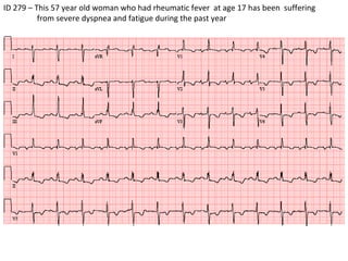
- 1. ID 279 – This 57 year old woman who had rheumatic fever at age 17 has been suffering from severe dyspnea and fatigue during the past year
- 2. ID 279 – Normal sinus rhythm, 80/min Yes: The P waves originate from the sinus node– The rhythm is regular , the rate is 80/min. – Each P is followed by a QRS - The PR interval is normal – NORMAL SINUS RHYTHM, 80/min
- 3. ID 279 – Normal sinus rhythm, 80/min – Left atrial enlargement There are signs of left atrial enlargement
- 4. Let’s now look at the QRS complexes: There is Right axis deviation ID 279 – Normal sinus rhythm, 80/min – Left atrial enlargement – Right axis deviation
- 5. Let’s now look at the QRS complexes: There is Right axis deviation ID 279 – Normal sinus rhythm, 80/min – Left atrial enlargement – Right axis deviation
- 6. The QRS duration is normal : There is no right bundle branch block, left bundle branch block or non-specific block ID 279 – Normal sinus rhythm, 80/min – Left atrial enlargement – Right axis deviation
- 7. There is right ventricular hypertrophy ID 279 – Normal sinus rhythm, 80/min – Left atrial enlargement – Right axis deviation Right ventricular hypertrophy
- 8. There are no QRS signs of myocardial infarction ID 279 – Normal sinus rhythm, 80/min – Left atrial enlargement – Right axis deviation Right ventricular hypertrophy
- 9. There is ST depression in II, III, aVF and the right chest leads (V1-V3) may be due to RVH – There are negative T waves in V4-V6. Diffuse T changes are not uncommon in patients who are in heart failure. They may be due to ischemia ID 279 – Normal sinus rhythm, 80/min – Left atrial enlargement – Right axis deviation Right ventricular hypertrophy
- 10. There is ST depression in II, III, aVF and the right chest leads (V1-V3) may be due to RVH – There are negative T waves in V4-V6. Diffuse T changes are not uncommon in patients who are in heart failure. They may be due to ischemia ID 279 – Final diagnosis: Normal sinus rhythm, 80/min – Left atrial enlargement – Right axis deviation Right ventricular hypertrophy with ST-T abnormalities