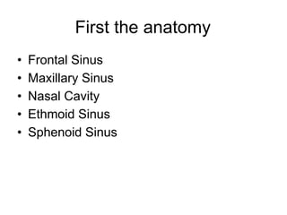
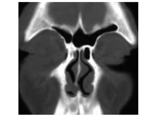
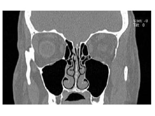
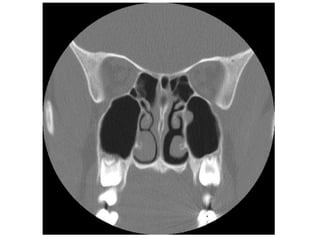
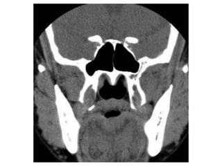
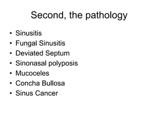
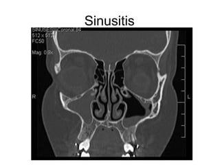
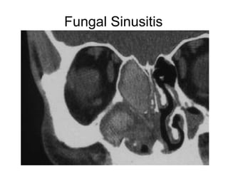
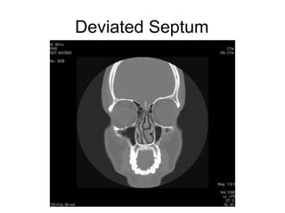
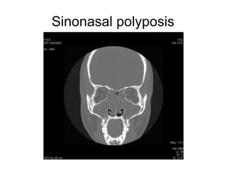
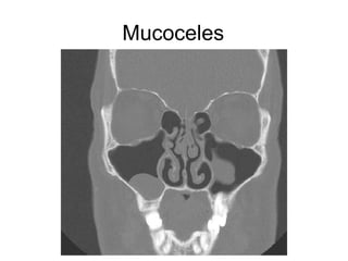
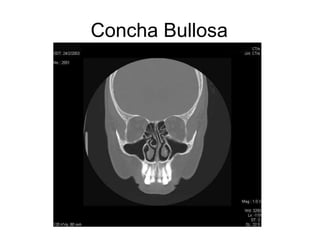
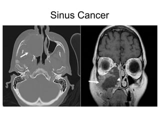
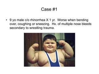
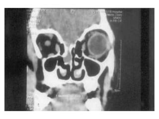
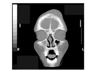
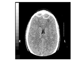
This document discusses sinus imaging using CT scans. It first describes the different sinus cavities in the anatomy: frontal, maxillary, nasal, ethmoid and sphenoid. Second, it lists common sinus pathologies seen on CT such as sinusitis, fungal sinusitis, deviated septum, sinus polyps, mucoceles, concha bullosa and sinus cancer. Finally, it presents two case studies and asks questions to test understanding of sinus conditions and anatomy.