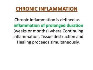
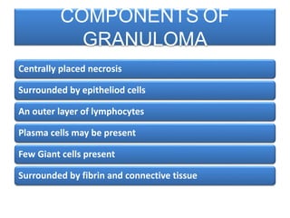
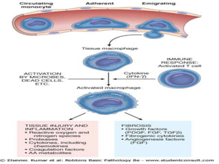
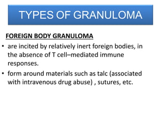
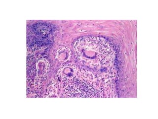
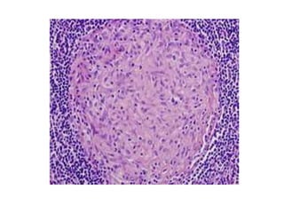
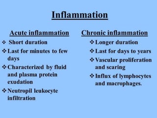
COURSE CODE: 301
Dr. UZMA NAZ
INFLAMMATION,
ACUTE INFLAMMATION :
VASCULAR AND
CELLULAR EVENTS - II
CHEMICAL MEDIATORS
OF INFLAMMATION
CHRONIC
INFLAMMATION
Chronic inflammation is defined as
inflammation of prolonged duration
(weeks or months) where Continuing
inflammation, Tissue destruction and
Healing proceeds simultaneously.
CHRONIC INFLAMMATION
MORPHOLOGIC FEATURES
• Infiltration with mononuclear cells:
- Macrophages
- T- lymphocytes
- Plasma cells
• Tissue destruction  by persistent offending
agent or by inflammatory cells.
• Attempts at healing  by connective tissue
replacement of damaged tissue
MACROPHAGES
• Macrophages are tissue cells derived from
hematopoietic stem cells in the bone marrow
and from progenitors in the embryonic yolk
sac and fetal liver during early development.
• Circulating cells of this lineage are known as
monocytes.
• They are found in specific locations in organs
such as the:
• LIVER (where they are called KUPFFER CELLS),
• SPLEEN and LYMPH NODES (called SINUS
HISTIOCYTES),
• CENTRAL NERVOUS SYSTEM (MICROGLIAL
CELLS),
• LUNGS (ALVEOLAR MACROPHAGES).
LYMPHOCYTES
• Mobilized to the setting of any specific immune
stimulus as well as non-immune-mediated
inflammation.
EOSINOPHILS
• Characteristically found in inflammatory Sites
around parasitic infections
Or
As part of immune reactions mediated by IgE,
typically associated with allergies and parasitic
infections.
MAST CELLS
• These are widely distributed in connective
tissues throughout the body,
• Participate in both acute and chronic
inflammatory responses
• “Armed" with IgE antibody specific for certain
environmental antigens.
• Response occurs during allergic reactions to
foods, insect venom, or drugs, etc.
• Distinctive pattern of chronic inflammation
characterized by formation of granulomas.
• Characterized by presence of
– Activated macrophages
– Lymphocytes
– Occasional plasma cells
• CLASSICAL EXAMPLE IS TUBERCULOSIS
GRANULOMATOUS INFLAMMATION
Centrally placed necrosis
Surrounded by epitheliod cells
An outer layer of lymphocytes
Plasma cells may be present
Few Giant cells present
Surrounded by fibrin and connective tissue
COMPONENTS OF
GRANULOMA
FOREIGN BODY GRANULOMA
• are incited by relatively inert foreign bodies, in
the absence of T cell–mediated immune
responses.
• form around materials such as talc (associated
with intravenous drug abuse) , sutures, etc.
TYPES OF GRANULOMA
• The foreign material can usually be identified
in the center of the granuloma, particularly if
viewed with polarized light, in which it
appears refractile.
IMMUNE GRANULOMAS
• caused by a variety of agents that are capable
of inducing a persistent T cell–mediated
immune response.
• This type of immune response produces
granulomas usually when the inciting agent is
difficult to eradicate, such as a persistent
microbe or a self antigen.
• In such responses, macrophages activate T
cells to produce cytokines and other
mediators which activates the macrophages
1. Bacteria
Tuberculosis
Leprosy
2. Parasites
Schistosomiasis
3. Fungi
Histoplasmosis
Blastomycosis
INFECTIOUS CAUSES
PRIMARY TUBERCULOSIS
• Extensive necrosis
drains out the necrotic
core
• Leads to cavity
formation
CAVITATORY LESION
• Involvement of lymph
nodes gives them a
matted appearance
• The centres are filled
with cheesy necrotic
material
TB LYMPH NODE
• Caseous necrosis
– Pink structureless centre
– Surrounded by lymphocytes
• Epitheliod cells
– Activated macrophages
– Pink granular cytoplasm
– Slipper shaped nucleus
• Giant cells
– Large mass of cytoplasm
– Numerous nuclei
TB- MICROSCOPY
SYSTEMIC EFFECTS OF
INFLAMMATION
• They are collectively called the acute-phase
reaction, or the systemic inflammatory
response syndrome.
• The cytokines produced by leukocytes TNF, IL-
1, and IL-6 are the most important mediators,
leading to systemic effects.
• IL-6 stimulates the hepatic synthesis of a
number of plasma proteins.
SYSTEMIC EFFECTS OF ACUTE
INFLALMMATION
• Elevation of body temperature usually by 1oC
to 4oC
• Most important manifestation especially when
inflammation is associated with infection.
• Substances that induce fever are known as
pyrogens
FEVER or PYREXIA
Neutrophilia: Increased number of
neutrophils, indicating bacterial infection.
Lymphocytosis: Increased number of
lymphocytes, indicating viral infection.
Eosinophilia: in allergy or parasitic
infection
LEUKOCYTOSIS
These are plasma proteins whose plasma
concentration may increase several hundred-
fold as part of the response to inflammatory
stimuli.
• Fibrinogen
• CRP
• SAA ( Serum amyloid A)
leads to increased ESR
Acute Phase Protein Production
In Liver
• Increased erythrocyte sedimentation rate
due to increased production of acute phase
proteins and reactants.
• In the presence of acute phase reactants
(fibrinogen) erythrocytes aggregate due to
loss of their negative charge resulting in
increased sedimentation.
Increased ESR
SHS.301.Lect-07.pptx

SHS.301.Lect-07.pptx

  • 1.
  • 2.
  • 5.
  • 8.
  • 9.
    Chronic inflammation isdefined as inflammation of prolonged duration (weeks or months) where Continuing inflammation, Tissue destruction and Healing proceeds simultaneously. CHRONIC INFLAMMATION
  • 10.
    MORPHOLOGIC FEATURES • Infiltrationwith mononuclear cells: - Macrophages - T- lymphocytes - Plasma cells • Tissue destruction  by persistent offending agent or by inflammatory cells. • Attempts at healing  by connective tissue replacement of damaged tissue
  • 11.
    MACROPHAGES • Macrophages aretissue cells derived from hematopoietic stem cells in the bone marrow and from progenitors in the embryonic yolk sac and fetal liver during early development. • Circulating cells of this lineage are known as monocytes.
  • 12.
    • They arefound in specific locations in organs such as the: • LIVER (where they are called KUPFFER CELLS), • SPLEEN and LYMPH NODES (called SINUS HISTIOCYTES), • CENTRAL NERVOUS SYSTEM (MICROGLIAL CELLS), • LUNGS (ALVEOLAR MACROPHAGES).
  • 13.
    LYMPHOCYTES • Mobilized tothe setting of any specific immune stimulus as well as non-immune-mediated inflammation.
  • 14.
    EOSINOPHILS • Characteristically foundin inflammatory Sites around parasitic infections Or As part of immune reactions mediated by IgE, typically associated with allergies and parasitic infections.
  • 15.
    MAST CELLS • Theseare widely distributed in connective tissues throughout the body, • Participate in both acute and chronic inflammatory responses • “Armed" with IgE antibody specific for certain environmental antigens. • Response occurs during allergic reactions to foods, insect venom, or drugs, etc.
  • 16.
    • Distinctive patternof chronic inflammation characterized by formation of granulomas. • Characterized by presence of – Activated macrophages – Lymphocytes – Occasional plasma cells • CLASSICAL EXAMPLE IS TUBERCULOSIS GRANULOMATOUS INFLAMMATION
  • 17.
    Centrally placed necrosis Surroundedby epitheliod cells An outer layer of lymphocytes Plasma cells may be present Few Giant cells present Surrounded by fibrin and connective tissue COMPONENTS OF GRANULOMA
  • 19.
    FOREIGN BODY GRANULOMA •are incited by relatively inert foreign bodies, in the absence of T cell–mediated immune responses. • form around materials such as talc (associated with intravenous drug abuse) , sutures, etc. TYPES OF GRANULOMA
  • 20.
    • The foreignmaterial can usually be identified in the center of the granuloma, particularly if viewed with polarized light, in which it appears refractile.
  • 22.
    IMMUNE GRANULOMAS • causedby a variety of agents that are capable of inducing a persistent T cell–mediated immune response. • This type of immune response produces granulomas usually when the inciting agent is difficult to eradicate, such as a persistent microbe or a self antigen.
  • 23.
    • In suchresponses, macrophages activate T cells to produce cytokines and other mediators which activates the macrophages
  • 26.
    1. Bacteria Tuberculosis Leprosy 2. Parasites Schistosomiasis 3.Fungi Histoplasmosis Blastomycosis INFECTIOUS CAUSES
  • 27.
  • 28.
    • Extensive necrosis drainsout the necrotic core • Leads to cavity formation CAVITATORY LESION
  • 29.
    • Involvement oflymph nodes gives them a matted appearance • The centres are filled with cheesy necrotic material TB LYMPH NODE
  • 30.
    • Caseous necrosis –Pink structureless centre – Surrounded by lymphocytes • Epitheliod cells – Activated macrophages – Pink granular cytoplasm – Slipper shaped nucleus • Giant cells – Large mass of cytoplasm – Numerous nuclei TB- MICROSCOPY
  • 31.
  • 33.
    • They arecollectively called the acute-phase reaction, or the systemic inflammatory response syndrome. • The cytokines produced by leukocytes TNF, IL- 1, and IL-6 are the most important mediators, leading to systemic effects. • IL-6 stimulates the hepatic synthesis of a number of plasma proteins. SYSTEMIC EFFECTS OF ACUTE INFLALMMATION
  • 34.
    • Elevation ofbody temperature usually by 1oC to 4oC • Most important manifestation especially when inflammation is associated with infection. • Substances that induce fever are known as pyrogens FEVER or PYREXIA
  • 36.
    Neutrophilia: Increased numberof neutrophils, indicating bacterial infection. Lymphocytosis: Increased number of lymphocytes, indicating viral infection. Eosinophilia: in allergy or parasitic infection LEUKOCYTOSIS
  • 37.
    These are plasmaproteins whose plasma concentration may increase several hundred- fold as part of the response to inflammatory stimuli. • Fibrinogen • CRP • SAA ( Serum amyloid A) leads to increased ESR Acute Phase Protein Production In Liver
  • 38.
    • Increased erythrocytesedimentation rate due to increased production of acute phase proteins and reactants. • In the presence of acute phase reactants (fibrinogen) erythrocytes aggregate due to loss of their negative charge resulting in increased sedimentation. Increased ESR