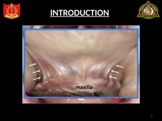
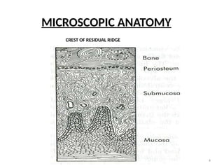
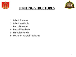
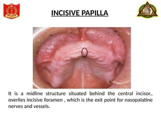
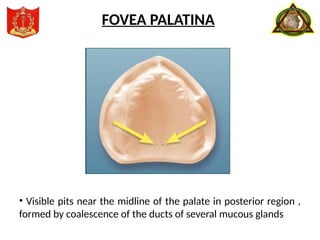
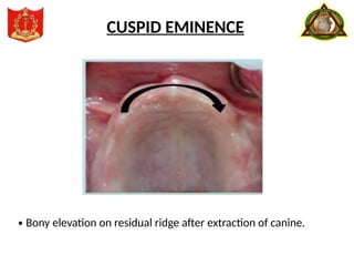
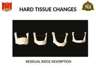
1
PRESENTED BY MODERATOR
MAJ MUNSHI KAMRUL HASAN MAJ RANDHIR SINGH
JR-1(PROSTHODONTICS)
BIOLOGICAL CONSIDERATIONS
OF MAXILLARY IMPRESSIONS
2
CONTENTS
• Introduction
• Microscopic Anatomy
• Anatomy of limiting structures.
• Behavior of the mucosa under stress.
• Tissue changes in complete denture wearers.
• Summary
• References.
3
INTRODUCTION
4
MICROSCOPIC ANATOMY
CREST OF RESIDUAL RIDGE
5
MICROSCOPIC ANATOMY
SLOPE OF RIDGE:
Loosely attached mucous membrane.
Epithelium - non keratinized.
Submucosa – has loose connective tissue so cannot
withstand the forces of mastication as well as crest of
the ridge.
6
MICROSCOPIC ANATOMY
ANTEROLATERAL AND POSTEROLATERAL PART OF THE HARD PALATE
7
MAXILLARY EDENTULOUS ARCH
LIMITING STRUCTURES SUPPORTING STRUCTURES
STRESS BEARING AREAS RELIEF AREAS
PRIMARY SECONDARY
1. Labial Frenum
2. Labial Vestibule
3. Buccal Frenum
4. Buccal Vestibule
5. Hamular Notch
6. Posterior Palatal
Seal Area 1. Hard Palate on
either side of Palatal
Raphe
2. Maxillary tuberosity
1. Rugae
2. Alveolar ridge
1. Incisive
Papilla
2. Cuspid
Eminence
3. Mid-Palatine
Raphe
4. Fovea
Palatina
9
LIMITING STRUCTURES
1. Labial Frenum
2. Labial Vestibule
3. Buccal Frenum
4. Buccal Vestibule
5. Hamular Notch
6. Posterior Palatal Seal Area
Its a fold of mucous membrane at the median line.
LABIAL FRENUM
The labial vestibule extends from buccal frenum on one side to
buccal frenum on other side and is divided into a left and right labial
vestibule by the labial frenum.
LABIAL VESTIBULE
• Its a fold of mucous membrane
• Separates labial and buccal vestibule.
BUCCAL FRENUM
It can be single fold, double, broad or fan shaped.
BUCCAL FRENUM
1. The levator anguli oris
(caninus) muscle
2. The buccinator
3. Orbicularis oris
MUSCLE ATTACHMENT
The portion of oral cavity that is bounded on one side by the teeth,
gingiva and alveolar ridge (residual alveolar ridge) on the lateral side
by the cheek posterior to the buccal frenum”
BUCCAL VESTIBULE
Size variation
• Contraction of the buccinator muscle
• Position of the mandible
• Amount of bone loss
BUCCAL VESTIBULE
Depression that lies between maxillary tuberosity & hamular
process of the medial pterygoid plate.
HAMULAR NOTCH
POSTERIOR PALATAL SEAL AREA
Effect of type of soft palate on posterior palatal seal area
POSTERIOR PALATAL SEAL AREA
SUPPORTING STRUCTURES
1. Hard Palate on
either side of Palatal Raphe
2. Residual Alveolar Ridge
1. Rugae
2. Maxillary tuberosity
PRIMARY STRESS BEARING
AREA
SECONDARY STRESS BEARING
AREA
20
HARD PALATE
The hard palate is formed by palatine process of maxillary bone &
horizontal plate of palatine bones.
21
• Epithelium is well keratinized stratified squamous epithelium.
• Anterolateral Area:Fatty zone
• Posterolateral Area: Glandular zone.
• Mid Palatine Raphe: Submucosa is extremely thin and
non resilient.
HARD PALATE
• It is a bulbuos extension of residual ridge in second and third molar
region
MAXILLARY TUBEROSITY
23
The portion of the alveolar ridge and its soft tissue covering which
remains following the removal of teeth.
RESIDUAL RIDGE
24
• Raised area of dense connective tissue in ant 1/3 of palate
• Mucosa is keratinized and the submucosa is fibrous and contains fatty
tissue
RUGAE
RELIEF AREAS
1. Incisive Papilla
2. Cuspid Eminence
3. Mid-Palatine Raphe
4. Fovea Palatina
It is a midline structure situated behind the central incisor.,
overlies incisive foramen , which is the exit point for nasopalatine
nerves and vessels.
INCISIVE PAPILLA
• Visible pits near the midline of the palate in posterior region ,
formed by coalescence of the ducts of several mucous glands
FOVEA PALATINA
· Bony elevation on residual ridge after extraction of canine.
CUSPID EMINENCE
BEHAVIOUR
OF
ORAL MUCOSA UNDER STRESS
• Mean denture bearing area
Maxilla -22.96cm2
Mandible-12.25cm2
• Area of periodontal ligament -45cm2
• Masticatory loads during chewing
For natural teeth- 44lbs(20 kg)
For artificial teeth- 13-16lbs(6-8 kgs)
• With complete dentures , the mucous membrane is forced to serve
the same purpose as the periodontal ligament that provide for the
natural teeth.
BEHAVIOUR
OF
ORAL MUCOSA UNDER STRESS
SOFT TISSUE
AND
HARD TISSUE CHANGES
• Tolerance of the person to injury differs from individual to
individual.
Denture wearer
Tolerance of mucosal tissues
If less If more
Injury & inflammation. Fibrous growth
Flabby hyperplastic
tissue
HARD TISSUE CHANGES
RESIDUAL RIDGE RESORPTION
CONCLUSION
• The scientific knowledge of denture supporting and influencing
struture forms an integral part of denture fabrication.
• The macroscopic anotomy helps in the meticulous replacement of
missing strutures.
• The microscopic level enhances the preservation of what remains.
thus the health of the tissues can be preserved.
REFERENCES
1. Zarb. Bolender:Prosthodontic Treatment for Edentulous
patients.13th
edn.the c.v.mossby co.,2004.
2. Charles M. Heartwell: Textbook of complete dentures. 5th
edition
3. Sheldon Winkler. Essentials of complete denture prosthodontics.
2nd
edition
4. Orbans:oral histology and embryology
5. Kapur,k:Effect of complete dentures on alveolar mucosa.
J.prosthet dent13:1030-1037,1963
6. Judson.C.Hickey, Charles.l.Bolender.:Bouchers Prosthodontic
Treatment for Edentulous Patients.9th
edn.The c. v. Mossy
Co,1985
7. Tencate:oral histology, development struture and function

SeminMMMMMMMMMMMMMMMMMMMMMMMMMMar-01.pptx

  • 1.
    1 PRESENTED BY MODERATOR MAJMUNSHI KAMRUL HASAN MAJ RANDHIR SINGH JR-1(PROSTHODONTICS) BIOLOGICAL CONSIDERATIONS OF MAXILLARY IMPRESSIONS
  • 2.
    2 CONTENTS • Introduction • MicroscopicAnatomy • Anatomy of limiting structures. • Behavior of the mucosa under stress. • Tissue changes in complete denture wearers. • Summary • References.
  • 3.
  • 4.
  • 5.
    5 MICROSCOPIC ANATOMY SLOPE OFRIDGE: Loosely attached mucous membrane. Epithelium - non keratinized. Submucosa – has loose connective tissue so cannot withstand the forces of mastication as well as crest of the ridge.
  • 6.
    6 MICROSCOPIC ANATOMY ANTEROLATERAL ANDPOSTEROLATERAL PART OF THE HARD PALATE
  • 7.
  • 8.
    MAXILLARY EDENTULOUS ARCH LIMITINGSTRUCTURES SUPPORTING STRUCTURES STRESS BEARING AREAS RELIEF AREAS PRIMARY SECONDARY 1. Labial Frenum 2. Labial Vestibule 3. Buccal Frenum 4. Buccal Vestibule 5. Hamular Notch 6. Posterior Palatal Seal Area 1. Hard Palate on either side of Palatal Raphe 2. Maxillary tuberosity 1. Rugae 2. Alveolar ridge 1. Incisive Papilla 2. Cuspid Eminence 3. Mid-Palatine Raphe 4. Fovea Palatina
  • 9.
    9 LIMITING STRUCTURES 1. LabialFrenum 2. Labial Vestibule 3. Buccal Frenum 4. Buccal Vestibule 5. Hamular Notch 6. Posterior Palatal Seal Area
  • 10.
    Its a foldof mucous membrane at the median line. LABIAL FRENUM
  • 11.
    The labial vestibuleextends from buccal frenum on one side to buccal frenum on other side and is divided into a left and right labial vestibule by the labial frenum. LABIAL VESTIBULE
  • 12.
    • Its afold of mucous membrane • Separates labial and buccal vestibule. BUCCAL FRENUM
  • 13.
    It can besingle fold, double, broad or fan shaped. BUCCAL FRENUM 1. The levator anguli oris (caninus) muscle 2. The buccinator 3. Orbicularis oris MUSCLE ATTACHMENT
  • 14.
    The portion oforal cavity that is bounded on one side by the teeth, gingiva and alveolar ridge (residual alveolar ridge) on the lateral side by the cheek posterior to the buccal frenum” BUCCAL VESTIBULE
  • 15.
    Size variation • Contractionof the buccinator muscle • Position of the mandible • Amount of bone loss BUCCAL VESTIBULE
  • 16.
    Depression that liesbetween maxillary tuberosity & hamular process of the medial pterygoid plate. HAMULAR NOTCH
  • 17.
  • 18.
    Effect of typeof soft palate on posterior palatal seal area POSTERIOR PALATAL SEAL AREA
  • 19.
    SUPPORTING STRUCTURES 1. HardPalate on either side of Palatal Raphe 2. Residual Alveolar Ridge 1. Rugae 2. Maxillary tuberosity PRIMARY STRESS BEARING AREA SECONDARY STRESS BEARING AREA
  • 20.
    20 HARD PALATE The hardpalate is formed by palatine process of maxillary bone & horizontal plate of palatine bones.
  • 21.
    21 • Epithelium iswell keratinized stratified squamous epithelium. • Anterolateral Area:Fatty zone • Posterolateral Area: Glandular zone. • Mid Palatine Raphe: Submucosa is extremely thin and non resilient. HARD PALATE
  • 22.
    • It isa bulbuos extension of residual ridge in second and third molar region MAXILLARY TUBEROSITY
  • 23.
    23 The portion ofthe alveolar ridge and its soft tissue covering which remains following the removal of teeth. RESIDUAL RIDGE
  • 24.
    24 • Raised areaof dense connective tissue in ant 1/3 of palate • Mucosa is keratinized and the submucosa is fibrous and contains fatty tissue RUGAE
  • 25.
    RELIEF AREAS 1. IncisivePapilla 2. Cuspid Eminence 3. Mid-Palatine Raphe 4. Fovea Palatina
  • 26.
    It is amidline structure situated behind the central incisor., overlies incisive foramen , which is the exit point for nasopalatine nerves and vessels. INCISIVE PAPILLA
  • 27.
    • Visible pitsnear the midline of the palate in posterior region , formed by coalescence of the ducts of several mucous glands FOVEA PALATINA
  • 28.
    · Bony elevationon residual ridge after extraction of canine. CUSPID EMINENCE
  • 29.
    BEHAVIOUR OF ORAL MUCOSA UNDERSTRESS • Mean denture bearing area Maxilla -22.96cm2 Mandible-12.25cm2 • Area of periodontal ligament -45cm2
  • 30.
    • Masticatory loadsduring chewing For natural teeth- 44lbs(20 kg) For artificial teeth- 13-16lbs(6-8 kgs) • With complete dentures , the mucous membrane is forced to serve the same purpose as the periodontal ligament that provide for the natural teeth. BEHAVIOUR OF ORAL MUCOSA UNDER STRESS
  • 31.
    SOFT TISSUE AND HARD TISSUECHANGES • Tolerance of the person to injury differs from individual to individual. Denture wearer Tolerance of mucosal tissues If less If more Injury & inflammation. Fibrous growth Flabby hyperplastic tissue
  • 32.
  • 33.
    CONCLUSION • The scientificknowledge of denture supporting and influencing struture forms an integral part of denture fabrication. • The macroscopic anotomy helps in the meticulous replacement of missing strutures. • The microscopic level enhances the preservation of what remains. thus the health of the tissues can be preserved.
  • 34.
    REFERENCES 1. Zarb. Bolender:ProsthodonticTreatment for Edentulous patients.13th edn.the c.v.mossby co.,2004. 2. Charles M. Heartwell: Textbook of complete dentures. 5th edition 3. Sheldon Winkler. Essentials of complete denture prosthodontics. 2nd edition 4. Orbans:oral histology and embryology 5. Kapur,k:Effect of complete dentures on alveolar mucosa. J.prosthet dent13:1030-1037,1963 6. Judson.C.Hickey, Charles.l.Bolender.:Bouchers Prosthodontic Treatment for Edentulous Patients.9th edn.The c. v. Mossy Co,1985 7. Tencate:oral histology, development struture and function

Editor's Notes

  • #21 10% used by salles et al who disagree with present study