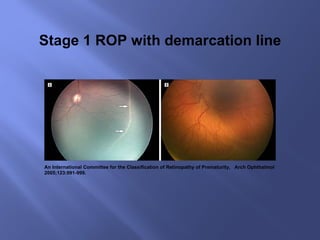
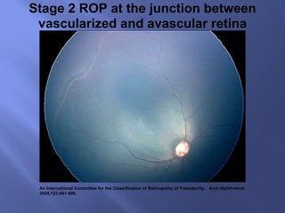
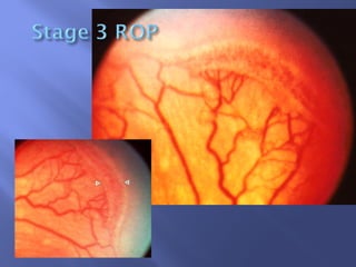
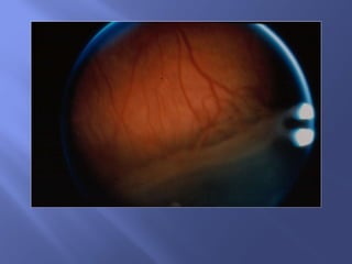
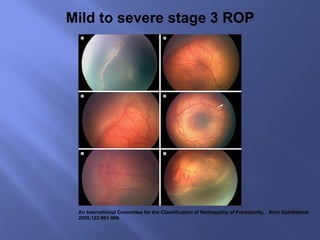
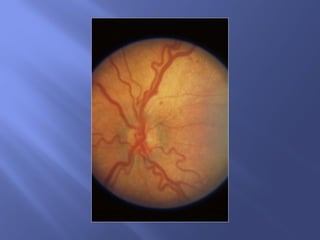
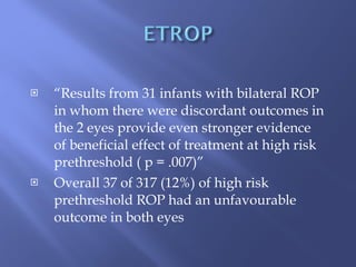
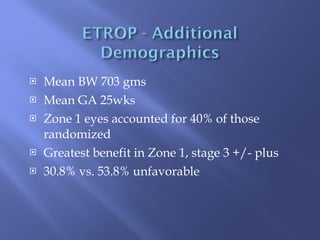
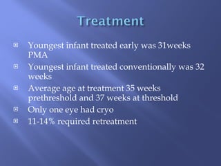
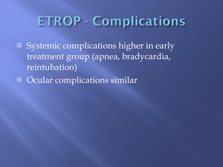
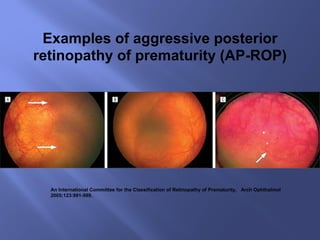
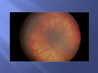
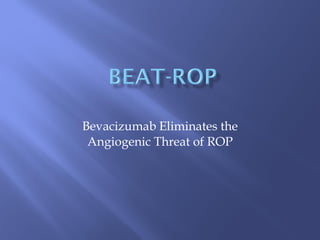
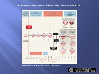
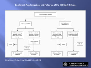
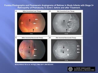
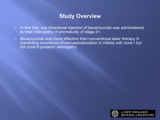
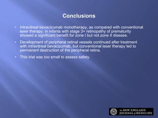
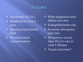
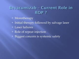
Dr Kathryn McCreery March 2011 RVEEH


An International Committee for the Classification of Retinopathy of Prematurity,  Arch Ophthalmol 2005;123:991-999. Stage 1 ROP with demarcation line
An International Committee for the Classification of Retinopathy of Prematurity,  Arch Ophthalmol 2005;123:991-999. Stage 2 ROP at the junction between vascularized and avascular retina
ICROP – Staging Level of abnormal vascular response observed


An International Committee for the Classification of Retinopathy of Prematurity,  Arch Ophthalmol 2005;123:991-999. Mild to severe stage 3 ROP


Stage 4. Subtotal retinal detachment  4a. Extrafoveal 4b. Retinal detachment involving fovea Stage 5.  Total retinal detachment Funnel:  Anterior Posterior Open   Open Narrow Narrow Open Narrow Narrow
An International Committee for the Classification of Retinopathy of Prematurity,  Arch Ophthalmol 2005;123:991-999. Stage 5 retinopathy of prematurity
 
All infants born in 26 centers Oct 2000 – Sept 2002 BW <1250g Survived 28 days First exam at @ 6 weeks Weekly exams Zone 1 or Zone 2 stage 2
Zone 1, any stage < threshold Zone 2, stage 2, + plus Zone 2 , stage 3, - plus Zone 2, stage 3, + plus < threshold
Data entered to risk programme based on natural history data from Cryo ROP Stratified into high risk and low risk prethreshold Risk > 15% - second masked confirming exam Randomized to early or conventional treatment at threshold (within 48hrs)
 
401 Randomized 317 Bilateral Cases 44 Asymmetric Cases (Treated) 40 Asymmetric Cases  ( Controls)
9 month grating (Teller) VA by masked tester Also a 6 and 9 month structural exam Amblyopia /refractive error treated between 6 and 9 month exam
Favorable Normal 3.70 cycles/degree Below normal 1.85 – 3.70 cycles/degree Unfavorable Poor <1.8 cycles but measurable Blind (NLP/LP/LV)
Posterior fold involving macula RD involving macula Retrolental tissue Any eye that had vitrectomy or scleral buckle
96% of those who survived  Unfavorable outcome reduced form 19.5% to 14.5% with early intervention  P < 0.01
Early treatment reduced risk of unfavorable structural outcome 15.9% (conventional) to 9.1%(early) P < 0.001
“ Results from 31 infants with bilateral ROP in whom there were discordant outcomes in the 2 eyes provide even stronger evidence of beneficial effect of treatment at high risk prethreshold ( p = .007)” Overall 37 of 317 (12%) of high risk prethreshold ROP had an unfavourable outcome in both eyes
Mean BW 703 gms Mean GA 25wks Zone 1 eyes accounted for 40% of those randomized Greatest benefit in Zone 1, stage 3 +/- plus 30.8% vs. 53.8% unfavorable
 
Youngest infant treated early was 31weeks PMA Youngest infant treated conventionally was 32 weeks Average age at treatment 35 weeks prethreshold and 37 weeks at threshold Only one eye had cryo 11-14% required retreatment
Cryo ROP 6% required treatment ETROP 9% will need treatment
Systemic complications higher in early treatment group (apnea, bradycardia, reintubation) Ocular complications similar
Zone 1, any stage, + plus Zone 1, stage 3, - plus Zone 2, stage 2 or 3, + plus
An International Committee for the Classification of Retinopathy of Prematurity,  Arch Ophthalmol 2005;123:991-999. Examples of aggressive posterior retinopathy of prematurity (AP-ROP)
 
Bevacizumab Eliminates the Angiogenic Threat of ROP
Pathogenesis and Therapy of Retinopathy of Prematurity (ROP). Mintz-Hittner HA et al. N Engl J Med 2011;364:603-615
1500 gm and 30 wks PCA March 2008 – August 2010 Stage 3+ ROP Zone 1 or Posterior Zone 2 Retcam  photos Confirming evaluation of photos by second ophthalmologist Both eyes randomized to injection or laser
0.625mg bevacizumab in 0.025ml Indirect laser
Enrollment, Randomization, and Follow-up of the 150 Study Infants. Mintz-Hittner HA et al. N Engl J Med 2011;364:603-615
Photos at 1 week, 1 month and 54 weeks Photos sent to BEAT ROP reading centre Photos cropped to remove peripheral retina to assess structural outcome If at week one there were skip areas in the laser group they received supplemental laser which were not considered to be recurrences
Significantly less for injection (6%) vs laser (26%) Recurrence significantly higher with laser than injection in Zone 1 disease  42% vs 6% Only 2 of 70 eyes injected developed RD ( both unilateral)
Risk Factors and Other Characteristics of Infants with Zone I Retinopathy of Prematurity (ROP) or Zone II Posterior ROP, According to Treatment Group. Mintz-Hittner HA et al. N Engl J Med 2011;364:603-615
Ocular Outcomes in the 143 Survivors at 54 Weeks' Postmenstrual Age. Mintz-Hittner HA et al. N Engl J Med 2011;364:603-615
Characteristics of Infants Who Died. Mintz-Hittner HA et al. N Engl J Med 2011;364:603-615
Fundus Photographs and Fluorescein Angiograms of Retinas in Study Infants with Stage 3+ Retinopathy of Prematurity in Zone I, before and after Treatment. Mintz-Hittner HA et al. N Engl J Med 2011;364:603-615
In this trial, one intravitreal injection of bevacizumab was administered to treat retinopathy of prematurity of stage 3+. Bevacizumab was more effective than conventional laser therapy in preventing recurrence of neovascularization in infants with zone I but not zone II posterior retinopathy. Study Overview
Intravitreal bevacizumab monotherapy, as compared with conventional laser therapy, in infants with stage 3+ retinopathy of prematurity showed a significant benefit for zone I but not zone II disease. Development of peripheral retinal vessels continued after treatment with intravitreal bevacizumab, but conventional laser therapy led to permanent destruction of the peripheral retina. This trial was too small to assess safety. Conclusions
Avoidance of GA’s Beneficial in Zone 1  eyes Sparing of peripheral field Normal retinal  vascularization What happened after failure not clear Endophthalmitis risk Systemic absorption and risks Recurrence occurs later 19 vs 6 wks in zone 1 disease Visual outcomes?
Monotherapy Initial therapy followed by salvage laser Laser failures Role of repeat injection Biggest concern is systemic safety
 

Rop – emerging therapies march 2011

  • 1.
    Dr Kathryn McCreeryMarch 2011 RVEEH
  • 2.
  • 3.
  • 4.
  • 5.
    An International Committeefor the Classification of Retinopathy of Prematurity, Arch Ophthalmol 2005;123:991-999. Stage 1 ROP with demarcation line
  • 6.
    An International Committeefor the Classification of Retinopathy of Prematurity, Arch Ophthalmol 2005;123:991-999. Stage 2 ROP at the junction between vascularized and avascular retina
  • 7.
    ICROP – StagingLevel of abnormal vascular response observed
  • 8.
  • 9.
  • 10.
  • 11.
    An International Committeefor the Classification of Retinopathy of Prematurity, Arch Ophthalmol 2005;123:991-999. Mild to severe stage 3 ROP
  • 12.
  • 13.
  • 14.
  • 15.
    Stage 4. Subtotalretinal detachment 4a. Extrafoveal 4b. Retinal detachment involving fovea Stage 5. Total retinal detachment Funnel: Anterior Posterior Open Open Narrow Narrow Open Narrow Narrow
  • 16.
    An International Committeefor the Classification of Retinopathy of Prematurity, Arch Ophthalmol 2005;123:991-999. Stage 5 retinopathy of prematurity
  • 17.
  • 18.
    All infants bornin 26 centers Oct 2000 – Sept 2002 BW <1250g Survived 28 days First exam at @ 6 weeks Weekly exams Zone 1 or Zone 2 stage 2
  • 19.
    Zone 1, anystage < threshold Zone 2, stage 2, + plus Zone 2 , stage 3, - plus Zone 2, stage 3, + plus < threshold
  • 20.
    Data entered torisk programme based on natural history data from Cryo ROP Stratified into high risk and low risk prethreshold Risk > 15% - second masked confirming exam Randomized to early or conventional treatment at threshold (within 48hrs)
  • 21.
  • 22.
    401 Randomized 317Bilateral Cases 44 Asymmetric Cases (Treated) 40 Asymmetric Cases ( Controls)
  • 23.
    9 month grating(Teller) VA by masked tester Also a 6 and 9 month structural exam Amblyopia /refractive error treated between 6 and 9 month exam
  • 24.
    Favorable Normal 3.70cycles/degree Below normal 1.85 – 3.70 cycles/degree Unfavorable Poor <1.8 cycles but measurable Blind (NLP/LP/LV)
  • 25.
    Posterior fold involvingmacula RD involving macula Retrolental tissue Any eye that had vitrectomy or scleral buckle
  • 26.
    96% of thosewho survived Unfavorable outcome reduced form 19.5% to 14.5% with early intervention P < 0.01
  • 27.
    Early treatment reducedrisk of unfavorable structural outcome 15.9% (conventional) to 9.1%(early) P < 0.001
  • 28.
    “ Results from31 infants with bilateral ROP in whom there were discordant outcomes in the 2 eyes provide even stronger evidence of beneficial effect of treatment at high risk prethreshold ( p = .007)” Overall 37 of 317 (12%) of high risk prethreshold ROP had an unfavourable outcome in both eyes
  • 29.
    Mean BW 703gms Mean GA 25wks Zone 1 eyes accounted for 40% of those randomized Greatest benefit in Zone 1, stage 3 +/- plus 30.8% vs. 53.8% unfavorable
  • 30.
  • 31.
    Youngest infant treatedearly was 31weeks PMA Youngest infant treated conventionally was 32 weeks Average age at treatment 35 weeks prethreshold and 37 weeks at threshold Only one eye had cryo 11-14% required retreatment
  • 32.
    Cryo ROP 6%required treatment ETROP 9% will need treatment
  • 33.
    Systemic complications higherin early treatment group (apnea, bradycardia, reintubation) Ocular complications similar
  • 34.
    Zone 1, anystage, + plus Zone 1, stage 3, - plus Zone 2, stage 2 or 3, + plus
  • 35.
    An International Committeefor the Classification of Retinopathy of Prematurity, Arch Ophthalmol 2005;123:991-999. Examples of aggressive posterior retinopathy of prematurity (AP-ROP)
  • 36.
  • 37.
    Bevacizumab Eliminates theAngiogenic Threat of ROP
  • 38.
    Pathogenesis and Therapyof Retinopathy of Prematurity (ROP). Mintz-Hittner HA et al. N Engl J Med 2011;364:603-615
  • 39.
    1500 gm and30 wks PCA March 2008 – August 2010 Stage 3+ ROP Zone 1 or Posterior Zone 2 Retcam photos Confirming evaluation of photos by second ophthalmologist Both eyes randomized to injection or laser
  • 40.
    0.625mg bevacizumab in0.025ml Indirect laser
  • 41.
    Enrollment, Randomization, andFollow-up of the 150 Study Infants. Mintz-Hittner HA et al. N Engl J Med 2011;364:603-615
  • 42.
    Photos at 1week, 1 month and 54 weeks Photos sent to BEAT ROP reading centre Photos cropped to remove peripheral retina to assess structural outcome If at week one there were skip areas in the laser group they received supplemental laser which were not considered to be recurrences
  • 43.
    Significantly less forinjection (6%) vs laser (26%) Recurrence significantly higher with laser than injection in Zone 1 disease 42% vs 6% Only 2 of 70 eyes injected developed RD ( both unilateral)
  • 44.
    Risk Factors andOther Characteristics of Infants with Zone I Retinopathy of Prematurity (ROP) or Zone II Posterior ROP, According to Treatment Group. Mintz-Hittner HA et al. N Engl J Med 2011;364:603-615
  • 45.
    Ocular Outcomes inthe 143 Survivors at 54 Weeks' Postmenstrual Age. Mintz-Hittner HA et al. N Engl J Med 2011;364:603-615
  • 46.
    Characteristics of InfantsWho Died. Mintz-Hittner HA et al. N Engl J Med 2011;364:603-615
  • 47.
    Fundus Photographs andFluorescein Angiograms of Retinas in Study Infants with Stage 3+ Retinopathy of Prematurity in Zone I, before and after Treatment. Mintz-Hittner HA et al. N Engl J Med 2011;364:603-615
  • 48.
    In this trial,one intravitreal injection of bevacizumab was administered to treat retinopathy of prematurity of stage 3+. Bevacizumab was more effective than conventional laser therapy in preventing recurrence of neovascularization in infants with zone I but not zone II posterior retinopathy. Study Overview
  • 49.
    Intravitreal bevacizumab monotherapy,as compared with conventional laser therapy, in infants with stage 3+ retinopathy of prematurity showed a significant benefit for zone I but not zone II disease. Development of peripheral retinal vessels continued after treatment with intravitreal bevacizumab, but conventional laser therapy led to permanent destruction of the peripheral retina. This trial was too small to assess safety. Conclusions
  • 50.
    Avoidance of GA’sBeneficial in Zone 1 eyes Sparing of peripheral field Normal retinal vascularization What happened after failure not clear Endophthalmitis risk Systemic absorption and risks Recurrence occurs later 19 vs 6 wks in zone 1 disease Visual outcomes?
  • 51.
    Monotherapy Initial therapyfollowed by salvage laser Laser failures Role of repeat injection Biggest concern is systemic safety
  • 52.

Editor's Notes

  • #39 Figure 1 Pathogenesis and Therapy of Retinopathy of Prematurity (ROP). Phases 1 and 2 of ROP are associated with different levels of vascular endothelial growth factor (VEGF), oxygen, and neovascular activity. ROP stages 0 to 5 are shown, as are the outcomes, when therapy is successful, of cryotherapy (established in a clinical trial in 1988),10 laser therapy (established in clinical trial in 2003),12 and intravitreal bevacizumab (this study). Cryotherapy involves scarring of the full ocular thickness, laser therapy scarring of the retinal thickness, and intravitreal bevacizumab scarring with a needle near the limbus. Also shown are the postmenstrual ages at which infants are at high risk for ROP, the appropriate postmenstrual age for the first ocular screening examination for ROP, and the mean postmenstrual ages at the onset of aggressive posterior ROP (APROP) (type 2 ROP) and stages 1, 2, and 3 (type 1 ROP).
  • #42 Figure 2 Enrollment, Randomization, and Follow-up of the 150 Study Infants.
  • #45 Table 1 Risk Factors and Other Characteristics of Infants with Zone I Retinopathy of Prematurity (ROP) or Zone II Posterior ROP, According to Treatment Group.
  • #46 Table 2 Ocular Outcomes in the 143 Survivors at 54 Weeks&apos; Postmenstrual Age.
  • #47 Table 3 Characteristics of Infants Who Died.
  • #48 Figure 3 Fundus Photographs and Fluorescein Angiograms of Retinas in Study Infants with Stage 3+ Retinopathy of Prematurity in Zone I, before and after Treatment. Panels A and B show the left retina of an infant before conventional laser therapy (at approximately 2 months of age, or 33.1 weeks&apos; postmenstrual age) and after therapy (at 13 months&apos; postmenstrual age), respectively. The infant was born at 24 weeks&apos; gestational age, with a birth weight of 760 g. The post-treatment photograph shows destruction of the full thickness of the peripheral retina, with only choroidal vessels (not retinal vessels) visible in the lasered area. Panels C and D show the left retina in another infant before intravitreal bevacizumab therapy (at approximately 3 months of age, or 35.6 weeks&apos; postmenstrual age) and after therapy (at 13 months&apos; postmenstrual age), respectively. The infant was born at 23 weeks&apos; gestational age, with a birth weight of 495 g. The post-treatment photograph shows continued vascularization of the peripheral retina. In all four panels, black arrows indicate identical retinal points for comparison before and after treatment, and thin white arrows indicate the extent of vascularization at each time point; the wide white arrows in Panel D indicate the extent of vascularization at the time of treatment with bevacizumab.