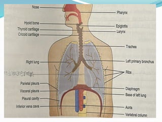
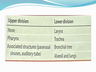
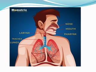
RESPIRATORY
SYSTEM
Ms. SHIVANGI R MISTRY
M.Sc. NURSING
INTRODUCTION
 The respiratory system functions to
supply oxygen from atmosphere to the
body cells and transport carbon dioxide
produced by the body cells to the
atmosphere.
 It is also involved in sound production
and helps in expulsive acts such as
micturition and defaecation.
 All the parts of the respiratory system
up to terminal bronchi conduct air.
 The parts beyond terminal bronchi
perform the respiratory function,
which is exchange of gases.
 The respiratory system can be divided
into upper and lower divisions
NOSE
The nose consists of external nose and
nasal cavities.
 External nose: It is pyramidal. It has a
root and a free tip, two lateral surfaces
and median dorsum nasi. It is formed
of nasal bones at the root and cartilage
and fibroareolar tissue at the nostrils.
 Nasal cavity The two nasal cavities are
separated by nasal septum. Each nasal
cavity has the following parts:-
 The anterior (external) nasal apertures
(nostrils). are piriform, extending 2 cm
anteroposteriorly and 2.5 cm
transversely.
 The posterior (internal) nasal apertures
(choanae) are oval (2.5 cm vertically, 2
cm transversely). They open into the
nasopharynx.
 The roof of the cavity is formed by
cribriform plate of the ethmoid
(through which pass olfactory nerves)
and frontal, nasal and sphenoid bones.
 The floor of the cavity is the hard palate
formed by maxillae and palatine bones.
MEDIAL WALL OF NOSE
LATERAL WALL OF NOSE
1. Superior concha
2. Middle concha
3. Inferior concha
Openings in lateral wall of nose
Nasal Mucous Membrane
 The nasal mucous membrane is
respiratory type (pseu-dostratified
ciliated columnar epithelium with
goblet cells), except on the inner side of
nostrils (lined by skin with hair) and
olfactory mucosa over the upper part
of the septum and superior concha.
 The arterial supply of the nose is by
branches from ophthalmic, maxillary
and facial arteries.
 The venous drainage is into the facial
vein from the anterior part, and into
the pharyngeal and pterygoid plexus
of veins from the posterior part of the
nose.
 The lymphatic drainage occurs into
submandibular nodes from the anterior
part and deep cervical nodes (via
retropharyngeal nodes) from the posterior
part of the nose.
 The nerve supply is by branches of
ophthalmic and maxillary nerves. The
olfactory nerves start from the olfactory
mucosa close to the roof of the nose.
Vessels and nerve of nasal septum
Vessels and nerve of lateral wall of nose
Blood and nerve supply
Functions of the Nose
 Filtering of the incoming air by hair
and mucus
 Warming and moistening of inhaled
air
 Sensation of smell by olfactory mucosa
 Vocal resonance and diminution of
auditory feedback
Paranasal Sinuses
 They are the air cavities in frontal,
ethmoidal, maxillary and sphenoid
bones.
 They are lined by the respiratory
mucosa that continues with the nasal
mucosa through the openings.
 The air sinuses are rudimentary at
birth and develop with growth of the
face and eruption of teeth.
Functions of Paranasal
Sinuses
 The paranasal sinuses lighten the
skull.
 They give resonance to the voice.
 They provide additional area for
warming and humidifying the air
 They provide mucus, which entraps
foreign particles in the inspired air.
Maxillary Air Sinus (Antrum of
Highmore)
 It is the largest air sinus and is pyramidal
in shape.
Boundaries
 The roof is formed by floor of the orbit
with infraorbital nerve and vessels on it.
 The floor is formed by the alveolar
process of maxilla. Roots of second and
third molars may reach up to the floor.
 The anterior and posterior walls are
formed by maxilla.
 Canine fossa is the thinnest part of the
anterior wall.
 The apex is directed to the zygomatic
process of the maxilla.
 The base is directed to the lateral wall
of nose.
Frontal Air Sinuses
 They are very variable. The frontal air
sinuses are in the frontal bone.
 Each sinus is triangular and lies above
the roof of the nose and orbit, posteriorly
related to anterior cranial fossa.
 The sinus drains by frontonasal duct into
the anterior part of the middle meatus of
nose.
Ethmoidal Air Sinuses
 Ethmoid bone It contains air cells
forming labyrinth. The cells are grouped
to form anterior, middle and posterior
ethmoidal sinuses.
 Anterior ethmoidal sinus drains into the
middle meatus, middle ethmoidal sinus
opens on the bulla ethmoidalis and
posterior sinus drains into the superior
meatus of nose.
 Ethmoidal sinusitis is prone to
complications as the ethmoidal air
sinuses are closely related to the eye
and anterior cranial fossa.
Sphenoidal Air Sinuses
 They are located in the body of the
sphenoid in the middle cranial fossa.
 Sinuses are usually asymmetrical.
 They are related above to the
hypophysis and optic chiasma, and on
each side to the internal carotid artery
and cavernous sinus.
Larynx
 The larynx (voice box) is an organ of
phonation(proces of producing sounds by
vibrating vocal folds) and a respiratory
passage, extending from the root of the
tongue to the trachea.
Relations
 Anterior : Skin, fascia, muscles
attached to the thyroid cartilage
 Posterior: Laryngopharynx, three to
six cervical vertebrae
 Superior: Opens into the pharynx
through the inlet of larynx
 Inferior: Continues with trachea
Measurements
 The size of the larynx does not differ in males
and females till puberty.
 At puberty, the larynx increases in size in males
and the thyroid cartilage projects considerably
forwards (Adam's apple) and may lead to
change in voice.
Structure of the Larynx
It consists of cartilages, ligaments, membranes and
muscles.
Cartilages
 Cartilages are paired and unpaired.
 Thyroid cartilage (thyroid = shield shaped). It is
the largest laryngeal cartilage. It has two laminae,
which fuse anteriorly to form laryngeal
prominence (Adam's apple).
 Cricoid cartilage It completely encircles the
airway.It is shaped like a signet ring with a
narrow anterior arch and broad posterior lamina.
 Epiglottis It is a leaf-like elastic cartilage. Its upper
free margin is broad and lies behind the tongue.
 Arytenoid cartilages The paired arytenoid cartilages
are pyramidal in shape.
 Corniculate cartilages They are conical in shape and
are attached to the tip of arytenoid cartilages.
 Cuneiform cartilages They are club-shaped, superior
to the corniculate cartilage in aryepiglottic folds.
 All the cartilages are lined by respiratory mucosa
except major part of the epiglottis and upper half of
the inner aspect of the thyroid cartilage covered by
stratified squamous epithelium.
Front view of larynx
Larynx view from behind
Interior of larynx
Ligaments and Membranes
 Ligaments and membranes of larynx They
are fibroelastic and link the cartilages of the
larynx.
 Its upper border is attached to the thyroid
cartilage in the front and vocal processes of
arytenoid cartilages behind.
 The free margin, in between, forms vocal
ligaments, which are covered by mucosa to
form true vocal folds (vocal cords) of the
larynx.
Cavity of Larynx
 Supraglottic part or vestibule It is present
between the inlet of larynx and the
vestibular folds (false vocal folds or cords).
 Glottic part or ventricle or sinus of larynx It
is present between vestibular and vocal
folds on each side.
 Infraglottic part It is present below true
vocal folds. It extends up to the cricoid
cartilage and continues with the trachea
below.-
o Rima vestibuli: It is a triangular opening
between two vestibular folds.
o Rima glottis: It is a narrower, triangular
opening X between two vocal folds.
Muscles of the Larynx
Extrinsic muscles They move the larynx up and
down during deglutition(process of swallowing).
The vocal cords are adducted during phonation
and deglutition; rima glottis is a narrow slit.
During forced breathing, the vocal cords abduct
and glottis becomes triangular
Abducted and Adducted vocal
cords
Functions of the Larynx
 Phonation: During phonation, the vocal cords
and arytenoid cartilages are adducted and air
is forced through the closed glottis during
expiration.
 The vocal cords vibrate and sound is
produced. The sound is modified by upper
respiratory passages and tongue. The longer
vocal cords produce a lower-pitch sound (as
in males). The short, tense cords produce a
high-pitch sound. The forceful vibrations of
cords produce a louder sound. The partial
abduction of the vocal cords produces a
 Respiration The larynx acts as a respiratory
passage.
 Protection of lower respiratory parts The
larynx moves upwards during deglutition and
the inlet of the larynx is closed. This prevents
the food from entering the respiratory tract
 Complete closure of glottis It is done to
increase the intra-abdominal pressure as in
lifting of heavy weight.
 Filtering of air The mucus engulfs particles
from air and the cilia push them upwards.
Arterial Supply
 The arterial supply is by superior laryngeal
artery (branch of superior thyroid artery)
and inferior laryngeal artery (branch of
inferior thyroid artery).
Venous Drainage
 Superior and inferior laryngeal veins join
the correspanding thyroid veins.
Lymphatic Drainage
 Lymphatic drainage is through deep cervical
nodes
Nerve Supply
 The sensory supply is by internal and
recurrent laryngeal branches of the vagus
nerve (parasympathetic). The sympathetic
supply is from the superior cervical
ganglion. The motor supply is described in
the section Muscles of the larynx.
TRACHEA
 The trachea (wind pipe) is a continuation of
the larynx downwards.
 It extends from the sixth cervical vertebra to
the fifth thoracic vertebra, where it bifurcates
into two principal bronchi.
Measurement
 The length is 10-11 cm, and diameter 12 mm
in the living.
Relation of trachea
Course and relations
 It lies in the lower part of the neck (cervical part)
and superior mediastinum (thoracic part).
Cervical part- Relations
 Anterior-sternothyroid, sternohyoid muscles,
isthmus of the thyroid gland at two to four
tracheal rings
 Posterior-oesophagus, vertebral column,
recurrent laryngeal nerves in the tracheo-
oesophageal groove
 Lateral-lobes of thyroid, common carotid artery
Thoracic part- Relations
 Anterior-sternum, left brachiocephalic vein,
arch of aorta
 Posterior-oesophagus, vertebral column
 Lateral-right lung and superior vena cava
(SVC), azygos vein on the right side, arch of
aorta, left recurrent laryngeal nerve, left
common carotid and left subclavian arteries
on the left side
 Arterial supply It is through inferior thyroid
arteries.
 Venous drainage It is through inferior
thyroid veins.
 Lymphatic drainage It is through pretracheal
and paratracheal nodes.
 Nerve supply Vagus and recurrent laryngeal
nerves supply parasympathetic fibres.
 Sympathetic fibres are derived from the
sympathetic trunk.
Structure
 The trachea has 10-12 incomplete 'C'-shaped
cartilaginous rings, which keep it permanently
open.The gap between the ends of the cartilage
is filled by smooth muscle (trachealis,
posteriorly, which allows distension of the
oesophagus during swallowing.
 The cartilages and muscles are covered by
areolar tissue outside.
 The trachea is lined by pseudostratified ciliated
columnar epithelium with goblet cells.
Trachea and bronchial
tree
BRONCHIAL TREE
 The trachea divides into two principal
(primary) bronchi, which supply the lungs.
 Their repeated branching in the lungs
produces successively smaller air passages,
which end as alveoli.
 Right principal bronchus It is shorter (2.5 cm)
and more vertical. Therefore, the foreign-
bodies entering the trachea more often lodge in
the right lung.
 Left principal bronchus It is oblique and
longer (5 cm) and lies below the arch of aorta.
 Each primary bronchus divides to form lobar
(secondary) bronchi, supplying lobes of the
lungs.
 Each lobar bronchus branches into three to five
divisions to form segmental (tertiary) bronchi,
supplying bronchopulmonary segments
Structure
 The extrapulmonary parts of bronchi have
the structure similar to that of trachea. In the
intrapulmonary bronchi, the cartilage rings
are replaced by cartilage plates.
 The cartilage disappears in bronchioles. The
terminal bronchioles have a thick muscle.
 The epithelium progressively changes from
the respiratory type (in bronchi) to the
squamous type (in alveoli) and the walls
become thinner.
 The type I alveolar cells (pneumonocytes I)
carry out gaseous exchange and type Il
alveolar cells (pneumonocytes II) produce
the surfactant. It lowers the surface tension
and prevents the alveoli from collapsing
during expiration.
 It plays an important role in the breathing of
a newborn. An insufficient amount of
surfactant may produce respiratory
difficulties in premature babies.
PLEURA
 The pleura forms covering for the lungs
as the lungs invaginate the pleural sacs.
 Parietal pleura it lines the thoracic walls
 Visceral pleura it covers the lungs and
lines fissures (hence, surface projections
of visceral pleura and lungs are same). It
continues over the root of lung and
extends as pulmonary ligaments below
the root of lung.
 Pleural cavity It is a potential space
between parietal and visceral pleurae
filled with the thin film of serous fluid.
It allows expansion of the lungs and
movements of the two layers over each
other.
Pleural recesses
 They are parts of the pleural cavity, unoccupied
by the lungs during ordinary respiration.
 Blood supply: The parietal pleura is supplied
by branches from intercostal, internal
mammary and pericardiacophrenic arteries.
The veins drain into the azygos system of
veins. The visceral pleura is supplied by the
vessels of the lungs.
 Nerve supply: The parietal pleura has
somatic nerve supply (intercostals and
phrenic nerves). Therefore, it is pain
sensitive. The visceral pleura has autonomic
nerve supply and is not pain sensitive.
Mediastinum
 The mediastinum is the interval between two
pleural sacs. It is divided into the superior
mediastinum above the sternal angle and the
inferior mediastinum below it.
 The inferior mediastinum is further divided
into the anterior mediastinum in front of the
heart, middle mediastinum containing heart
and the great vessels and posterior
mediastinum behind the heart.
Lungs
 The lungs are the essential organs of
respiration.
Location
 The lungs lie in the thorax, separated by the
mediastinum.
External features
 The lungs are pink in newborn and grey with
black pigment (accumulation of carbon
particles that are inhaled) in an adult.
Each lung has the following parts:
 The apex is conical in shape and extends above
clavicle till the neck of the first rib.
 The base is semilunar, concave resting on the
dome of the diaphragm.
 The anterior border is sharp, separating costal
and medial surfaces.
 The inferior border separates the base from
other surfaces.
 The blunt posterior border separates costal and
medial surfaces.
 Right lung
(a) eparterial bronchus;
(b) pulmonary artery;
(c) hyparterial bronchus; and
(d) pulmonary vein
 Left lung
(a) pulmonary artery;
(b) bronchus; and
(c) pulmonary vein
Difference between right and left lungs
 Arterial supply Bronchial arteries, two for
the left lung and one for the right lung, arise
from thoracic aorta. They supply bronchi
and bronchiole inside the lung.
 Venous drainage The left bronchial veins
drain into the superior intercostal vein and
the right bronchial veins drain into the
azygos vein.
 Lymphatic drainage The lungs drain into the
nodes present at the hilus (hilar or
pulmonary nodes) that carry lymph to the
tracheal nodes.
 Nerve supply The sympathetic nerves (from
the sympathetic chain) are bronchodilators
and the parasympathetic nerves (vagus) are
bronchoconstrictors.
 Bronchopulmonary segments A part of the lung
tissue supplied by a tertiary bronchus and a
tertiary branch of the pulmonary artery is called
bronchopulmonary segment. The pulmonary
veins are, however, intersegmental. Therefore,
bronchopulmonary segments are not
bronchovascular segments.
There are external and internal respirations.
 External respiration It is concerned with
exchange of gases between atmosphere and
blood and their transport to and from the
tissues.
 Internal respiration It is utilisation of
oxygen by a cell for oxidation of nutrients
for obtaining energy, during which CO, and
water are formed.
FUNCTIONS OF RESPIRATORY
SYSTEM
 Exchange of gases between atmosphere and
blood O2 from the atmosphere enters the
blood and CO, from the blood is excreted into
the atmosphere.
 Maintenance of pH of body fluids This occurs
by changing the amount of CO, excreted.
 Excretion of water vapour It leads to loss of
heat from the body.
 The respiratory system enables excretion of
volatile substances such as acetone.
 Respiratory muscles are used in laughing,
sucking, singing, etc.
 Many vasoactive substances such as
bradykinin, serotonin and prostaglandins
are inactivated in lungs.
 Capillary endothelium of lung-capillaries
have angiotensin-converting enzyme (ACE)
that converts angiotensin I to angiotensin II
(which increases the blood pressure).
Lungs are attached to the body only at their hila
(hilum is the site where bronchus, vessels and
nerves pass in and out of the lung). The rest of the
lung surface is free to move.
Lungs contain elastic fibres that can stretch the
lung.
There is tidal respiration (normal breathing going
on unconsciously) and forceful respiration.
Thank you

RESPIRATORY SYSTEM RESPIRATORY SYSTEM PPT

  • 1.
  • 2.
    INTRODUCTION  The respiratorysystem functions to supply oxygen from atmosphere to the body cells and transport carbon dioxide produced by the body cells to the atmosphere.  It is also involved in sound production and helps in expulsive acts such as micturition and defaecation.
  • 5.
     All theparts of the respiratory system up to terminal bronchi conduct air.  The parts beyond terminal bronchi perform the respiratory function, which is exchange of gases.  The respiratory system can be divided into upper and lower divisions
  • 6.
    NOSE The nose consistsof external nose and nasal cavities.  External nose: It is pyramidal. It has a root and a free tip, two lateral surfaces and median dorsum nasi. It is formed of nasal bones at the root and cartilage and fibroareolar tissue at the nostrils.
  • 7.
     Nasal cavityThe two nasal cavities are separated by nasal septum. Each nasal cavity has the following parts:-  The anterior (external) nasal apertures (nostrils). are piriform, extending 2 cm anteroposteriorly and 2.5 cm transversely.  The posterior (internal) nasal apertures (choanae) are oval (2.5 cm vertically, 2 cm transversely). They open into the nasopharynx.
  • 8.
     The roofof the cavity is formed by cribriform plate of the ethmoid (through which pass olfactory nerves) and frontal, nasal and sphenoid bones.  The floor of the cavity is the hard palate formed by maxillae and palatine bones.
  • 9.
  • 10.
    LATERAL WALL OFNOSE 1. Superior concha 2. Middle concha 3. Inferior concha
  • 11.
    Openings in lateralwall of nose
  • 13.
    Nasal Mucous Membrane The nasal mucous membrane is respiratory type (pseu-dostratified ciliated columnar epithelium with goblet cells), except on the inner side of nostrils (lined by skin with hair) and olfactory mucosa over the upper part of the septum and superior concha.
  • 14.
     The arterialsupply of the nose is by branches from ophthalmic, maxillary and facial arteries.  The venous drainage is into the facial vein from the anterior part, and into the pharyngeal and pterygoid plexus of veins from the posterior part of the nose.
  • 15.
     The lymphaticdrainage occurs into submandibular nodes from the anterior part and deep cervical nodes (via retropharyngeal nodes) from the posterior part of the nose.  The nerve supply is by branches of ophthalmic and maxillary nerves. The olfactory nerves start from the olfactory mucosa close to the roof of the nose.
  • 16.
    Vessels and nerveof nasal septum
  • 17.
    Vessels and nerveof lateral wall of nose
  • 18.
  • 19.
  • 20.
    Functions of theNose  Filtering of the incoming air by hair and mucus  Warming and moistening of inhaled air  Sensation of smell by olfactory mucosa  Vocal resonance and diminution of auditory feedback
  • 21.
    Paranasal Sinuses  Theyare the air cavities in frontal, ethmoidal, maxillary and sphenoid bones.  They are lined by the respiratory mucosa that continues with the nasal mucosa through the openings.  The air sinuses are rudimentary at birth and develop with growth of the face and eruption of teeth.
  • 23.
    Functions of Paranasal Sinuses The paranasal sinuses lighten the skull.  They give resonance to the voice.  They provide additional area for warming and humidifying the air  They provide mucus, which entraps foreign particles in the inspired air.
  • 24.
    Maxillary Air Sinus(Antrum of Highmore)  It is the largest air sinus and is pyramidal in shape. Boundaries  The roof is formed by floor of the orbit with infraorbital nerve and vessels on it.  The floor is formed by the alveolar process of maxilla. Roots of second and third molars may reach up to the floor.
  • 25.
     The anteriorand posterior walls are formed by maxilla.  Canine fossa is the thinnest part of the anterior wall.  The apex is directed to the zygomatic process of the maxilla.  The base is directed to the lateral wall of nose.
  • 27.
    Frontal Air Sinuses They are very variable. The frontal air sinuses are in the frontal bone.  Each sinus is triangular and lies above the roof of the nose and orbit, posteriorly related to anterior cranial fossa.  The sinus drains by frontonasal duct into the anterior part of the middle meatus of nose.
  • 28.
    Ethmoidal Air Sinuses Ethmoid bone It contains air cells forming labyrinth. The cells are grouped to form anterior, middle and posterior ethmoidal sinuses.  Anterior ethmoidal sinus drains into the middle meatus, middle ethmoidal sinus opens on the bulla ethmoidalis and posterior sinus drains into the superior meatus of nose.
  • 29.
     Ethmoidal sinusitisis prone to complications as the ethmoidal air sinuses are closely related to the eye and anterior cranial fossa.
  • 30.
    Sphenoidal Air Sinuses They are located in the body of the sphenoid in the middle cranial fossa.  Sinuses are usually asymmetrical.  They are related above to the hypophysis and optic chiasma, and on each side to the internal carotid artery and cavernous sinus.
  • 31.
    Larynx  The larynx(voice box) is an organ of phonation(proces of producing sounds by vibrating vocal folds) and a respiratory passage, extending from the root of the tongue to the trachea.
  • 33.
    Relations  Anterior :Skin, fascia, muscles attached to the thyroid cartilage  Posterior: Laryngopharynx, three to six cervical vertebrae  Superior: Opens into the pharynx through the inlet of larynx  Inferior: Continues with trachea
  • 34.
    Measurements  The sizeof the larynx does not differ in males and females till puberty.  At puberty, the larynx increases in size in males and the thyroid cartilage projects considerably forwards (Adam's apple) and may lead to change in voice.
  • 35.
    Structure of theLarynx It consists of cartilages, ligaments, membranes and muscles. Cartilages  Cartilages are paired and unpaired.  Thyroid cartilage (thyroid = shield shaped). It is the largest laryngeal cartilage. It has two laminae, which fuse anteriorly to form laryngeal prominence (Adam's apple).  Cricoid cartilage It completely encircles the airway.It is shaped like a signet ring with a narrow anterior arch and broad posterior lamina.
  • 36.
     Epiglottis Itis a leaf-like elastic cartilage. Its upper free margin is broad and lies behind the tongue.  Arytenoid cartilages The paired arytenoid cartilages are pyramidal in shape.  Corniculate cartilages They are conical in shape and are attached to the tip of arytenoid cartilages.  Cuneiform cartilages They are club-shaped, superior to the corniculate cartilage in aryepiglottic folds.  All the cartilages are lined by respiratory mucosa except major part of the epiglottis and upper half of the inner aspect of the thyroid cartilage covered by stratified squamous epithelium.
  • 38.
  • 39.
  • 40.
  • 41.
    Ligaments and Membranes Ligaments and membranes of larynx They are fibroelastic and link the cartilages of the larynx.  Its upper border is attached to the thyroid cartilage in the front and vocal processes of arytenoid cartilages behind.  The free margin, in between, forms vocal ligaments, which are covered by mucosa to form true vocal folds (vocal cords) of the larynx.
  • 42.
    Cavity of Larynx Supraglottic part or vestibule It is present between the inlet of larynx and the vestibular folds (false vocal folds or cords).  Glottic part or ventricle or sinus of larynx It is present between vestibular and vocal folds on each side.
  • 43.
     Infraglottic partIt is present below true vocal folds. It extends up to the cricoid cartilage and continues with the trachea below.- o Rima vestibuli: It is a triangular opening between two vestibular folds. o Rima glottis: It is a narrower, triangular opening X between two vocal folds.
  • 44.
    Muscles of theLarynx Extrinsic muscles They move the larynx up and down during deglutition(process of swallowing). The vocal cords are adducted during phonation and deglutition; rima glottis is a narrow slit. During forced breathing, the vocal cords abduct and glottis becomes triangular
  • 45.
  • 46.
    Functions of theLarynx  Phonation: During phonation, the vocal cords and arytenoid cartilages are adducted and air is forced through the closed glottis during expiration.  The vocal cords vibrate and sound is produced. The sound is modified by upper respiratory passages and tongue. The longer vocal cords produce a lower-pitch sound (as in males). The short, tense cords produce a high-pitch sound. The forceful vibrations of cords produce a louder sound. The partial abduction of the vocal cords produces a
  • 47.
     Respiration Thelarynx acts as a respiratory passage.  Protection of lower respiratory parts The larynx moves upwards during deglutition and the inlet of the larynx is closed. This prevents the food from entering the respiratory tract  Complete closure of glottis It is done to increase the intra-abdominal pressure as in lifting of heavy weight.  Filtering of air The mucus engulfs particles from air and the cilia push them upwards.
  • 48.
    Arterial Supply  Thearterial supply is by superior laryngeal artery (branch of superior thyroid artery) and inferior laryngeal artery (branch of inferior thyroid artery). Venous Drainage  Superior and inferior laryngeal veins join the correspanding thyroid veins.
  • 49.
    Lymphatic Drainage  Lymphaticdrainage is through deep cervical nodes Nerve Supply  The sensory supply is by internal and recurrent laryngeal branches of the vagus nerve (parasympathetic). The sympathetic supply is from the superior cervical ganglion. The motor supply is described in the section Muscles of the larynx.
  • 50.
    TRACHEA  The trachea(wind pipe) is a continuation of the larynx downwards.  It extends from the sixth cervical vertebra to the fifth thoracic vertebra, where it bifurcates into two principal bronchi. Measurement  The length is 10-11 cm, and diameter 12 mm in the living.
  • 51.
  • 52.
    Course and relations It lies in the lower part of the neck (cervical part) and superior mediastinum (thoracic part). Cervical part- Relations  Anterior-sternothyroid, sternohyoid muscles, isthmus of the thyroid gland at two to four tracheal rings  Posterior-oesophagus, vertebral column, recurrent laryngeal nerves in the tracheo- oesophageal groove  Lateral-lobes of thyroid, common carotid artery
  • 53.
    Thoracic part- Relations Anterior-sternum, left brachiocephalic vein, arch of aorta  Posterior-oesophagus, vertebral column  Lateral-right lung and superior vena cava (SVC), azygos vein on the right side, arch of aorta, left recurrent laryngeal nerve, left common carotid and left subclavian arteries on the left side
  • 54.
     Arterial supplyIt is through inferior thyroid arteries.  Venous drainage It is through inferior thyroid veins.  Lymphatic drainage It is through pretracheal and paratracheal nodes.  Nerve supply Vagus and recurrent laryngeal nerves supply parasympathetic fibres.  Sympathetic fibres are derived from the sympathetic trunk.
  • 55.
    Structure  The tracheahas 10-12 incomplete 'C'-shaped cartilaginous rings, which keep it permanently open.The gap between the ends of the cartilage is filled by smooth muscle (trachealis, posteriorly, which allows distension of the oesophagus during swallowing.  The cartilages and muscles are covered by areolar tissue outside.  The trachea is lined by pseudostratified ciliated columnar epithelium with goblet cells.
  • 56.
  • 58.
    BRONCHIAL TREE  Thetrachea divides into two principal (primary) bronchi, which supply the lungs.  Their repeated branching in the lungs produces successively smaller air passages, which end as alveoli.
  • 60.
     Right principalbronchus It is shorter (2.5 cm) and more vertical. Therefore, the foreign- bodies entering the trachea more often lodge in the right lung.  Left principal bronchus It is oblique and longer (5 cm) and lies below the arch of aorta.  Each primary bronchus divides to form lobar (secondary) bronchi, supplying lobes of the lungs.  Each lobar bronchus branches into three to five divisions to form segmental (tertiary) bronchi, supplying bronchopulmonary segments
  • 61.
    Structure  The extrapulmonaryparts of bronchi have the structure similar to that of trachea. In the intrapulmonary bronchi, the cartilage rings are replaced by cartilage plates.  The cartilage disappears in bronchioles. The terminal bronchioles have a thick muscle.  The epithelium progressively changes from the respiratory type (in bronchi) to the squamous type (in alveoli) and the walls become thinner.
  • 62.
     The typeI alveolar cells (pneumonocytes I) carry out gaseous exchange and type Il alveolar cells (pneumonocytes II) produce the surfactant. It lowers the surface tension and prevents the alveoli from collapsing during expiration.  It plays an important role in the breathing of a newborn. An insufficient amount of surfactant may produce respiratory difficulties in premature babies.
  • 63.
    PLEURA  The pleuraforms covering for the lungs as the lungs invaginate the pleural sacs.  Parietal pleura it lines the thoracic walls  Visceral pleura it covers the lungs and lines fissures (hence, surface projections of visceral pleura and lungs are same). It continues over the root of lung and extends as pulmonary ligaments below the root of lung.
  • 64.
     Pleural cavityIt is a potential space between parietal and visceral pleurae filled with the thin film of serous fluid. It allows expansion of the lungs and movements of the two layers over each other.
  • 66.
    Pleural recesses  Theyare parts of the pleural cavity, unoccupied by the lungs during ordinary respiration.
  • 68.
     Blood supply:The parietal pleura is supplied by branches from intercostal, internal mammary and pericardiacophrenic arteries. The veins drain into the azygos system of veins. The visceral pleura is supplied by the vessels of the lungs.  Nerve supply: The parietal pleura has somatic nerve supply (intercostals and phrenic nerves). Therefore, it is pain sensitive. The visceral pleura has autonomic nerve supply and is not pain sensitive.
  • 69.
    Mediastinum  The mediastinumis the interval between two pleural sacs. It is divided into the superior mediastinum above the sternal angle and the inferior mediastinum below it.  The inferior mediastinum is further divided into the anterior mediastinum in front of the heart, middle mediastinum containing heart and the great vessels and posterior mediastinum behind the heart.
  • 71.
  • 72.
     The lungsare the essential organs of respiration. Location  The lungs lie in the thorax, separated by the mediastinum. External features  The lungs are pink in newborn and grey with black pigment (accumulation of carbon particles that are inhaled) in an adult.
  • 73.
    Each lung hasthe following parts:  The apex is conical in shape and extends above clavicle till the neck of the first rib.  The base is semilunar, concave resting on the dome of the diaphragm.  The anterior border is sharp, separating costal and medial surfaces.  The inferior border separates the base from other surfaces.  The blunt posterior border separates costal and medial surfaces.
  • 74.
     Right lung (a)eparterial bronchus; (b) pulmonary artery; (c) hyparterial bronchus; and (d) pulmonary vein  Left lung (a) pulmonary artery; (b) bronchus; and (c) pulmonary vein
  • 75.
  • 76.
     Arterial supplyBronchial arteries, two for the left lung and one for the right lung, arise from thoracic aorta. They supply bronchi and bronchiole inside the lung.  Venous drainage The left bronchial veins drain into the superior intercostal vein and the right bronchial veins drain into the azygos vein.  Lymphatic drainage The lungs drain into the nodes present at the hilus (hilar or pulmonary nodes) that carry lymph to the tracheal nodes.
  • 77.
     Nerve supplyThe sympathetic nerves (from the sympathetic chain) are bronchodilators and the parasympathetic nerves (vagus) are bronchoconstrictors.  Bronchopulmonary segments A part of the lung tissue supplied by a tertiary bronchus and a tertiary branch of the pulmonary artery is called bronchopulmonary segment. The pulmonary veins are, however, intersegmental. Therefore, bronchopulmonary segments are not bronchovascular segments.
  • 78.
    There are externaland internal respirations.  External respiration It is concerned with exchange of gases between atmosphere and blood and their transport to and from the tissues.  Internal respiration It is utilisation of oxygen by a cell for oxidation of nutrients for obtaining energy, during which CO, and water are formed.
  • 79.
    FUNCTIONS OF RESPIRATORY SYSTEM Exchange of gases between atmosphere and blood O2 from the atmosphere enters the blood and CO, from the blood is excreted into the atmosphere.  Maintenance of pH of body fluids This occurs by changing the amount of CO, excreted.  Excretion of water vapour It leads to loss of heat from the body.
  • 81.
     The respiratorysystem enables excretion of volatile substances such as acetone.  Respiratory muscles are used in laughing, sucking, singing, etc.  Many vasoactive substances such as bradykinin, serotonin and prostaglandins are inactivated in lungs.  Capillary endothelium of lung-capillaries have angiotensin-converting enzyme (ACE) that converts angiotensin I to angiotensin II (which increases the blood pressure).
  • 82.
    Lungs are attachedto the body only at their hila (hilum is the site where bronchus, vessels and nerves pass in and out of the lung). The rest of the lung surface is free to move. Lungs contain elastic fibres that can stretch the lung. There is tidal respiration (normal breathing going on unconsciously) and forceful respiration.
  • 103.