SlideShare a Scribd company logo
1 of 1
Human Serum Albumin: A Vehicle to Co-Deliver Fatty Acid-Modified MethotrexateHuman Serum Albumin: A Vehicle to Co-Deliver Fatty Acid-Modified Methotrexate
and a p53-Derived Peptide to Enhance Apoptosis in Cancer Therapyand a p53-Derived Peptide to Enhance Apoptosis in Cancer Therapy
Michelle R. Joshi, Nianhuan Yao, Song Chae, Zhiyu Li
Department of Pharmaceutical Sciences, Philadelphia College of Pharmacy, University of the Sciences, Philadelphia, PA 19104
INTRODUCTIONINTRODUCTION RESULTSRESULTS
CONCLUSIONSCONCLUSIONS
The E3 ubiquitin ligase, MDM2, negatively regulates the activity of p53 via two main mechanisms: (1) reduces
transcriptional activity by sequestering the N-terminal transactivation domain of p53 and (2) ubiquitylates lysines in
the C-terminal domain of p53, thus promoting p53 degradation by the proteosome. The structural basis for the
first mechanism has been well characterized and consists of a minimum binding sequence within 19-26p53 that
forms an amphiphilc α-helix containing three critical residues; Phe19, Trp23, and Leu26. These residues are
located within a hydrophobic cavity of MDM2. Previous in vitro studies by Bernal and colleagues have
demonstrated reactivation of the p53 tumor suppressor cascade following treatment with a small peptide bearing
the key interacting residues. We plan to use a solid phase peptide synthesis method to generate a 14 amino acid
length peptide (p53i) containing the minimum binding sequence necessary for MDM2 interaction.
In recent years, the advent of small peptide therapeutic agents has resulted in the ability to enhance target
specificity and blunt toxicity compared to small molecule drugs. Despite this, serum instability and rapid renal
clearance have plagued their widespread usage. Lipidation and enhanced plasma protein binding are two
strategies capable of extending the half life of prodrugs. Our plan is to combine these methods by synthesizing
p53i conjugated to FA (FA-p53i); creating a species capable of being incorporated and transported by HSA.
The application of the synthesis described above is to develop an efficient drug carrier system utilizing HSA for
the targeted co-delivery of methotrexate (MTX) and FA-p53i. Such a system offers the unique ability for one drug
formulation to simultaneously deliver two anti-cancer agents to a single cell and thus, promote a supra-additive
effect on apoptosis. MTX is a well described and widely used chemotherapeutic agent whose mechanism of
action relies on the inhibition of key enzymatic activities required for DNA synthesis. It will thus serve as a model
drug for the development of this platform technology.
BACKGROUNDBACKGROUNDBACKGROUNDBACKGROUND
Co-Delivery
Combination Therapy
Vs.
Co-Delivery Approach
• Most abundant plasma protein
• Molecular weight = 66.5 kDa
• Long half-life
• Solublizing agent for long chain FA’s
• Binds a number of drugs
• Proven lack of toxicity and immunogenicity1
• Accumulates in malignant and inflamed tissue2Nat Struct Biol. 5(9), 827-35 (1998).
1
METHODSMETHODS
2
1) Methotrexate
 MTX has been conjugated to:
• Albumin Clin. Cancer Res. 9, 1917-26 (2003).
• Gelatin Pharm. Res. 17, 1309-15 (2000).
• Fibrinogen Cancer Lett. 148, 189-95 (2000).
• Polyethylene glycol (PEG) Bioconjug Chem. 13, 773-85 (2002).
• Tumor-targeting Ab Nat. Biotechnol. 23, 1137-46 (2005).
 Conjugation has been shown to increase plasma retention and
enhance accumulation in tumor tissue
2) p53-Derived Peptide (p53i)
 Designed to inhibit native p53-MDM2
interaction
3) Design and construct a recombinant HSA C-terminal fusion protein
Caspase-3 Acl I p53i Spe I Nhe I
DEVDG ETF SDLWKLLPETAA TS AS Amino acid sequence
DNA sequence
Caspase-3 PMI ScaI SacII Nhe I
DEVDG TSFAETWALLSP PR AS Amino acid sequence
DNA sequence
wt p53
high
affinity
peptide
PMI
Wuyuan Lu et al. JBC, 398: 200-213 (2010)
alternative strategyalternative strategy
N-protected C-terminal amino acid residueN-protected C-terminal amino acid residue was anchored via its COOH group to the hydroxyl group of Wang resin. Sidewas anchored via its COOH group to the hydroxyl group of Wang resin. Side
chain functional groups of amino acids were masked with permanent protection groups. The N-terminal amino group waschain functional groups of amino acids were masked with permanent protection groups. The N-terminal amino group was
protected by a temporary moiety that can be removed for coupling to the next residue. Deprotection/coupling process wasprotected by a temporary moiety that can be removed for coupling to the next residue. Deprotection/coupling process was
repeated until desired sequence was complete. Peptide was then released from resin and side chain protecting groupsrepeated until desired sequence was complete. Peptide was then released from resin and side chain protecting groups
were removed. Resulting peptide was detected by ESI/MS and purified by HPLC.were removed. Resulting peptide was detected by ESI/MS and purified by HPLC.
Solid phase peptide synthesis
Synthesis of FA-Modified FITC, FA-Modified p53i, & FA-Modified MTX
Fmoc-Lys(Alloc) was first coupled to Wang resin. After Fmoc deprotection, palmitic acid was coupled to the a- amino
group of Lys. After Alloc deprotection, Fmoc-SS-linker and NHS-Fluorescein were coupled sequentially. The final product
was cleaved using TFA.
Construction of recombinant HSA fusion proteins
• Amplified rHSA-p53i DNA by PCR
• Ligated rHSA-p53i into pPICZα A vector (Invitrogen)
• Transformed competent E.Coli cells using plasmid DNA & select for zeocin resistance
• Isolated single colonies from overnight culture & grow up in selection media
• Purified plasmid DNA & confirmed DNA sequence by restriction digest
• Linearized plasmid DNA & transformed Pichia yeast cells
• Expressed protein in Pichia strain, isolated and purified recombinant protein
Albumin/Fatty Acid Mobility Shift Assay
For experiments designed to detect albumin/FA-p53i complex formation, 120 pmol albumin (dissolved in 1X PBS) wasFor experiments designed to detect albumin/FA-p53i complex formation, 120 pmol albumin (dissolved in 1X PBS) was
incubated +/- FITC-labeled FA-p53i at desired molar ratios. Experiments aimed at detecting displacement of FA-FITC-p53iincubated +/- FITC-labeled FA-p53i at desired molar ratios. Experiments aimed at detecting displacement of FA-FITC-p53i
by unlabeled FA included an initial incubation carried out as described above, but at a fixed albumin:FA-FITC-p53i molarby unlabeled FA included an initial incubation carried out as described above, but at a fixed albumin:FA-FITC-p53i molar
ratio. Following this incubation, unlabeled FA was added at increasing molar ratios up to 1:8 (albumin:unlabeled FA).ratio. Following this incubation, unlabeled FA was added at increasing molar ratios up to 1:8 (albumin:unlabeled FA).
Reactions were conducted in 20 µl of PBS under room temperature for 30 minutes. The products were separated usingReactions were conducted in 20 µl of PBS under room temperature for 30 minutes. The products were separated using
Tris-Boric polyacrylamide gel 12 mA for 20 minutes. The gel was visualized under 305 nm UV.Tris-Boric polyacrylamide gel 12 mA for 20 minutes. The gel was visualized under 305 nm UV.
Cytotoxicity Assays
Cells were plated in standard growth media at approximately 40% confluence and allowed to attach overnight. On day 2,Cells were plated in standard growth media at approximately 40% confluence and allowed to attach overnight. On day 2,
MTX, FA-MTX, HSA/MTX, HSA/FA-MTX complexes or HSA fusion proteins were added at the indicated concentrationsMTX, FA-MTX, HSA/MTX, HSA/FA-MTX complexes or HSA fusion proteins were added at the indicated concentrations
and allowed to incubate at 37ºC/5%COand allowed to incubate at 37ºC/5%CO22 for the indicated time period. Cell proliferation was then measured by CyQuantfor the indicated time period. Cell proliferation was then measured by CyQuant
assay (Invitrogen). Data represented three replicas at indicated concentrations.assay (Invitrogen). Data represented three replicas at indicated concentrations.
1 2 3 4 5 6 7 8 9 10
Figure 1: Recombinant HSA fusion
proteins are able to form complexes with
FA-FITC. Recombinant HSA fusion proteins
as well as wild type HSA were incubated at
the indicated molar ratios with FA-FITC. The
speed at which molecules move through the
gel is dependent on size and charge. The
upper band corresponds to the less mobile
HSA/FA complex, while the lower band
contains free unbound FA-FITC.
Figure 1
Carrier: Human Serum Albumin
Cargo: FA-Methotrexate & FA-p53i/PMI
Figure 2 Figure 2: FA-FITC-p53i forms a stable
complex with human serum albumin that
is not displaced by the presence of
unlabeled FA. HSA/FA-FITC-p53i
complexes were allowed to form at a 1:4
molar ratio (HSA:FA-FITC-p53i; 120
pmol:480 pmol) as described in Methods.
Next, samples were incubated with
increasing concentrations of unlabeled FA.
The absence of a lower band indicates FA-
FITC-p53i was not displaced by the
unlabeled FA even at the highest
concentration. The upper band corresponds
to the less mobile HSA/FA complex, while
the lower band contains unbound free FA-
FITC or free FA-FITC-p53i.
1 2 3 4 5 6 7 8
FA-bound HSA
Free FA
HSA:FA-FITC-p53i (1:4)
unlabeled FA-FITC (pmol) 120 240 480360 960720--
120 pmol
FA-FITC-p53i
Benefits of Co-deliveryBenefits of Co-delivery
Both anti-cancer agents reach
target
Can use 2 species with
complimentary mechanisms to
promote robust apoptotic
response
Ease of formulation and
administration
One agent → multiple effects
Enhanced therapeutic effect at
lower doses
Targeted co-delivery reduces side
effects due to toxicity in normal
tissues
0.00
10.00
20.00
30.00
40.00
50.00
60.00
70.00
80.00
90.00
100.00
rHSA-p53i rHSA-PMI
Treatment Conditions
%CellDeath(RelativetoControl)
5 µM
10 µM
Figure 3
Figure 3: Recombinant HSA fusion proteins (A), FA-p53, FA-PMI and HSA-WT/FA-peptides (B) promote
cytotoxicity in SJSA-1 cells. SJSA-1 cells were plated in 96-well plates in standard growth media and allowed
to attach overnight. rHSA fusion proteins were added at the indicated concentrations in RPMI media containing
1% FBS + 0.05% DMSO and allowed to incubate x 24 hrs. DNA content was quantitated using the CyQuant
Assay (Invitrogen).
0
10
20
30
40
50
60
p53 PMI
Peptide
DNAcontent(as%ofControl)
FA-peptide (50 uM)
HSA-WT + FA-
peptide (50 uM)
AA BB
Figure 5
IP: MDM2
WB: MDM2
H
SA-W
T
HSA-p53i
HSA-PM
I
Figure 5: Biotinylated rHSA-p53i and rHSA-PMI bind MDM2 as
determined by co-immunoprecipitation using an SJSA-1 whole cell
lysate. To detect MDM2/rHSA-p53i &MDM2/rHSA-PMI interaction,
biotinylated HSA-WT, rHSA-p53i, or rHSA-PMI were added to an SJSA-1
lysate and incubated at RT x 1 hr. MDM2 antibody (Santa Cruz) was added
to each sample and allowed to incubate O/N at 4°C. Following a 1 hr RT
incubation with protein A/G-coupled agarose beads, immune complexes
were eluted and analyzed by SDS-PAGE followed by western blot analysis
to verify the identity of the antigen.
Figure 4: Recombinant
HSA fusion proteins
promote apoptosis via
caspase activation.
SJSA-1 cells were plated
in 96-well plates in
standard growth media
and allowed to attach
O/N. Recombinant HSA
fusion proteins were
Figure 4
0.00
1.00
2.00
3.00
4.00
5.00
6.00
7.00
8.00
rHSA-p53i rHSA-PMI
Treatment Conditions
FoldChangeRelativetoControl
5 µM
10 µM
AA BB CC DD
EE FF
added at the indicated concentrations in RPMI media containing 1% FBS + 0.05% DMSO and allowed to
incubate x 24 hrs. Cells were then washed and caspase activation was quantitated using a fluorimetric
Homogeneous Caspase Assay (Roche)(A). Phase micrographs of 10 µM-treated wells: (B) untreated (C) nutlin
(D) FA-p53i (E) rHSA-p53i (F) rHSA-PMI.
Figure 7: Cytotoxicity of MTX, FA-MTX, HSA/MTX, and HSA/FA-MTX complexes. Cell proliferation was
measured by CyQuant assay (Invitrogen). Data represented three replicas at indicated concentrations.
Experiments were repeated twice. MTX and FA-MTX showed comparable cytotoxicity in MDA-MB-435 and
SKBR-3 cells. In MCF-7 cell, cytotoxicity of FA-MTX was about 3 times lower than that of MTX. FA
modification on MTX has no obvious effects on in vitro cytotoxicity.
Figure 7
p53p53
MDM2MDM2
GAPDHGAPDH
untreateduntreated
NutlinNutlin
rHSA-p53irHSA-p53i
rHSA-PMIrHSA-PMI
FA-p53iFA-p53i
Figure 5: FA-p53i, rHSA-p53i, and rHSA-PMI increase p53, but not MDM2 protein expression. This is in
contrast to the actions of the cis-imidazoline analog, nutlin. SJSA-1 cells were plated in standard growth
media and allowed to attach overnight. On day 2, 10 µM nutlin, rHSA-p53i, rHSA-PMI or FA-p53i were added in
RPMI media containing 1% FBS + 0.05% DMSO and allowed to incubate x 24 hrs. Cell monolayers were then
washed, lysed and immunoblotted for p53 and MDM2 (Santa Cruz). P53 protein expression increased by
approximately 60% in rHSA-p53i-treated cells and 30% in both rHSA-PMI and FA-p53i treatments (as
determined using Image J software), while MDM2 expression did not change relative to untreated wells.
Figure 6
Figure 8: Effect of MTX and FA-MTX on
H1299 cells xenografts. Fifteen mice were
randomly split into 3 groups for USP saline,
MTX, and FA-MTX treatment. Cancer cells
were injected as described in Methods and
tumor size was measured 2X per week. MTX
(25 mg/kg) and FA-MTX (equal to MTX 4.15
mg/kg) were administered i.p. once a week for
3 weeks. Based on the relative tumor volume,
FA-MTX with 1/6 dose of MTX showed
comparable or slightly better efficacy.
Figure 8
HSA
p53iMTX
FA-modification of MTX and p53i is a valid method to facilitate non-
covalent incorporation into HSA
Recombinant HSA fusion proteins already containing the integrated
p53i and PMI peptides are capable of delivering FA-MTX.
The increase in p53 protein expression by FA-p53i, rHSA-p53i and
rHSA-PMI confirms intracellular uptake and reactivation of p53.
rHSA fusion proteins already containing the integrated p53i and PMI
peptides are capable of promoting apoptosis via caspase activation.
Studies are currently underway to determine if rHSA fusion proteins
co-delivered with FA-MTX can enhance cytotoxicity compared to
single agent administration.
p53-GFP
p53
GAPDH
GAPDH
MDM2
31 4 5 76 98 11102
31 4 5 76 98 11102
1: PMI 5 µM
2: FA-p53i 50 µM
3: FA-p53i 5 µM
4: Nutlin 10 µM
5: untreated
6: PMI 50 µM
7: PMI 5 µM
8: FA-p53i 50 µM
9: FA-p53i 5 µM
10: Nutlin 10 µM
11: untreated
p53-GFP
transfected
untransfected

More Related Content

What's hot

PPAR alpha poster
PPAR alpha posterPPAR alpha poster
PPAR alpha posterEric Cobb
 
Sigma Xi Presentation
Sigma Xi PresentationSigma Xi Presentation
Sigma Xi PresentationSKU sxi
 
The Discovery and Development of HMG-CoA Reductase Inhibitors
The Discovery and Development of HMG-CoA Reductase InhibitorsThe Discovery and Development of HMG-CoA Reductase Inhibitors
The Discovery and Development of HMG-CoA Reductase InhibitorsSanam Parajuli
 
NUC050 PosterPresentations.com-36x72-Template-V5
NUC050 PosterPresentations.com-36x72-Template-V5NUC050 PosterPresentations.com-36x72-Template-V5
NUC050 PosterPresentations.com-36x72-Template-V5Richard Daifuku
 
PPAR alpha final research paper
PPAR alpha final research paperPPAR alpha final research paper
PPAR alpha final research paperEric Cobb
 
Drugs transporters
Drugs transportersDrugs transporters
Drugs transportersalaa essa
 
P glycoprotein
P glycoprotein P glycoprotein
P glycoprotein khaterehz
 
Intestinal transporters in drug absorption
Intestinal transporters in drug absorptionIntestinal transporters in drug absorption
Intestinal transporters in drug absorptionOmer Mustapha
 
V tubiashii - NSA 2009
V tubiashii - NSA 2009V tubiashii - NSA 2009
V tubiashii - NSA 2009sr320
 
Membrane transporters and drug response
Membrane transporters and drug responseMembrane transporters and drug response
Membrane transporters and drug responsebhagyamohod90
 
Different Therapeutic Aspects of Peroxisomes Proliferator-Activated Receptors
Different Therapeutic Aspects of Peroxisomes Proliferator-Activated ReceptorsDifferent Therapeutic Aspects of Peroxisomes Proliferator-Activated Receptors
Different Therapeutic Aspects of Peroxisomes Proliferator-Activated ReceptorsAI Publications
 
Alcohol induced metabolic alterations - A Case based discussion
Alcohol induced metabolic alterations - A Case based discussionAlcohol induced metabolic alterations - A Case based discussion
Alcohol induced metabolic alterations - A Case based discussionNamrata Chhabra
 

What's hot (20)

PPAR alpha poster
PPAR alpha posterPPAR alpha poster
PPAR alpha poster
 
Abc seminar
Abc seminarAbc seminar
Abc seminar
 
Drug interactions linked to transporters
Drug interactions linked to transportersDrug interactions linked to transporters
Drug interactions linked to transporters
 
Sigma Xi Presentation
Sigma Xi PresentationSigma Xi Presentation
Sigma Xi Presentation
 
The Discovery and Development of HMG-CoA Reductase Inhibitors
The Discovery and Development of HMG-CoA Reductase InhibitorsThe Discovery and Development of HMG-CoA Reductase Inhibitors
The Discovery and Development of HMG-CoA Reductase Inhibitors
 
NUC050 PosterPresentations.com-36x72-Template-V5
NUC050 PosterPresentations.com-36x72-Template-V5NUC050 PosterPresentations.com-36x72-Template-V5
NUC050 PosterPresentations.com-36x72-Template-V5
 
PPAR alpha final research paper
PPAR alpha final research paperPPAR alpha final research paper
PPAR alpha final research paper
 
Memb transporter
Memb transporterMemb transporter
Memb transporter
 
Drugs transporters
Drugs transportersDrugs transporters
Drugs transporters
 
P glycoprotein
P glycoprotein P glycoprotein
P glycoprotein
 
A novel PPAR pan-agonist, 2-(4-(5,6-methylenedioxybenzo[d]thiazol-2-yl)-2-met...
A novel PPAR pan-agonist, 2-(4-(5,6-methylenedioxybenzo[d]thiazol-2-yl)-2-met...A novel PPAR pan-agonist, 2-(4-(5,6-methylenedioxybenzo[d]thiazol-2-yl)-2-met...
A novel PPAR pan-agonist, 2-(4-(5,6-methylenedioxybenzo[d]thiazol-2-yl)-2-met...
 
p-glycoprotein
p-glycoproteinp-glycoprotein
p-glycoprotein
 
P gp
P gpP gp
P gp
 
SatAMMasterCynSterling
SatAMMasterCynSterlingSatAMMasterCynSterling
SatAMMasterCynSterling
 
Intestinal transporters in drug absorption
Intestinal transporters in drug absorptionIntestinal transporters in drug absorption
Intestinal transporters in drug absorption
 
V tubiashii - NSA 2009
V tubiashii - NSA 2009V tubiashii - NSA 2009
V tubiashii - NSA 2009
 
Membrane transporters and drug response
Membrane transporters and drug responseMembrane transporters and drug response
Membrane transporters and drug response
 
Different Therapeutic Aspects of Peroxisomes Proliferator-Activated Receptors
Different Therapeutic Aspects of Peroxisomes Proliferator-Activated ReceptorsDifferent Therapeutic Aspects of Peroxisomes Proliferator-Activated Receptors
Different Therapeutic Aspects of Peroxisomes Proliferator-Activated Receptors
 
Alcohol induced metabolic alterations - A Case based discussion
Alcohol induced metabolic alterations - A Case based discussionAlcohol induced metabolic alterations - A Case based discussion
Alcohol induced metabolic alterations - A Case based discussion
 
Transporters
TransportersTransporters
Transporters
 

Similar to Research Day Poster

Pharmacology Of Pain Essay
Pharmacology Of Pain EssayPharmacology Of Pain Essay
Pharmacology Of Pain EssayLeslie Lee
 
Inhibition of glutathione by buthionine sulfoximine enhanced the anti-cancer ...
Inhibition of glutathione by buthionine sulfoximine enhanced the anti-cancer ...Inhibition of glutathione by buthionine sulfoximine enhanced the anti-cancer ...
Inhibition of glutathione by buthionine sulfoximine enhanced the anti-cancer ...Ashujit
 
FineSydney LSSURP Poster
FineSydney LSSURP PosterFineSydney LSSURP Poster
FineSydney LSSURP PosterSydney Fine
 
Discovery and Mechanistic Study of Mycobacterium tuberculosis PafA Inhibitors...
Discovery and Mechanistic Study of Mycobacterium tuberculosis PafA Inhibitors...Discovery and Mechanistic Study of Mycobacterium tuberculosis PafA Inhibitors...
Discovery and Mechanistic Study of Mycobacterium tuberculosis PafA Inhibitors...Dr.Shuaib Ahmad
 
The Development of Heterobifunctional Molecules.pdf
The Development of Heterobifunctional Molecules.pdfThe Development of Heterobifunctional Molecules.pdf
The Development of Heterobifunctional Molecules.pdfDoriaFang
 
Simplified receptor based pharmacophore approach to retrieve potent ptp lar i...
Simplified receptor based pharmacophore approach to retrieve potent ptp lar i...Simplified receptor based pharmacophore approach to retrieve potent ptp lar i...
Simplified receptor based pharmacophore approach to retrieve potent ptp lar i...rajmaha9
 
Thrombin activable fibrinolysis inhibitor (tafi) role
Thrombin activable fibrinolysis inhibitor (tafi) roleThrombin activable fibrinolysis inhibitor (tafi) role
Thrombin activable fibrinolysis inhibitor (tafi) rolegargitignath12
 
Sprayable adhesive nanotherapeutics.
Sprayable  adhesive nanotherapeutics.Sprayable  adhesive nanotherapeutics.
Sprayable adhesive nanotherapeutics.UJJALNASKAR2
 
Pathways of intracellular signal transduction
Pathways of intracellular signal transductionPathways of intracellular signal transduction
Pathways of intracellular signal transductionMohamedEramHosen
 
Genetic Dna And Bioinformatics ( Accession No. Xp Essay
Genetic Dna And Bioinformatics ( Accession No. Xp EssayGenetic Dna And Bioinformatics ( Accession No. Xp Essay
Genetic Dna And Bioinformatics ( Accession No. Xp EssayJessica Deakin
 
Proteolysis-targeting chimeras as tools in drug development (i)
Proteolysis-targeting chimeras as tools in drug development (i)Proteolysis-targeting chimeras as tools in drug development (i)
Proteolysis-targeting chimeras as tools in drug development (i)creativebiolabs11
 
Accelrys UGM slides 2011
Accelrys UGM slides 2011Accelrys UGM slides 2011
Accelrys UGM slides 2011Sean Ekins
 
Activation of AMPK inhibits cervical cancer cell growth through AKT/FOXO3a/FO...
Activation of AMPK inhibits cervical cancer cell growth through AKT/FOXO3a/FO...Activation of AMPK inhibits cervical cancer cell growth through AKT/FOXO3a/FO...
Activation of AMPK inhibits cervical cancer cell growth through AKT/FOXO3a/FO...Enrique Moreno Gonzalez
 
Poster Filippa Pettersson ICDT 2010
Poster Filippa Pettersson ICDT 2010Poster Filippa Pettersson ICDT 2010
Poster Filippa Pettersson ICDT 2010FilippaP
 

Similar to Research Day Poster (20)

Pharmacology Of Pain Essay
Pharmacology Of Pain EssayPharmacology Of Pain Essay
Pharmacology Of Pain Essay
 
2016 JHU_NCI Poster
2016 JHU_NCI Poster 2016 JHU_NCI Poster
2016 JHU_NCI Poster
 
BIOS 5260 Term Paper
BIOS 5260 Term PaperBIOS 5260 Term Paper
BIOS 5260 Term Paper
 
Inhibition of glutathione by buthionine sulfoximine enhanced the anti-cancer ...
Inhibition of glutathione by buthionine sulfoximine enhanced the anti-cancer ...Inhibition of glutathione by buthionine sulfoximine enhanced the anti-cancer ...
Inhibition of glutathione by buthionine sulfoximine enhanced the anti-cancer ...
 
J med chem-2
J med chem-2J med chem-2
J med chem-2
 
FineSydney LSSURP Poster
FineSydney LSSURP PosterFineSydney LSSURP Poster
FineSydney LSSURP Poster
 
Omara-Opyene et al 2004
Omara-Opyene et al 2004Omara-Opyene et al 2004
Omara-Opyene et al 2004
 
Discovery and Mechanistic Study of Mycobacterium tuberculosis PafA Inhibitors...
Discovery and Mechanistic Study of Mycobacterium tuberculosis PafA Inhibitors...Discovery and Mechanistic Study of Mycobacterium tuberculosis PafA Inhibitors...
Discovery and Mechanistic Study of Mycobacterium tuberculosis PafA Inhibitors...
 
The Development of Heterobifunctional Molecules.pdf
The Development of Heterobifunctional Molecules.pdfThe Development of Heterobifunctional Molecules.pdf
The Development of Heterobifunctional Molecules.pdf
 
Trastuzumab
Trastuzumab Trastuzumab
Trastuzumab
 
Simplified receptor based pharmacophore approach to retrieve potent ptp lar i...
Simplified receptor based pharmacophore approach to retrieve potent ptp lar i...Simplified receptor based pharmacophore approach to retrieve potent ptp lar i...
Simplified receptor based pharmacophore approach to retrieve potent ptp lar i...
 
Thrombin activable fibrinolysis inhibitor (tafi) role
Thrombin activable fibrinolysis inhibitor (tafi) roleThrombin activable fibrinolysis inhibitor (tafi) role
Thrombin activable fibrinolysis inhibitor (tafi) role
 
Sprayable adhesive nanotherapeutics.
Sprayable  adhesive nanotherapeutics.Sprayable  adhesive nanotherapeutics.
Sprayable adhesive nanotherapeutics.
 
Pathways of intracellular signal transduction
Pathways of intracellular signal transductionPathways of intracellular signal transduction
Pathways of intracellular signal transduction
 
Genetic Dna And Bioinformatics ( Accession No. Xp Essay
Genetic Dna And Bioinformatics ( Accession No. Xp EssayGenetic Dna And Bioinformatics ( Accession No. Xp Essay
Genetic Dna And Bioinformatics ( Accession No. Xp Essay
 
Proteolysis-targeting chimeras as tools in drug development (i)
Proteolysis-targeting chimeras as tools in drug development (i)Proteolysis-targeting chimeras as tools in drug development (i)
Proteolysis-targeting chimeras as tools in drug development (i)
 
Accelrys UGM slides 2011
Accelrys UGM slides 2011Accelrys UGM slides 2011
Accelrys UGM slides 2011
 
Activation of AMPK inhibits cervical cancer cell growth through AKT/FOXO3a/FO...
Activation of AMPK inhibits cervical cancer cell growth through AKT/FOXO3a/FO...Activation of AMPK inhibits cervical cancer cell growth through AKT/FOXO3a/FO...
Activation of AMPK inhibits cervical cancer cell growth through AKT/FOXO3a/FO...
 
Poster Filippa Pettersson ICDT 2010
Poster Filippa Pettersson ICDT 2010Poster Filippa Pettersson ICDT 2010
Poster Filippa Pettersson ICDT 2010
 
PARP Poly ADP Ribose
PARP Poly ADP RibosePARP Poly ADP Ribose
PARP Poly ADP Ribose
 

Research Day Poster

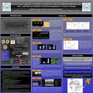
  • 1. Human Serum Albumin: A Vehicle to Co-Deliver Fatty Acid-Modified MethotrexateHuman Serum Albumin: A Vehicle to Co-Deliver Fatty Acid-Modified Methotrexate and a p53-Derived Peptide to Enhance Apoptosis in Cancer Therapyand a p53-Derived Peptide to Enhance Apoptosis in Cancer Therapy Michelle R. Joshi, Nianhuan Yao, Song Chae, Zhiyu Li Department of Pharmaceutical Sciences, Philadelphia College of Pharmacy, University of the Sciences, Philadelphia, PA 19104 INTRODUCTIONINTRODUCTION RESULTSRESULTS CONCLUSIONSCONCLUSIONS The E3 ubiquitin ligase, MDM2, negatively regulates the activity of p53 via two main mechanisms: (1) reduces transcriptional activity by sequestering the N-terminal transactivation domain of p53 and (2) ubiquitylates lysines in the C-terminal domain of p53, thus promoting p53 degradation by the proteosome. The structural basis for the first mechanism has been well characterized and consists of a minimum binding sequence within 19-26p53 that forms an amphiphilc α-helix containing three critical residues; Phe19, Trp23, and Leu26. These residues are located within a hydrophobic cavity of MDM2. Previous in vitro studies by Bernal and colleagues have demonstrated reactivation of the p53 tumor suppressor cascade following treatment with a small peptide bearing the key interacting residues. We plan to use a solid phase peptide synthesis method to generate a 14 amino acid length peptide (p53i) containing the minimum binding sequence necessary for MDM2 interaction. In recent years, the advent of small peptide therapeutic agents has resulted in the ability to enhance target specificity and blunt toxicity compared to small molecule drugs. Despite this, serum instability and rapid renal clearance have plagued their widespread usage. Lipidation and enhanced plasma protein binding are two strategies capable of extending the half life of prodrugs. Our plan is to combine these methods by synthesizing p53i conjugated to FA (FA-p53i); creating a species capable of being incorporated and transported by HSA. The application of the synthesis described above is to develop an efficient drug carrier system utilizing HSA for the targeted co-delivery of methotrexate (MTX) and FA-p53i. Such a system offers the unique ability for one drug formulation to simultaneously deliver two anti-cancer agents to a single cell and thus, promote a supra-additive effect on apoptosis. MTX is a well described and widely used chemotherapeutic agent whose mechanism of action relies on the inhibition of key enzymatic activities required for DNA synthesis. It will thus serve as a model drug for the development of this platform technology. BACKGROUNDBACKGROUNDBACKGROUNDBACKGROUND Co-Delivery Combination Therapy Vs. Co-Delivery Approach • Most abundant plasma protein • Molecular weight = 66.5 kDa • Long half-life • Solublizing agent for long chain FA’s • Binds a number of drugs • Proven lack of toxicity and immunogenicity1 • Accumulates in malignant and inflamed tissue2Nat Struct Biol. 5(9), 827-35 (1998). 1 METHODSMETHODS 2 1) Methotrexate  MTX has been conjugated to: • Albumin Clin. Cancer Res. 9, 1917-26 (2003). • Gelatin Pharm. Res. 17, 1309-15 (2000). • Fibrinogen Cancer Lett. 148, 189-95 (2000). • Polyethylene glycol (PEG) Bioconjug Chem. 13, 773-85 (2002). • Tumor-targeting Ab Nat. Biotechnol. 23, 1137-46 (2005).  Conjugation has been shown to increase plasma retention and enhance accumulation in tumor tissue 2) p53-Derived Peptide (p53i)  Designed to inhibit native p53-MDM2 interaction 3) Design and construct a recombinant HSA C-terminal fusion protein Caspase-3 Acl I p53i Spe I Nhe I DEVDG ETF SDLWKLLPETAA TS AS Amino acid sequence DNA sequence Caspase-3 PMI ScaI SacII Nhe I DEVDG TSFAETWALLSP PR AS Amino acid sequence DNA sequence wt p53 high affinity peptide PMI Wuyuan Lu et al. JBC, 398: 200-213 (2010) alternative strategyalternative strategy N-protected C-terminal amino acid residueN-protected C-terminal amino acid residue was anchored via its COOH group to the hydroxyl group of Wang resin. Sidewas anchored via its COOH group to the hydroxyl group of Wang resin. Side chain functional groups of amino acids were masked with permanent protection groups. The N-terminal amino group waschain functional groups of amino acids were masked with permanent protection groups. The N-terminal amino group was protected by a temporary moiety that can be removed for coupling to the next residue. Deprotection/coupling process wasprotected by a temporary moiety that can be removed for coupling to the next residue. Deprotection/coupling process was repeated until desired sequence was complete. Peptide was then released from resin and side chain protecting groupsrepeated until desired sequence was complete. Peptide was then released from resin and side chain protecting groups were removed. Resulting peptide was detected by ESI/MS and purified by HPLC.were removed. Resulting peptide was detected by ESI/MS and purified by HPLC. Solid phase peptide synthesis Synthesis of FA-Modified FITC, FA-Modified p53i, & FA-Modified MTX Fmoc-Lys(Alloc) was first coupled to Wang resin. After Fmoc deprotection, palmitic acid was coupled to the a- amino group of Lys. After Alloc deprotection, Fmoc-SS-linker and NHS-Fluorescein were coupled sequentially. The final product was cleaved using TFA. Construction of recombinant HSA fusion proteins • Amplified rHSA-p53i DNA by PCR • Ligated rHSA-p53i into pPICZα A vector (Invitrogen) • Transformed competent E.Coli cells using plasmid DNA & select for zeocin resistance • Isolated single colonies from overnight culture & grow up in selection media • Purified plasmid DNA & confirmed DNA sequence by restriction digest • Linearized plasmid DNA & transformed Pichia yeast cells • Expressed protein in Pichia strain, isolated and purified recombinant protein Albumin/Fatty Acid Mobility Shift Assay For experiments designed to detect albumin/FA-p53i complex formation, 120 pmol albumin (dissolved in 1X PBS) wasFor experiments designed to detect albumin/FA-p53i complex formation, 120 pmol albumin (dissolved in 1X PBS) was incubated +/- FITC-labeled FA-p53i at desired molar ratios. Experiments aimed at detecting displacement of FA-FITC-p53iincubated +/- FITC-labeled FA-p53i at desired molar ratios. Experiments aimed at detecting displacement of FA-FITC-p53i by unlabeled FA included an initial incubation carried out as described above, but at a fixed albumin:FA-FITC-p53i molarby unlabeled FA included an initial incubation carried out as described above, but at a fixed albumin:FA-FITC-p53i molar ratio. Following this incubation, unlabeled FA was added at increasing molar ratios up to 1:8 (albumin:unlabeled FA).ratio. Following this incubation, unlabeled FA was added at increasing molar ratios up to 1:8 (albumin:unlabeled FA). Reactions were conducted in 20 µl of PBS under room temperature for 30 minutes. The products were separated usingReactions were conducted in 20 µl of PBS under room temperature for 30 minutes. The products were separated using Tris-Boric polyacrylamide gel 12 mA for 20 minutes. The gel was visualized under 305 nm UV.Tris-Boric polyacrylamide gel 12 mA for 20 minutes. The gel was visualized under 305 nm UV. Cytotoxicity Assays Cells were plated in standard growth media at approximately 40% confluence and allowed to attach overnight. On day 2,Cells were plated in standard growth media at approximately 40% confluence and allowed to attach overnight. On day 2, MTX, FA-MTX, HSA/MTX, HSA/FA-MTX complexes or HSA fusion proteins were added at the indicated concentrationsMTX, FA-MTX, HSA/MTX, HSA/FA-MTX complexes or HSA fusion proteins were added at the indicated concentrations and allowed to incubate at 37ºC/5%COand allowed to incubate at 37ºC/5%CO22 for the indicated time period. Cell proliferation was then measured by CyQuantfor the indicated time period. Cell proliferation was then measured by CyQuant assay (Invitrogen). Data represented three replicas at indicated concentrations.assay (Invitrogen). Data represented three replicas at indicated concentrations. 1 2 3 4 5 6 7 8 9 10 Figure 1: Recombinant HSA fusion proteins are able to form complexes with FA-FITC. Recombinant HSA fusion proteins as well as wild type HSA were incubated at the indicated molar ratios with FA-FITC. The speed at which molecules move through the gel is dependent on size and charge. The upper band corresponds to the less mobile HSA/FA complex, while the lower band contains free unbound FA-FITC. Figure 1 Carrier: Human Serum Albumin Cargo: FA-Methotrexate & FA-p53i/PMI Figure 2 Figure 2: FA-FITC-p53i forms a stable complex with human serum albumin that is not displaced by the presence of unlabeled FA. HSA/FA-FITC-p53i complexes were allowed to form at a 1:4 molar ratio (HSA:FA-FITC-p53i; 120 pmol:480 pmol) as described in Methods. Next, samples were incubated with increasing concentrations of unlabeled FA. The absence of a lower band indicates FA- FITC-p53i was not displaced by the unlabeled FA even at the highest concentration. The upper band corresponds to the less mobile HSA/FA complex, while the lower band contains unbound free FA- FITC or free FA-FITC-p53i. 1 2 3 4 5 6 7 8 FA-bound HSA Free FA HSA:FA-FITC-p53i (1:4) unlabeled FA-FITC (pmol) 120 240 480360 960720-- 120 pmol FA-FITC-p53i Benefits of Co-deliveryBenefits of Co-delivery Both anti-cancer agents reach target Can use 2 species with complimentary mechanisms to promote robust apoptotic response Ease of formulation and administration One agent → multiple effects Enhanced therapeutic effect at lower doses Targeted co-delivery reduces side effects due to toxicity in normal tissues 0.00 10.00 20.00 30.00 40.00 50.00 60.00 70.00 80.00 90.00 100.00 rHSA-p53i rHSA-PMI Treatment Conditions %CellDeath(RelativetoControl) 5 µM 10 µM Figure 3 Figure 3: Recombinant HSA fusion proteins (A), FA-p53, FA-PMI and HSA-WT/FA-peptides (B) promote cytotoxicity in SJSA-1 cells. SJSA-1 cells were plated in 96-well plates in standard growth media and allowed to attach overnight. rHSA fusion proteins were added at the indicated concentrations in RPMI media containing 1% FBS + 0.05% DMSO and allowed to incubate x 24 hrs. DNA content was quantitated using the CyQuant Assay (Invitrogen). 0 10 20 30 40 50 60 p53 PMI Peptide DNAcontent(as%ofControl) FA-peptide (50 uM) HSA-WT + FA- peptide (50 uM) AA BB Figure 5 IP: MDM2 WB: MDM2 H SA-W T HSA-p53i HSA-PM I Figure 5: Biotinylated rHSA-p53i and rHSA-PMI bind MDM2 as determined by co-immunoprecipitation using an SJSA-1 whole cell lysate. To detect MDM2/rHSA-p53i &MDM2/rHSA-PMI interaction, biotinylated HSA-WT, rHSA-p53i, or rHSA-PMI were added to an SJSA-1 lysate and incubated at RT x 1 hr. MDM2 antibody (Santa Cruz) was added to each sample and allowed to incubate O/N at 4°C. Following a 1 hr RT incubation with protein A/G-coupled agarose beads, immune complexes were eluted and analyzed by SDS-PAGE followed by western blot analysis to verify the identity of the antigen. Figure 4: Recombinant HSA fusion proteins promote apoptosis via caspase activation. SJSA-1 cells were plated in 96-well plates in standard growth media and allowed to attach O/N. Recombinant HSA fusion proteins were Figure 4 0.00 1.00 2.00 3.00 4.00 5.00 6.00 7.00 8.00 rHSA-p53i rHSA-PMI Treatment Conditions FoldChangeRelativetoControl 5 µM 10 µM AA BB CC DD EE FF added at the indicated concentrations in RPMI media containing 1% FBS + 0.05% DMSO and allowed to incubate x 24 hrs. Cells were then washed and caspase activation was quantitated using a fluorimetric Homogeneous Caspase Assay (Roche)(A). Phase micrographs of 10 µM-treated wells: (B) untreated (C) nutlin (D) FA-p53i (E) rHSA-p53i (F) rHSA-PMI. Figure 7: Cytotoxicity of MTX, FA-MTX, HSA/MTX, and HSA/FA-MTX complexes. Cell proliferation was measured by CyQuant assay (Invitrogen). Data represented three replicas at indicated concentrations. Experiments were repeated twice. MTX and FA-MTX showed comparable cytotoxicity in MDA-MB-435 and SKBR-3 cells. In MCF-7 cell, cytotoxicity of FA-MTX was about 3 times lower than that of MTX. FA modification on MTX has no obvious effects on in vitro cytotoxicity. Figure 7 p53p53 MDM2MDM2 GAPDHGAPDH untreateduntreated NutlinNutlin rHSA-p53irHSA-p53i rHSA-PMIrHSA-PMI FA-p53iFA-p53i Figure 5: FA-p53i, rHSA-p53i, and rHSA-PMI increase p53, but not MDM2 protein expression. This is in contrast to the actions of the cis-imidazoline analog, nutlin. SJSA-1 cells were plated in standard growth media and allowed to attach overnight. On day 2, 10 µM nutlin, rHSA-p53i, rHSA-PMI or FA-p53i were added in RPMI media containing 1% FBS + 0.05% DMSO and allowed to incubate x 24 hrs. Cell monolayers were then washed, lysed and immunoblotted for p53 and MDM2 (Santa Cruz). P53 protein expression increased by approximately 60% in rHSA-p53i-treated cells and 30% in both rHSA-PMI and FA-p53i treatments (as determined using Image J software), while MDM2 expression did not change relative to untreated wells. Figure 6 Figure 8: Effect of MTX and FA-MTX on H1299 cells xenografts. Fifteen mice were randomly split into 3 groups for USP saline, MTX, and FA-MTX treatment. Cancer cells were injected as described in Methods and tumor size was measured 2X per week. MTX (25 mg/kg) and FA-MTX (equal to MTX 4.15 mg/kg) were administered i.p. once a week for 3 weeks. Based on the relative tumor volume, FA-MTX with 1/6 dose of MTX showed comparable or slightly better efficacy. Figure 8 HSA p53iMTX FA-modification of MTX and p53i is a valid method to facilitate non- covalent incorporation into HSA Recombinant HSA fusion proteins already containing the integrated p53i and PMI peptides are capable of delivering FA-MTX. The increase in p53 protein expression by FA-p53i, rHSA-p53i and rHSA-PMI confirms intracellular uptake and reactivation of p53. rHSA fusion proteins already containing the integrated p53i and PMI peptides are capable of promoting apoptosis via caspase activation. Studies are currently underway to determine if rHSA fusion proteins co-delivered with FA-MTX can enhance cytotoxicity compared to single agent administration. p53-GFP p53 GAPDH GAPDH MDM2 31 4 5 76 98 11102 31 4 5 76 98 11102 1: PMI 5 µM 2: FA-p53i 50 µM 3: FA-p53i 5 µM 4: Nutlin 10 µM 5: untreated 6: PMI 50 µM 7: PMI 5 µM 8: FA-p53i 50 µM 9: FA-p53i 5 µM 10: Nutlin 10 µM 11: untreated p53-GFP transfected untransfected