THE DESCENDING MOTORSYSTEMS
THE DESCENDING MOTOR SYSTEMS
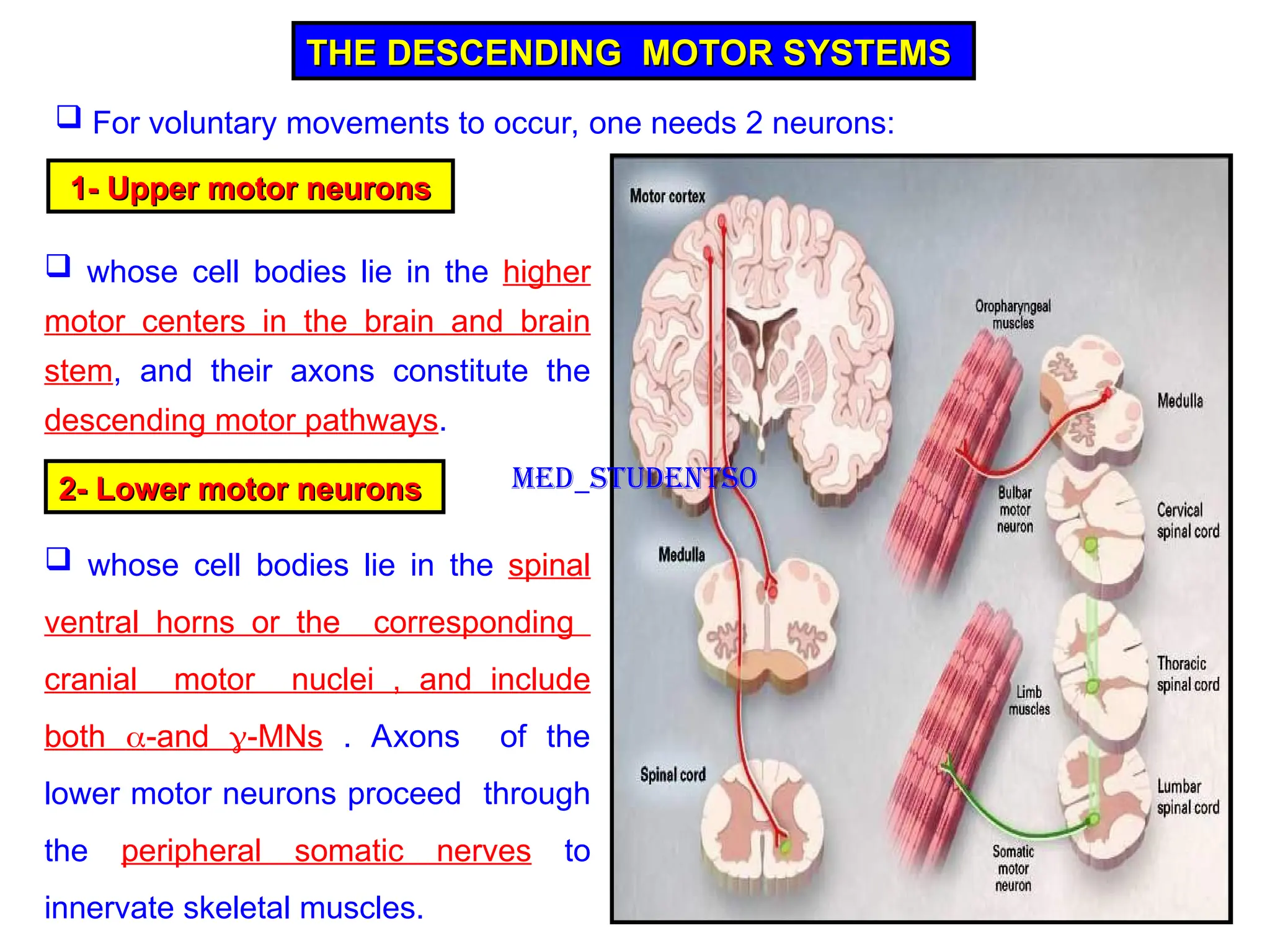
For voluntary movements to occur, one needs 2 neurons:
1-
1- Upper motor neurons
Upper motor neurons
whose cell bodies lie in the higher
motor centers in the brain and brain
stem, and their axons constitute the
descending motor pathways.
2- Lower motor neurons
2- Lower motor neurons
whose cell bodies lie in the spinal
ventral horns or the corresponding
cranial motor nuclei , and include
both -and -MNs . Axons of the
lower motor neurons proceed through
the peripheral somatic nerves to
innervate skeletal muscles.
Med_students0
3.
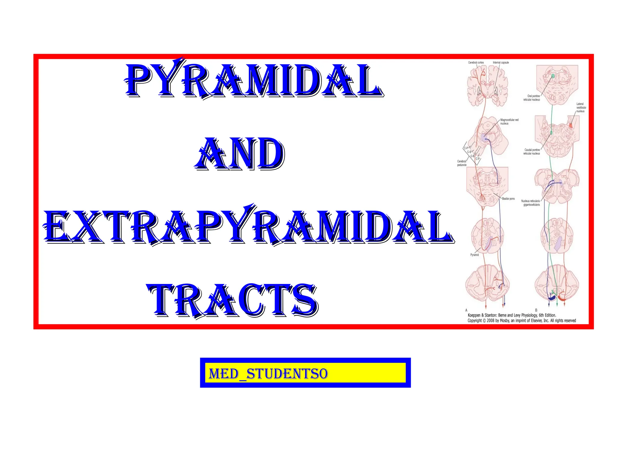
The descendingmotor pathways have commonly been divided into “pyramidal”
and “extrapyramidal” tracts.
This classification is based on the finding that the motor tract which originates
from the cerebral cortex and descends to the spinal cord (the corticospinal tract)
passes through the pyramids of the medulla, and therefore has been called the
“the pyramidal tract”.
The rest of the descending motor pathways do not travel through the medullary
pyramids, and are therefore collectively gathered under the heading: “the
extrapyramidal tracts”.
Classification of descending motor systems
Classification of descending motor systems
Med_students0
4.
An alternativeclassification of the descending motor tracts is
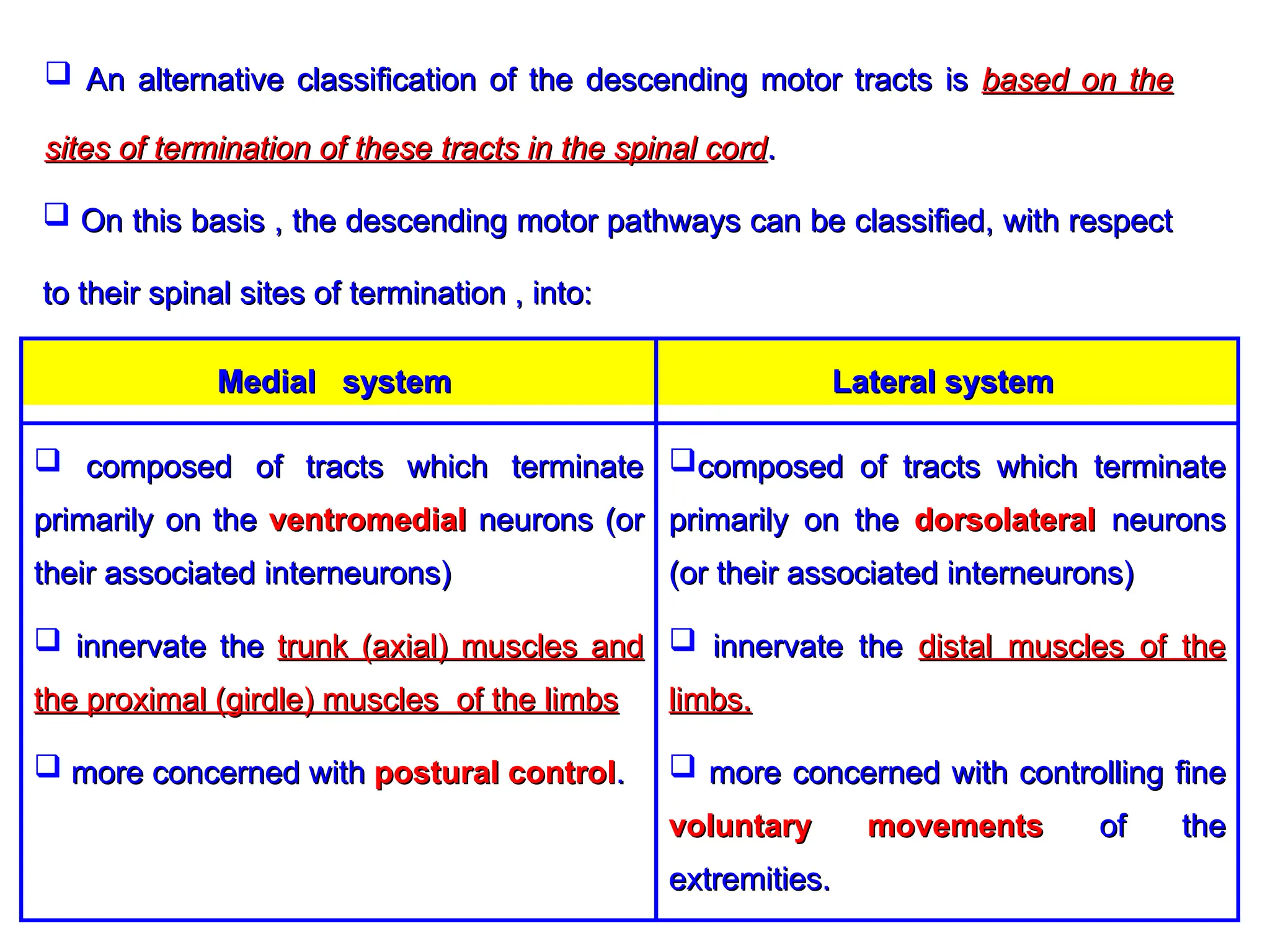
An alternative classification of the descending motor tracts is based on the
based on the
sites of termination of these tracts in the spinal cord
sites of termination of these tracts in the spinal cord.
.
On this basis , the descending motor pathways can be classified, with respect
On this basis , the descending motor pathways can be classified, with respect
to their spinal sites of termination , into:
to their spinal sites of termination , into:
composed of tracts which terminate
composed of tracts which terminate
primarily on the
primarily on the dorsolateral
dorsolateral neurons
neurons
(or their associated interneurons)
(or their associated interneurons)
innervate the
innervate the distal muscles of the
distal muscles of the
limbs.
limbs.
more concerned with controlling fine
more concerned with controlling fine
voluntary movements
voluntary movements of the
of the
extremities.
extremities.
composed of tracts which terminate
composed of tracts which terminate
primarily on the
primarily on the ventromedial
ventromedial neurons (or
neurons (or
their associated interneurons)
their associated interneurons)
innervate the
innervate the trunk (axial) muscles and
trunk (axial) muscles and
the proximal (girdle) muscles of the limbs
the proximal (girdle) muscles of the limbs
more concerned with
more concerned with postural control
postural control.
.
Lateral system
Lateral system
Medial system
Medial system
5.
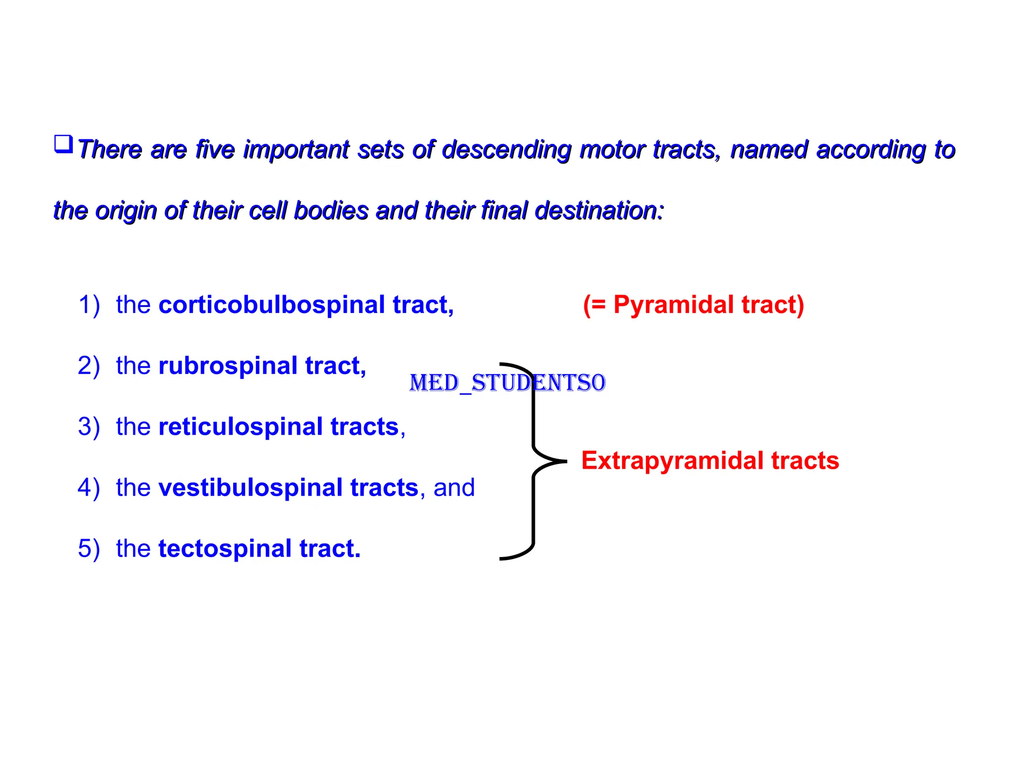
There are fiveimportant sets of descending motor tracts, named according to
There are five important sets of descending motor tracts, named according to
the origin of their cell bodies and their final destination:
the origin of their cell bodies and their final destination:
1) the corticobulbospinal tract, (= Pyramidal tract)
2) the rubrospinal tract,
3) the reticulospinal tracts,
4) the vestibulospinal tracts, and
5) the tectospinal tract.
Extrapyramidal tracts
Med_students0
6.
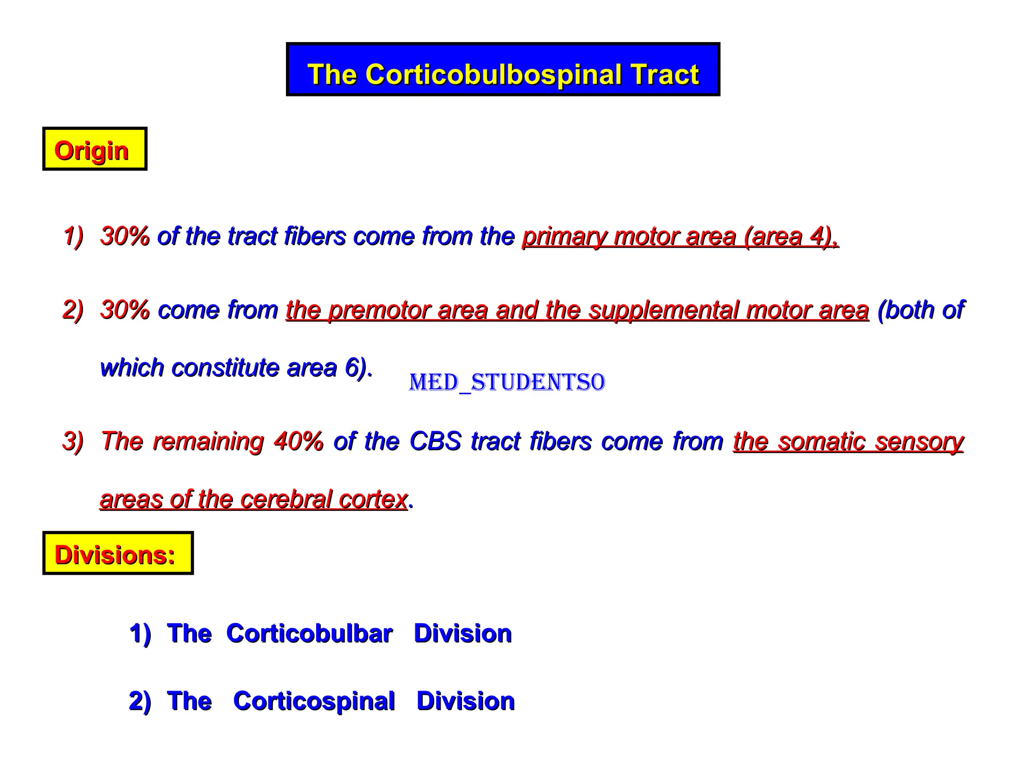
The Corticobulbospinal Tract
TheCorticobulbospinal Tract
Origin
Origin
1)
1) 30%
30% of the tract fibers come from the
of the tract fibers come from the primary motor area (area 4),
primary motor area (area 4),
2)
2) 30%
30% come from
come from the premotor area and the supplemental motor area
the premotor area and the supplemental motor area (both of
(both of
which constitute area 6).
which constitute area 6).
3)
3) The remaining 40%
The remaining 40% of the CBS tract fibers come from
of the CBS tract fibers come from the somatic sensory
the somatic sensory
areas of the cerebral cortex
areas of the cerebral cortex.
.
Divisions:
Divisions:
1)
1) The Corticobulbar Division
The Corticobulbar Division
2)
2) The Corticospinal Division
The Corticospinal Division
Med_students0
7.
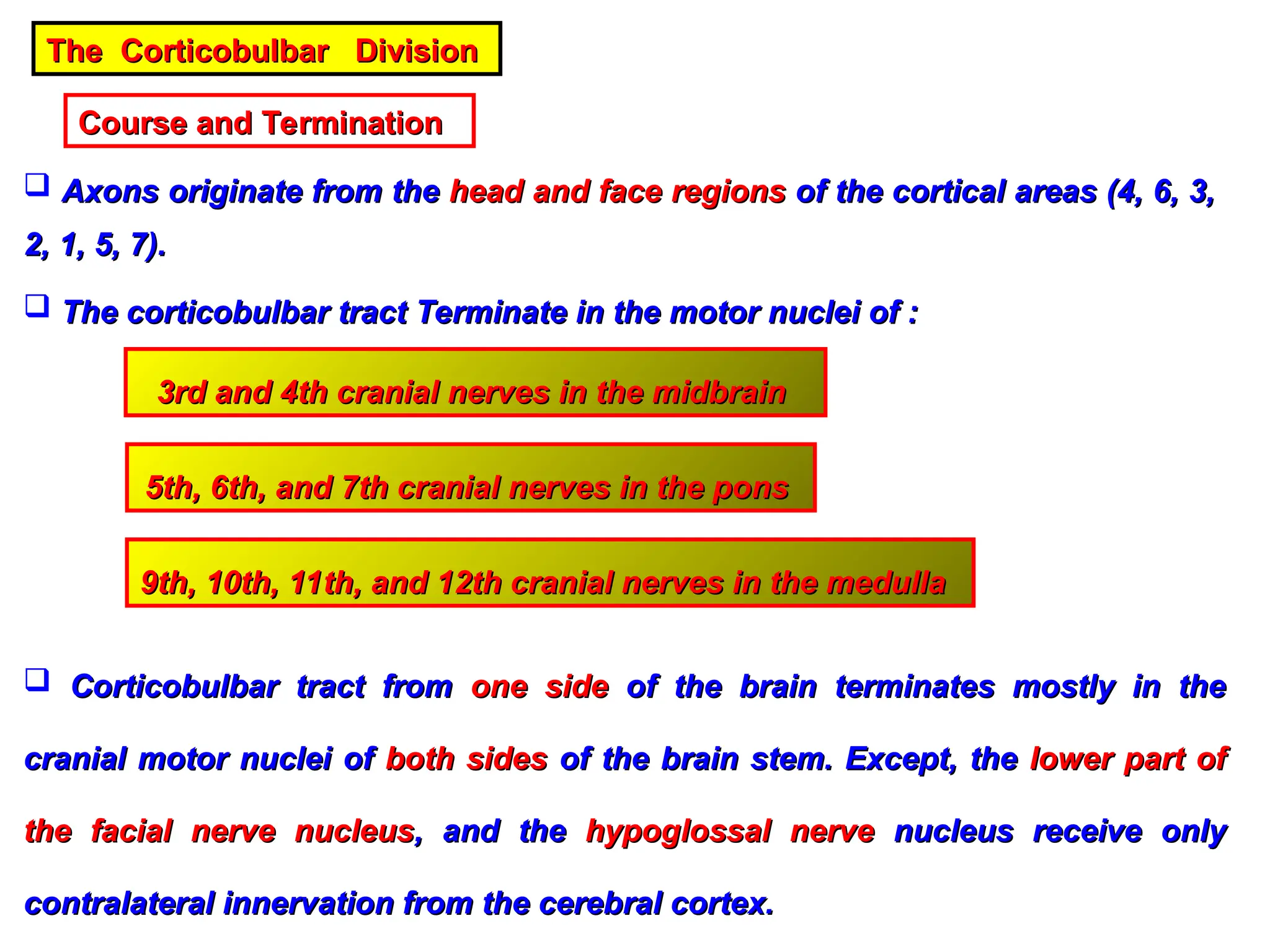
The Corticobulbar Division
TheCorticobulbar Division
Course and Termination
Course and Termination
Axons originate from the
Axons originate from the head and face regions
head and face regions of the cortical areas (4, 6, 3,
of the cortical areas (4, 6, 3,
2, 1, 5, 7).
2, 1, 5, 7).
The corticobulbar tract Terminate in the motor nuclei of :
The corticobulbar tract Terminate in the motor nuclei of :
3rd and 4th cranial nerves in the midbrain
3rd and 4th cranial nerves in the midbrain
5th, 6th, and 7th cranial nerves in the pons
5th, 6th, and 7th cranial nerves in the pons
9th, 10th, 11th, and 12th cranial nerves in the medulla
9th, 10th, 11th, and 12th cranial nerves in the medulla
Corticobulbar tract from
Corticobulbar tract from one side
one side of the brain terminates mostly in the
of the brain terminates mostly in the
cranial motor nuclei of
cranial motor nuclei of both sides
both sides of the brain stem. Except, the
of the brain stem. Except, the lower part of
lower part of
the facial nerve nucleus
the facial nerve nucleus, and the
, and the hypoglossal nerve
hypoglossal nerve nucleus receive only
nucleus receive only
contralateral innervation from the cerebral cortex.
contralateral innervation from the cerebral cortex.
8.
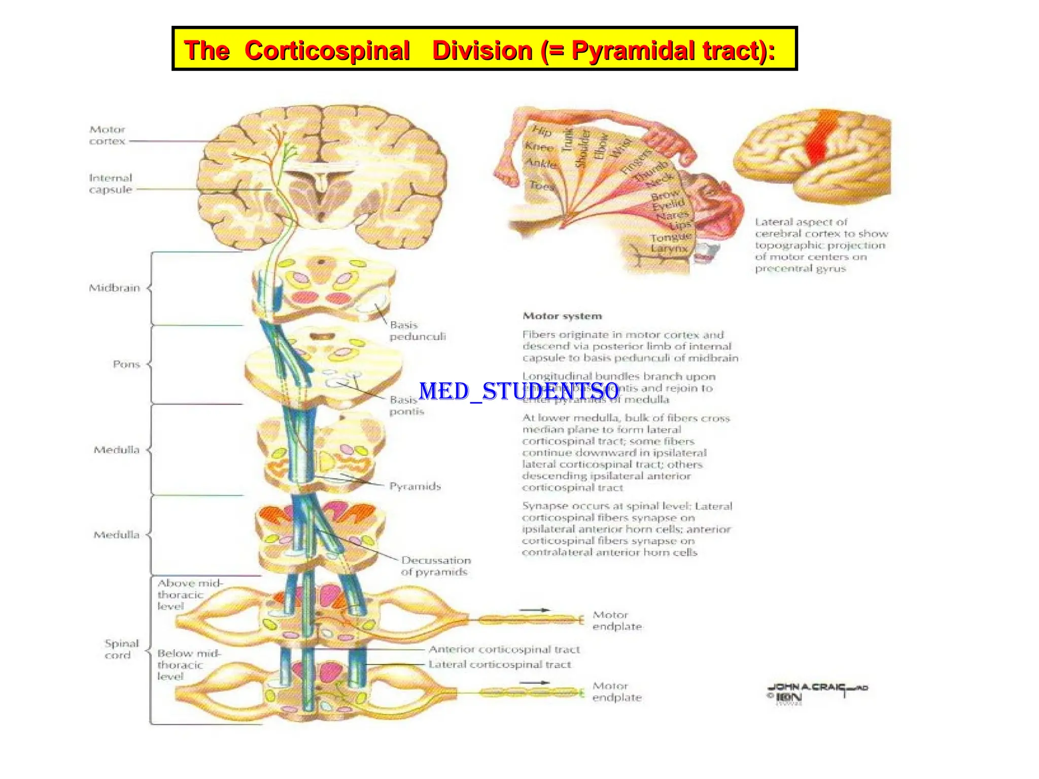
The Corticospinal Division(= Pyramidal tract):
The Corticospinal Division (= Pyramidal tract):
Med_students0
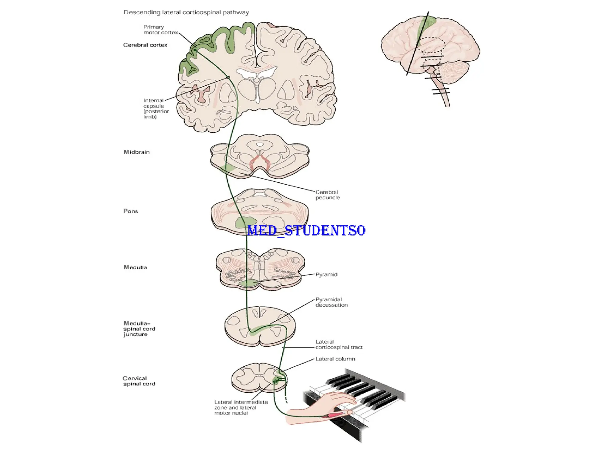
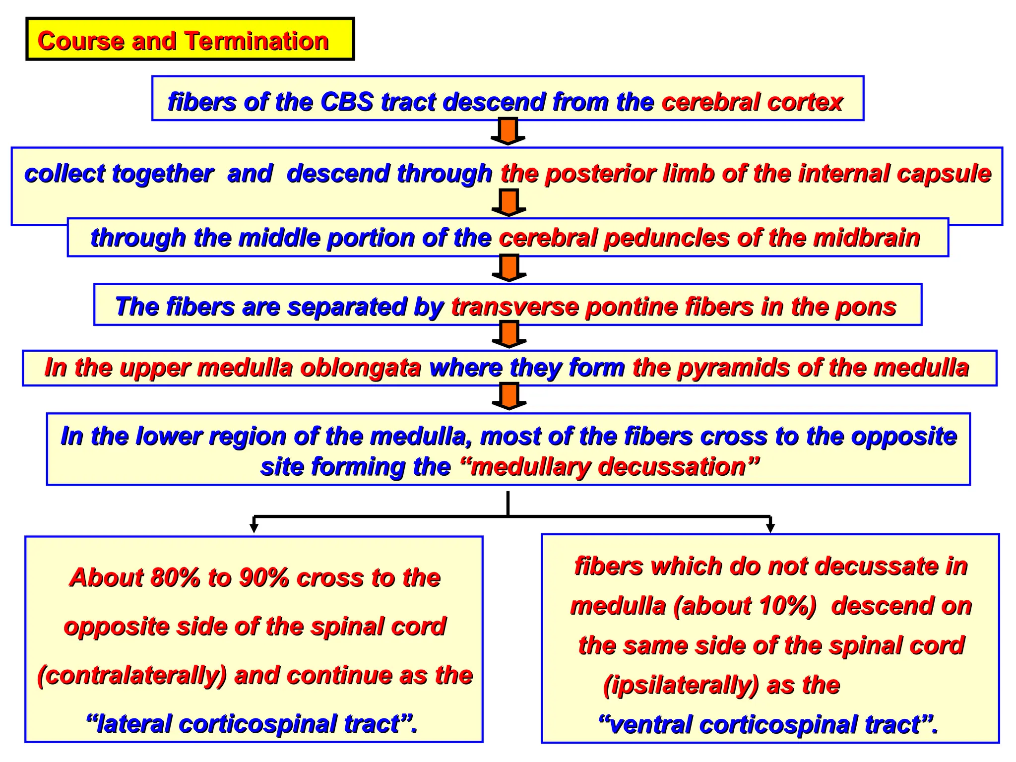
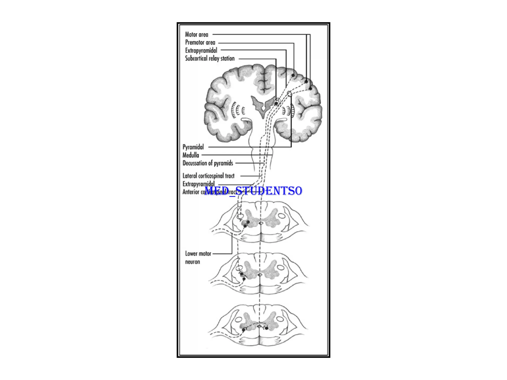
Course and Termination
Courseand Termination
fibers of the CBS tract descend from the
fibers of the CBS tract descend from the cerebral cortex
cerebral cortex
collect together and descend through
collect together and descend through the posterior limb of the internal capsule
the posterior limb of the internal capsule
through the middle portion of the
through the middle portion of the cerebral peduncles of the midbrain
cerebral peduncles of the midbrain
The fibers are separated by
The fibers are separated by transverse pontine fibers in the pons
transverse pontine fibers in the pons
In the upper medulla oblongata
In the upper medulla oblongata where they form
where they form the pyramids of the medulla
the pyramids of the medulla
About 80% to 90% cross to the
About 80% to 90% cross to the
opposite side of the spinal cord
opposite side of the spinal cord
(contralaterally) and continue as the
(contralaterally) and continue as the
“lateral corticospinal tract”.
“lateral corticospinal tract”.
fibers which do not decussate in
fibers which do not decussate in
medulla (about 10%) descend on
medulla (about 10%) descend on
the same side of the spinal cord
the same side of the spinal cord
(ipsilaterally) as the
(ipsilaterally) as the
“ventral corticospinal tract”.
“ventral corticospinal tract”.
In the lower region of the medulla, most of the fibers cross to the opposite
In the lower region of the medulla, most of the fibers cross to the opposite
site forming the
site forming the “medullary decussation”
“medullary decussation”
11.
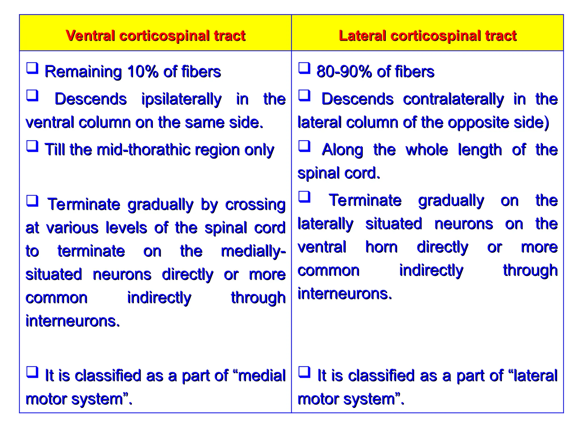
Lateral corticospinal tract
Lateralcorticospinal tract
Ventral corticospinal tract
Ventral corticospinal tract
80-90% of fibers
80-90% of fibers
Descends contralaterally in the
Descends contralaterally in the
lateral column of the opposite side)
lateral column of the opposite side)
Along the whole length of the
Along the whole length of the
spinal cord.
spinal cord.
Terminate gradually on the
Terminate gradually on the
laterally situated neurons on the
laterally situated neurons on the
ventral horn directly or more
ventral horn directly or more
common indirectly through
common indirectly through
interneurons.
interneurons.
It is classified as a part of “lateral
It is classified as a part of “lateral
motor system”.
motor system”.
Remaining 10% of fibers
Remaining 10% of fibers
Descends ipsilaterally in the
Descends ipsilaterally in the
ventral column on the same side.
ventral column on the same side.
Till the mid-thorathic region only
Till the mid-thorathic region only
Terminate gradually by crossing
Terminate gradually by crossing
at various levels of the spinal cord
at various levels of the spinal cord
to terminate on the medially-
to terminate on the medially-
situated neurons directly or more
situated neurons directly or more
common indirectly through
common indirectly through
interneurons.
interneurons.
It is classified as a part of “medial
It is classified as a part of “medial
motor system”.
motor system”.
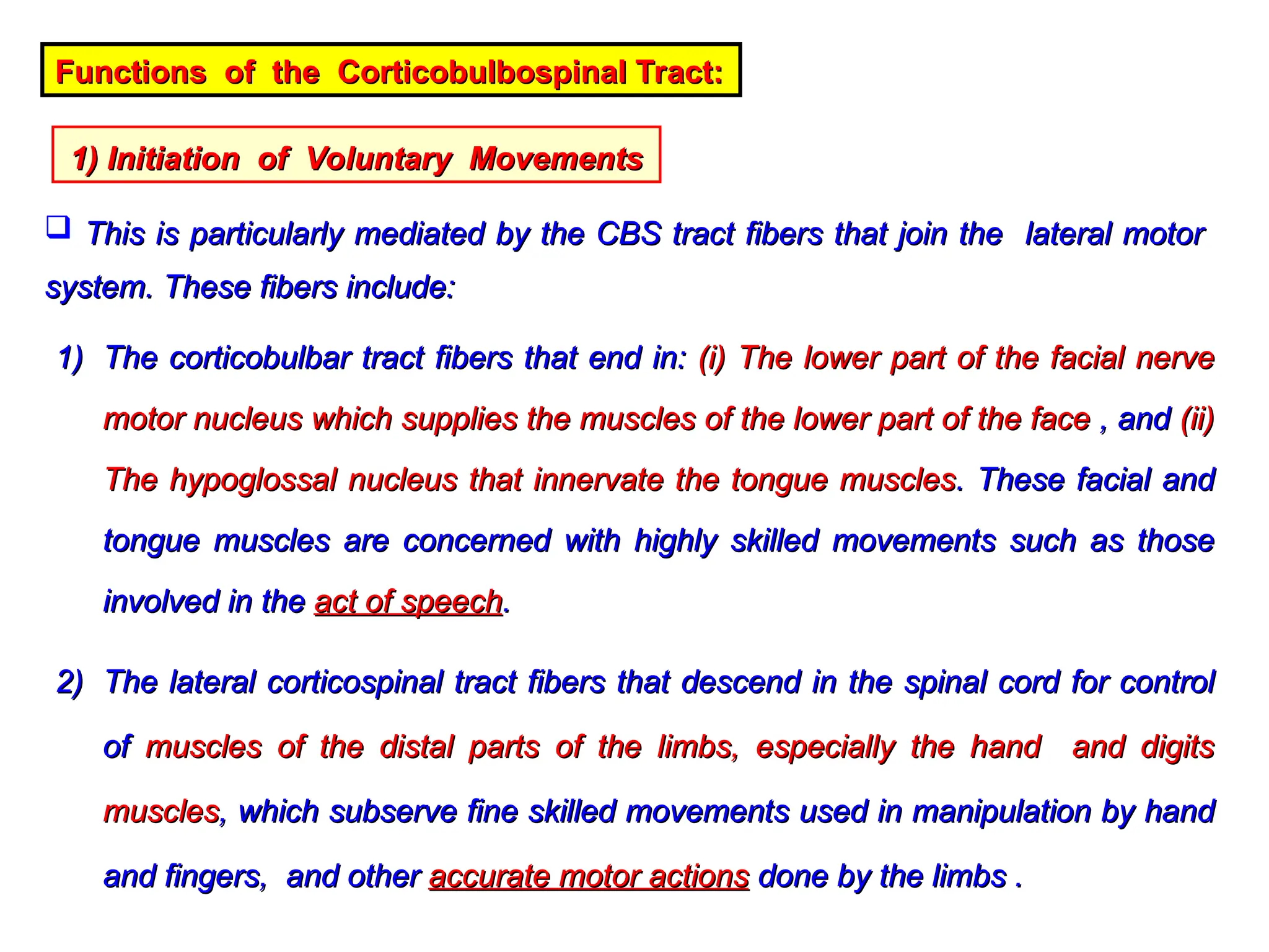
Functions of theCorticobulbospinal Tract:
Functions of the Corticobulbospinal Tract:
1) Initiation of Voluntary Movements
1) Initiation of Voluntary Movements
This is particularly mediated by the CBS tract fibers that join the lateral motor
This is particularly mediated by the CBS tract fibers that join the lateral motor
system. These fibers include:
system. These fibers include:
1)
1) The corticobulbar tract fibers that end in:
The corticobulbar tract fibers that end in: (i) The lower part of the facial nerve
(i) The lower part of the facial nerve
motor nucleus which supplies the muscles of the lower part of the face
motor nucleus which supplies the muscles of the lower part of the face , and
, and (ii)
(ii)
The hypoglossal nucleus that innervate the tongue muscles
The hypoglossal nucleus that innervate the tongue muscles. These facial and
. These facial and
tongue muscles are concerned with highly skilled movements such as those
tongue muscles are concerned with highly skilled movements such as those
involved in the
involved in the act of speech
act of speech.
.
2)
2) The lateral corticospinal tract fibers that descend in the spinal cord for control
The lateral corticospinal tract fibers that descend in the spinal cord for control
of
of muscles of the distal parts of the limbs, especially the hand and digits
muscles of the distal parts of the limbs, especially the hand and digits
muscles
muscles, which subserve fine skilled movements used in manipulation by hand
, which subserve fine skilled movements used in manipulation by hand
and fingers, and other
and fingers, and other accurate motor actions
accurate motor actions done by the limbs .
done by the limbs .
14.
2) Role inAutomatic and Postural Movements:
2) Role in Automatic and Postural Movements:
This is mediated by small portion of the CBS tract fibers that join the medial
This is mediated by small portion of the CBS tract fibers that join the medial
motor system. These fibers include:
motor system. These fibers include:
1)
1) Part of these fibers innervates some cranial nerve motor nuclei which
Part of these fibers innervates some cranial nerve motor nuclei which supply
supply
muscles of the head
muscles of the head involved in such activities as
involved in such activities as closure of the eye lids,
closure of the eye lids,
chewing, swallowing, and phonation
chewing, swallowing, and phonation which often occur automatically.
which often occur automatically.
2)
2) Another part descends in the ventral corticospinal tract which exerts some
Another part descends in the ventral corticospinal tract which exerts some
control on
control on axial and girdle muscles
axial and girdle muscles, that are concerned mainly with
, that are concerned mainly with postural
postural
adjustments.
adjustments.
3) Facilitation of the muscle tone:
3) Facilitation of the muscle tone:
CBS facilitates motor neurons, especially those innervating the distal flexor
CBS facilitates motor neurons, especially those innervating the distal flexor
muscles of the limbs.
muscles of the limbs.
15.
The Rubrospinal Tract
TheRubrospinal Tract
Origin
Origin red nucleus of the midbrain
red nucleus of the midbrain
Afferent
Afferent receives afferent connections from:
receives afferent connections from:
1)
1) Ipsilateral cortical motor areas (corticorubral pathway),
Ipsilateral cortical motor areas (corticorubral pathway),
2)
2) Contralateral side of the cerebellum,
Contralateral side of the cerebellum,
3)
3) Basal ganglia.
Basal ganglia.
Course and Termination
Course and Termination
On leaving the red nucleus, the fibers of the rubrospinal tract cross to the
On leaving the red nucleus, the fibers of the rubrospinal tract cross to the
opposite side -------> descend contralaterally through the brain stem and
opposite side -------> descend contralaterally through the brain stem and
the lateral column of the spinal white matter, very close to the lateral
the lateral column of the spinal white matter, very close to the lateral
corticospinal tract .
corticospinal tract .
16.
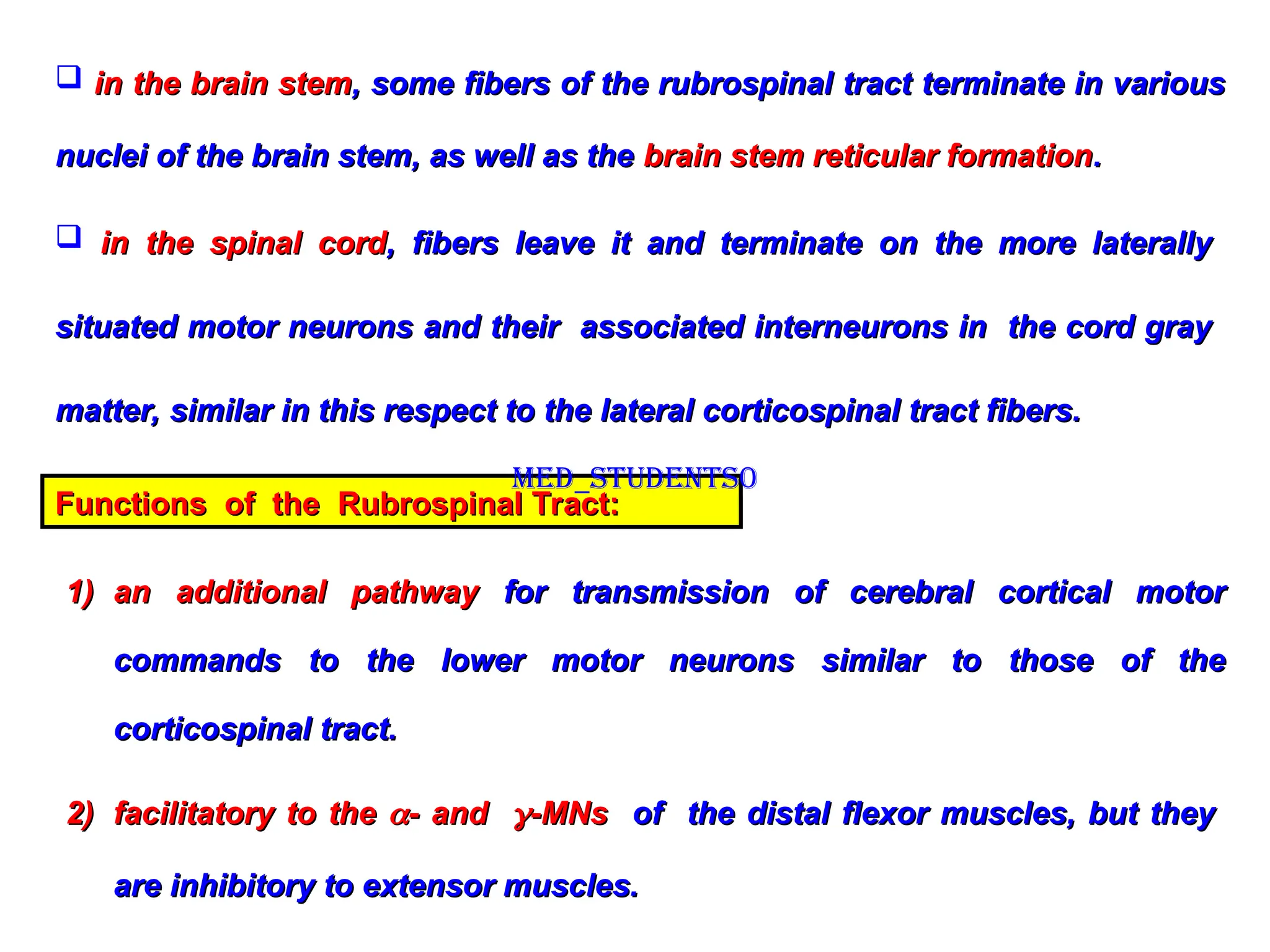
in thebrain stem
in the brain stem, some fibers of the rubrospinal tract terminate in various
, some fibers of the rubrospinal tract terminate in various
nuclei of the brain stem, as well as the
nuclei of the brain stem, as well as the brain stem reticular formation
brain stem reticular formation.
.
in the spinal cord
in the spinal cord, fibers leave it and terminate on the more laterally
, fibers leave it and terminate on the more laterally
situated motor neurons and their associated interneurons in the cord gray
situated motor neurons and their associated interneurons in the cord gray
matter, similar in this respect to the lateral corticospinal tract fibers.
matter, similar in this respect to the lateral corticospinal tract fibers.
Functions of the Rubrospinal Tract:
Functions of the Rubrospinal Tract:
1)
1) an additional pathway
an additional pathway for transmission of cerebral cortical motor
for transmission of cerebral cortical motor
commands to the lower motor neurons similar to those of the
commands to the lower motor neurons similar to those of the
corticospinal tract.
corticospinal tract.
2)
2) facilitatory to the
facilitatory to the
- and
- and
-MNs
-MNs of the distal flexor muscles, but they
of the distal flexor muscles, but they
are inhibitory to extensor muscles.
are inhibitory to extensor muscles.
Med_students0
17.
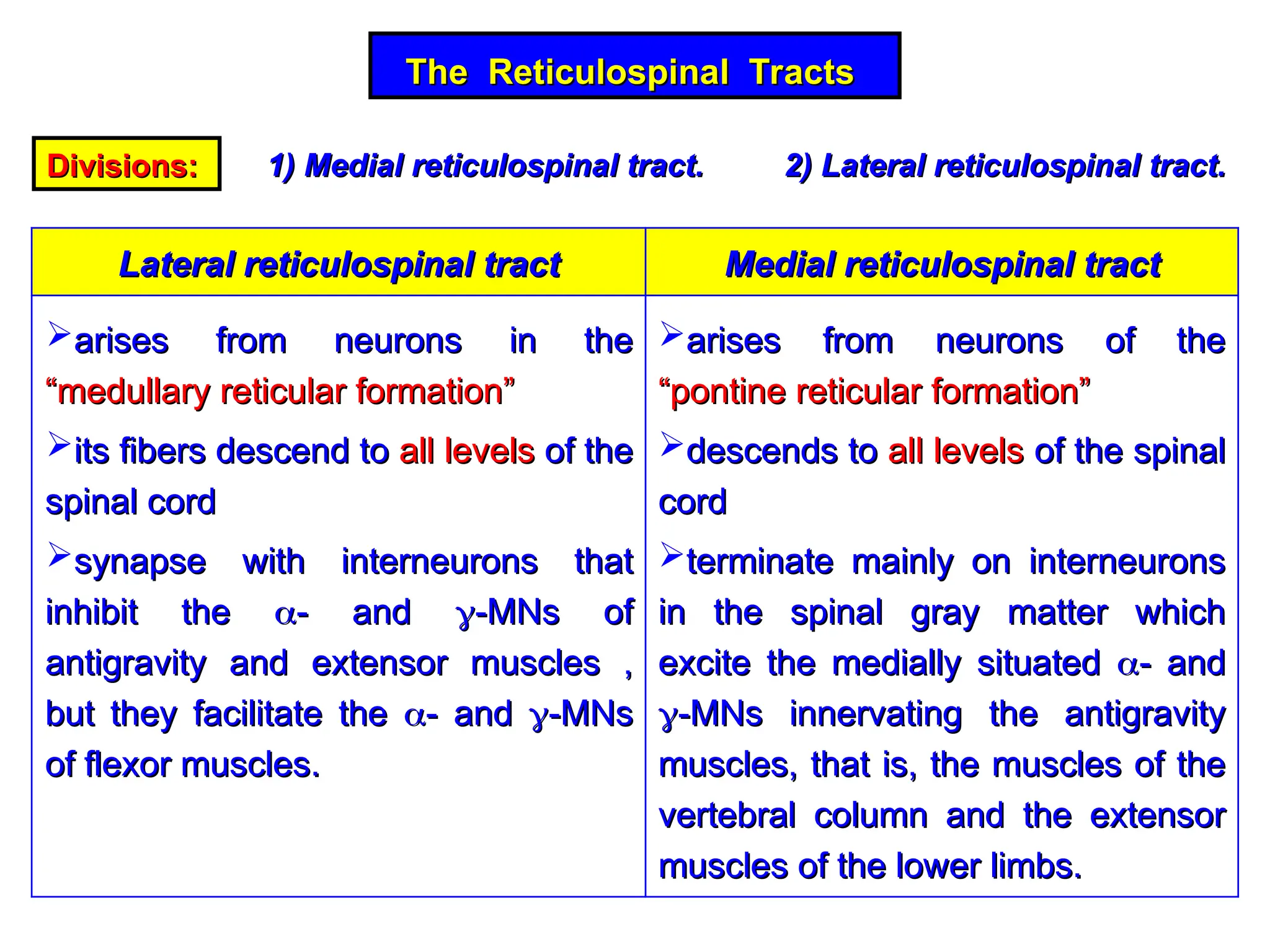
The Reticulospinal Tracts
TheReticulospinal Tracts
Divisions:
Divisions: 1) Medial reticulospinal tract. 2) Lateral reticulospinal tract.
1) Medial reticulospinal tract. 2) Lateral reticulospinal tract.
Medial reticulospinal tract
Medial reticulospinal tract
Lateral reticulospinal tract
Lateral reticulospinal tract
arises from neurons of the
arises from neurons of the
“pontine reticular formation”
“pontine reticular formation”
descends to
descends to all levels
all levels of the spinal
of the spinal
cord
cord
terminate mainly on interneurons
terminate mainly on interneurons
in the spinal gray matter which
in the spinal gray matter which
excite the medially situated
excite the medially situated
- and
- and
-MNs innervating the antigravity
-MNs innervating the antigravity
muscles, that is, the muscles of the
muscles, that is, the muscles of the
vertebral column and the extensor
vertebral column and the extensor
muscles of the lower limbs.
muscles of the lower limbs.
arises from neurons in the
arises from neurons in the
“medullary reticular formation”
“medullary reticular formation”
its fibers descend to
its fibers descend to all levels
all levels of the
of the
spinal cord
spinal cord
synapse with interneurons that
synapse with interneurons that
inhibit the
inhibit the
- and
- and
-MNs of
-MNs of
antigravity and extensor muscles ,
antigravity and extensor muscles ,
but they facilitate the
but they facilitate the
- and
- and
-MNs
-MNs
of flexor muscles.
of flexor muscles.
18.
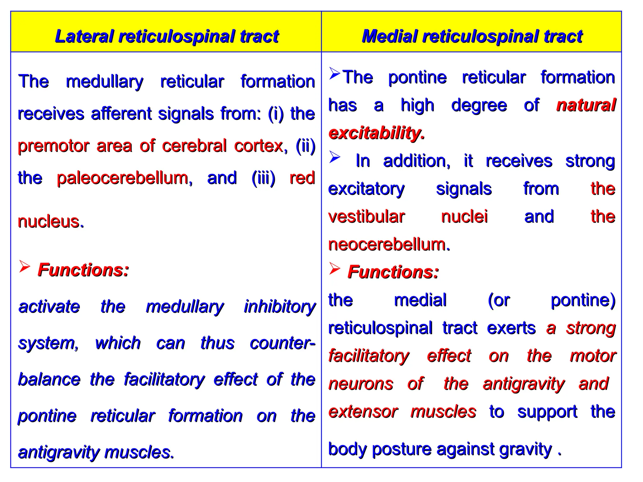
Medial reticulospinal tract
Medialreticulospinal tract
Lateral reticulospinal tract
Lateral reticulospinal tract
The pontine reticular formation
The pontine reticular formation
has a high degree of
has a high degree of natural
natural
excitability.
excitability.
In addition, it receives strong
In addition, it receives strong
excitatory signals from
excitatory signals from the
the
vestibular nuclei
vestibular nuclei and
and the
the
neocerebellum
neocerebellum.
.
Functions:
Functions:
the medial (or pontine)
the medial (or pontine)
reticulospinal tract exerts
reticulospinal tract exerts a strong
a strong
facilitatory effect on the motor
facilitatory effect on the motor
neurons of the antigravity and
neurons of the antigravity and
extensor muscles
extensor muscles to support the
to support the
body posture against gravity .
body posture against gravity .
The medullary reticular formation
The medullary reticular formation
receives afferent signals from: (i) the
receives afferent signals from: (i) the
premotor area of cerebral cortex
premotor area of cerebral cortex, (ii)
, (ii)
the
the paleocerebellum
paleocerebellum, and (iii)
, and (iii) red
red
nucleus
nucleus.
.
Functions:
Functions:
activate the medullary inhibitory
activate the medullary inhibitory
system, which can thus counter-
system, which can thus counter-
balance the facilitatory effect of the
balance the facilitatory effect of the
pontine reticular formation on the
pontine reticular formation on the
antigravity muscles.
antigravity muscles.
19.
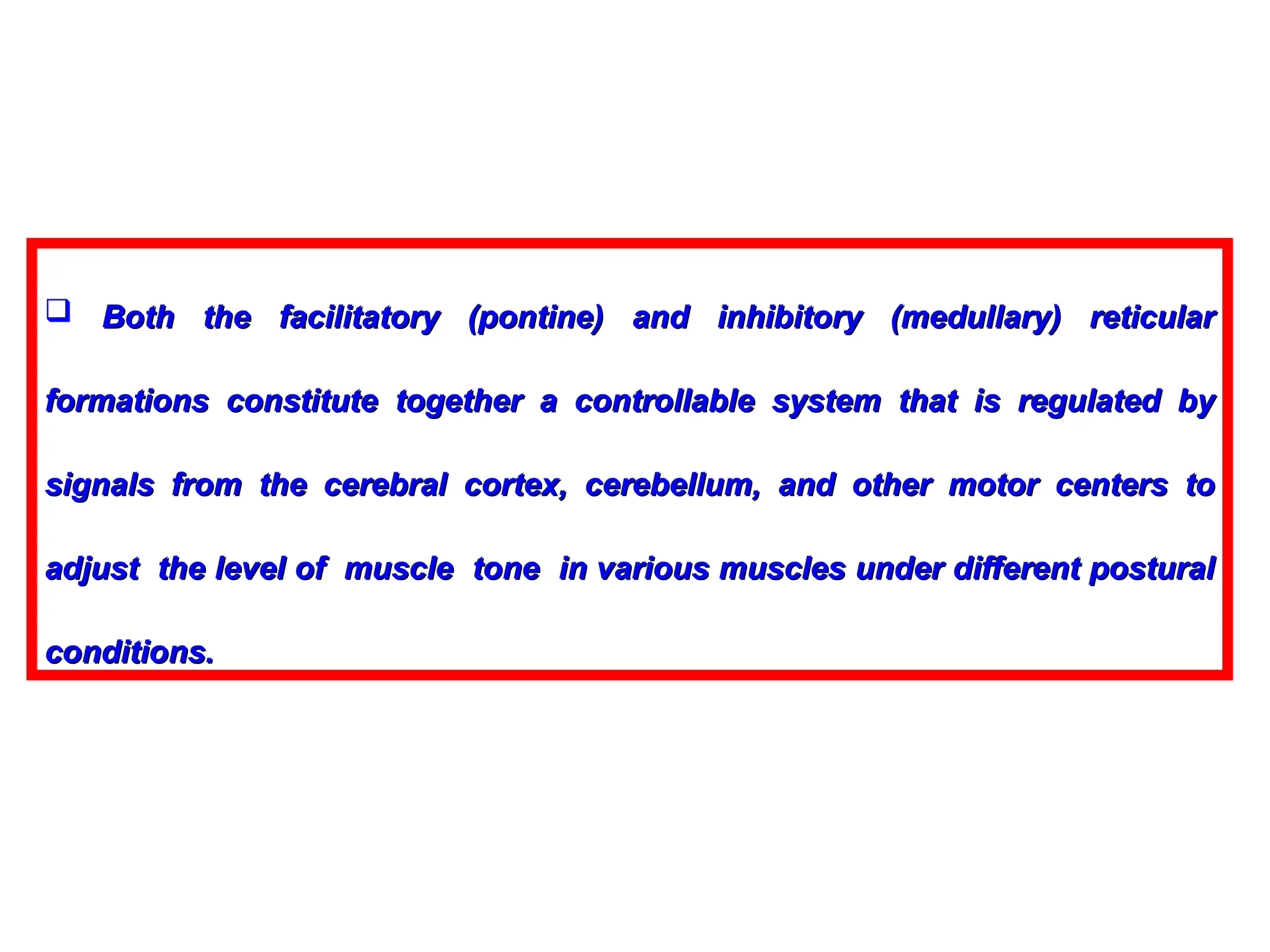
Both thefacilitatory (pontine) and inhibitory (medullary) reticular
Both the facilitatory (pontine) and inhibitory (medullary) reticular
formations constitute together a controllable system that is regulated by
formations constitute together a controllable system that is regulated by
signals from the cerebral cortex, cerebellum, and other motor centers to
signals from the cerebral cortex, cerebellum, and other motor centers to
adjust the level of muscle tone in various muscles under different postural
adjust the level of muscle tone in various muscles under different postural
conditions.
conditions.
20.
The Vestibulospinal Tracts
TheVestibulospinal Tracts
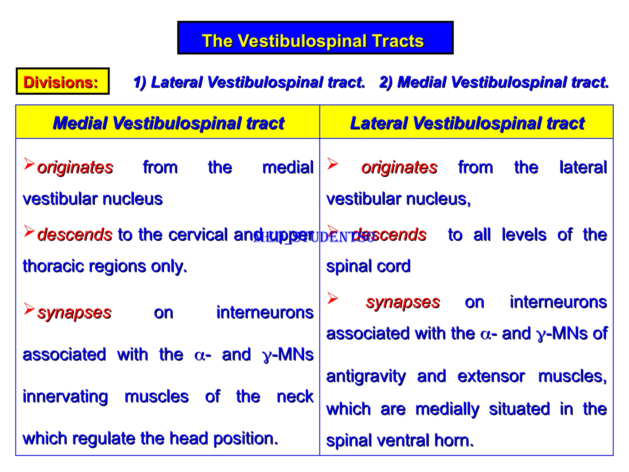
Divisions:
Divisions: 1) Lateral Vestibulospinal tract. 2) Medial Vestibulospinal tract.
1) Lateral Vestibulospinal tract. 2) Medial Vestibulospinal tract.
Lateral Vestibulospinal tract
Lateral Vestibulospinal tract
Medial Vestibulospinal tract
Medial Vestibulospinal tract
originates
originates from the lateral
from the lateral
vestibular nucleus,
vestibular nucleus,
descends
descends to all levels of the
to all levels of the
spinal cord
spinal cord
synapses
synapses on interneurons
on interneurons
associated with the
associated with the
- and
- and
-MNs of
-MNs of
antigravity and extensor
antigravity and extensor muscles,
muscles,
which are medially situated in the
which are medially situated in the
spinal ventral horn.
spinal ventral horn.
originates
originates from the medial
from the medial
vestibular nucleus
vestibular nucleus
descends
descends to the cervical and upper
to the cervical and upper
thoracic regions only.
thoracic regions only.
synapses
synapses on interneurons
on interneurons
associated with the
associated with the
- and
- and
-MNs
-MNs
innervating muscles of the neck
innervating muscles of the neck
which regulate the head position.
which regulate the head position.
Med_students0
21.
Functions
Functions
Change of headposition in relation to
Change of head position in relation to
the earth’s gravity
the earth’s gravity
Exposure to acceleratory
Exposure to acceleratory
forces
forces
Vestibular nuclei
Vestibular nuclei
Vestibulospinal Tract
Vestibulospinal Tract
1) adjusting the tone and contraction of
1) adjusting the tone and contraction of antigravity muscles
antigravity muscles to
to
maintain the body posture and equilibrium by
maintain the body posture and equilibrium by LVST
LVST .
.
2) regulating the position of the
2) regulating the position of the head and upper limbs
head and upper limbs during
during
exposure to acceleration by
exposure to acceleration by MVST
MVST.
.
Med_students0
22.
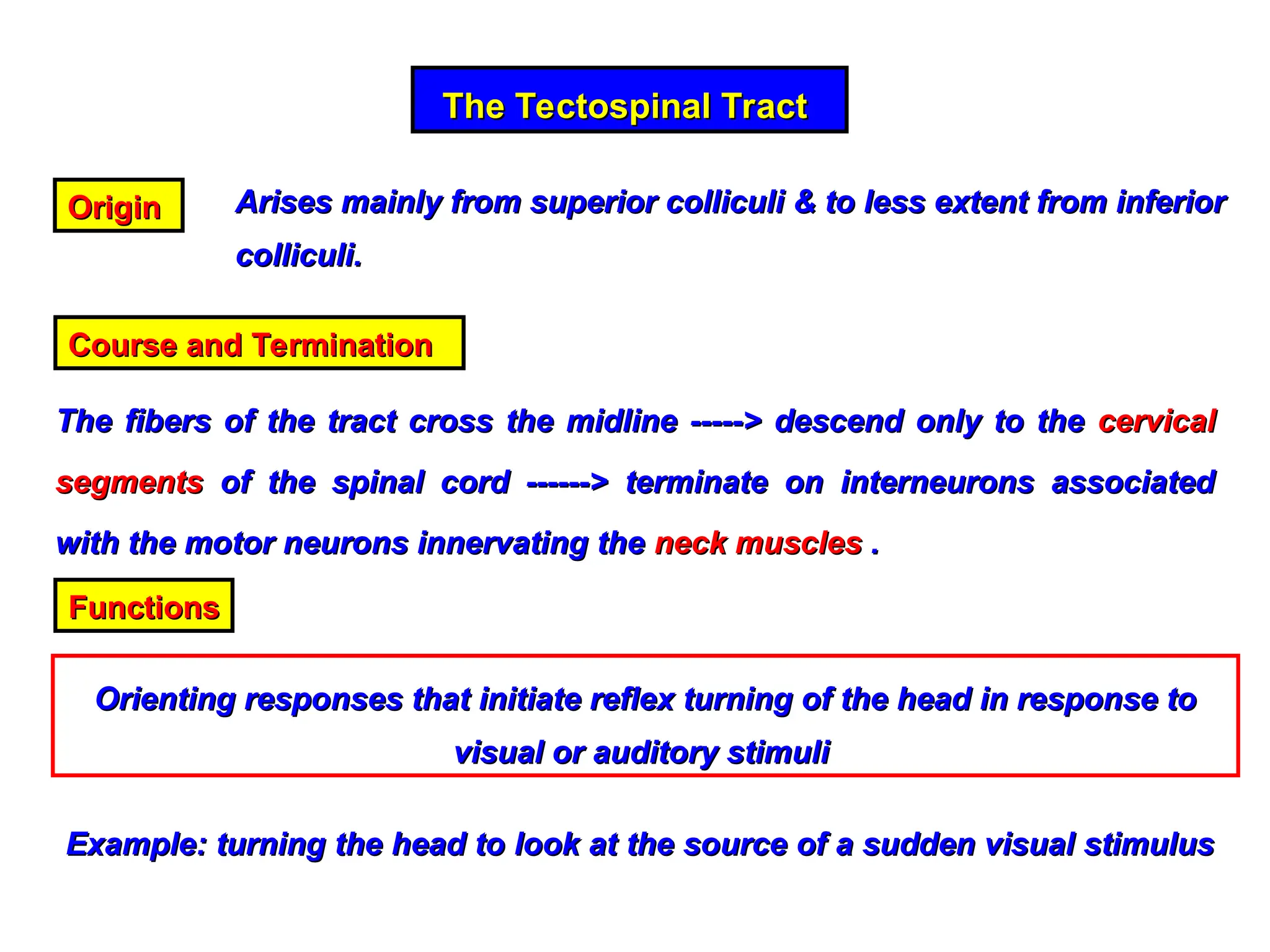
The Tectospinal Tract
TheTectospinal Tract
Origin
Origin Arises mainly from superior colliculi & to less extent from inferior
Arises mainly from superior colliculi & to less extent from inferior
colliculi.
colliculi.
Course and Termination
Course and Termination
The fibers of the tract cross the midline -----> descend only to the
The fibers of the tract cross the midline -----> descend only to the cervical
cervical
segments
segments of the spinal cord ------> terminate on interneurons associated
of the spinal cord ------> terminate on interneurons associated
with the motor neurons innervating the
with the motor neurons innervating the neck muscles
neck muscles .
.
Functions
Functions
Orienting responses that initiate reflex turning of the head in response to
Orienting responses that initiate reflex turning of the head in response to
visual or auditory stimuli
visual or auditory stimuli
Example: turning the head to look at the source of a sudden visual stimulus
Example: turning the head to look at the source of a sudden visual stimulus
23.
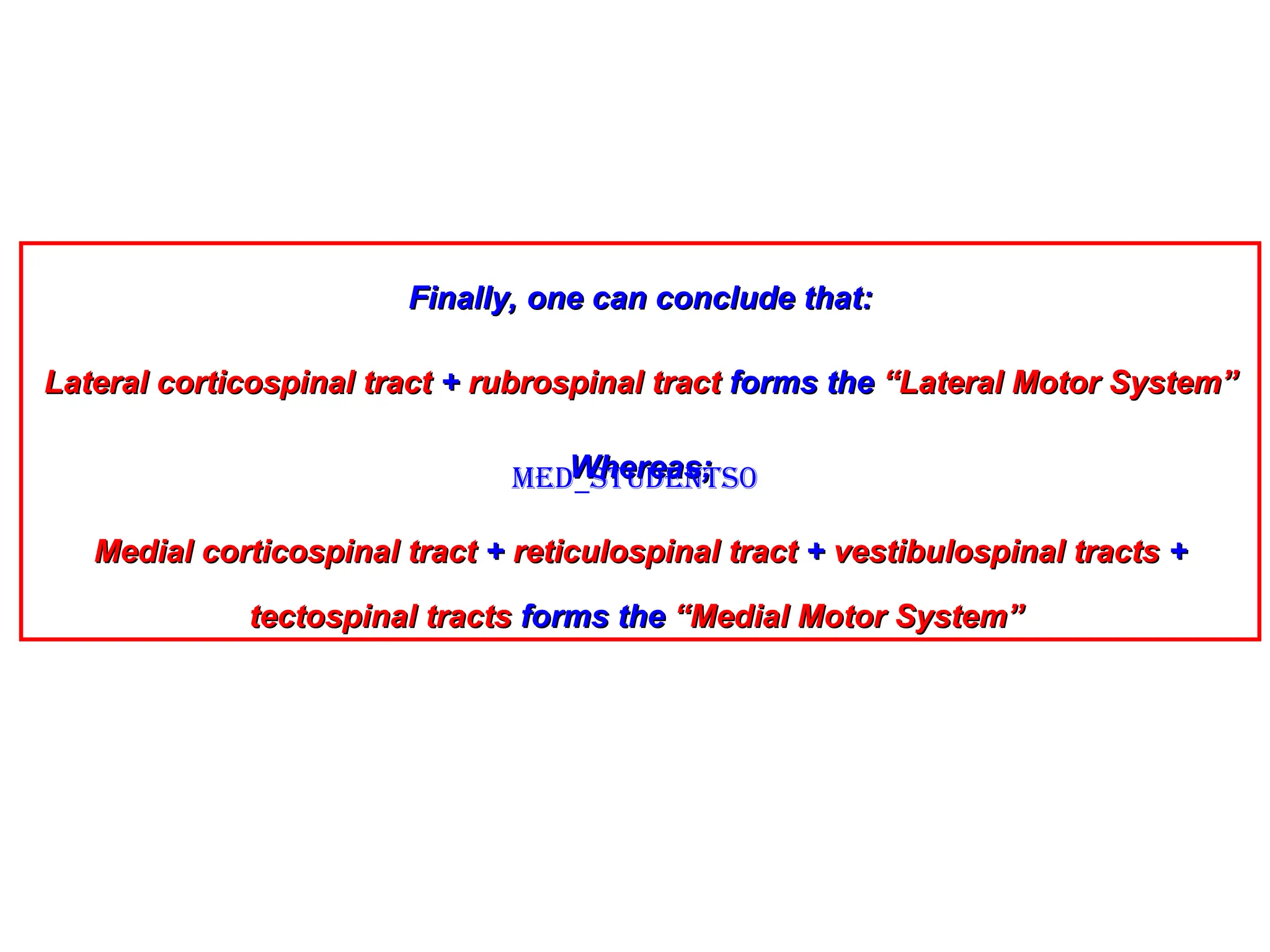
Finally, one canconclude that:
Finally, one can conclude that:
Lateral corticospinal tract
Lateral corticospinal tract +
+ rubrospinal tract
rubrospinal tract forms the
forms the “Lateral Motor System”
“Lateral Motor System”
Whereas;
Whereas;
Medial corticospinal tract
Medial corticospinal tract +
+ reticulospinal tract
reticulospinal tract +
+ vestibulospinal tracts
vestibulospinal tracts +
+
tectospinal tracts
tectospinal tracts forms the
forms the “Medial Motor System”
“Medial Motor System”
Med_students0
24.
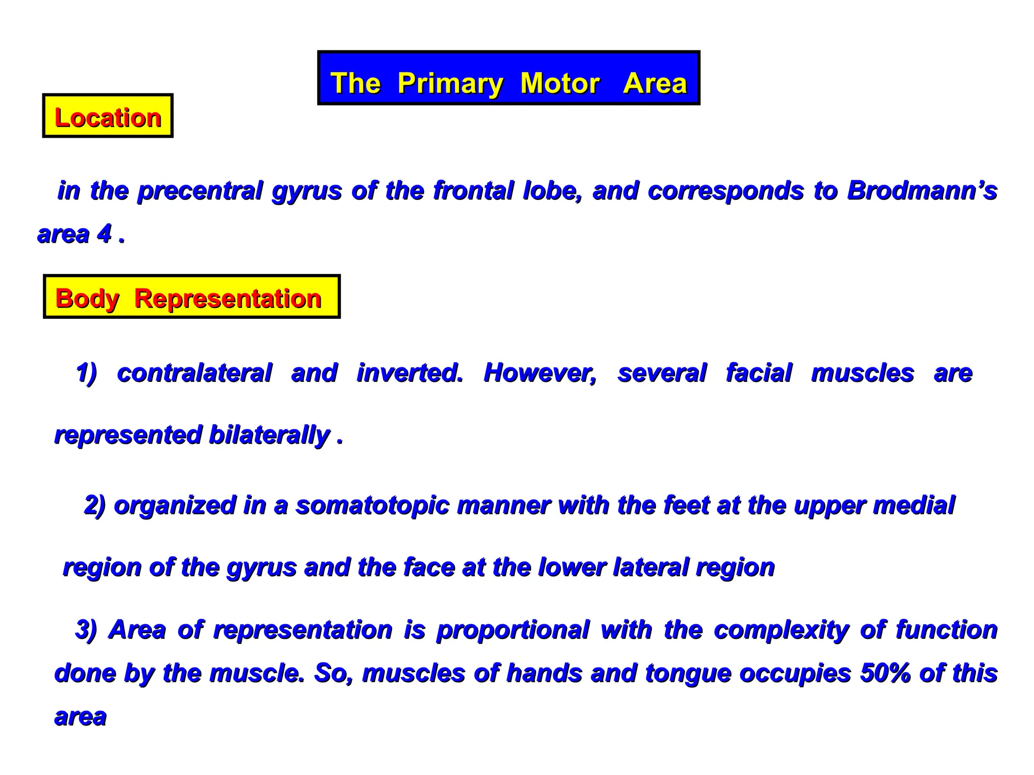
The Primary MotorArea
The Primary Motor Area
Location
Location
in the precentral gyrus of the frontal lobe, and corresponds to Brodmann’s
in the precentral gyrus of the frontal lobe, and corresponds to Brodmann’s
area 4 .
area 4 .
Body Representation
Body Representation
1) contralateral and inverted. However, several facial muscles are
1) contralateral and inverted. However, several facial muscles are
represented bilaterally .
represented bilaterally .
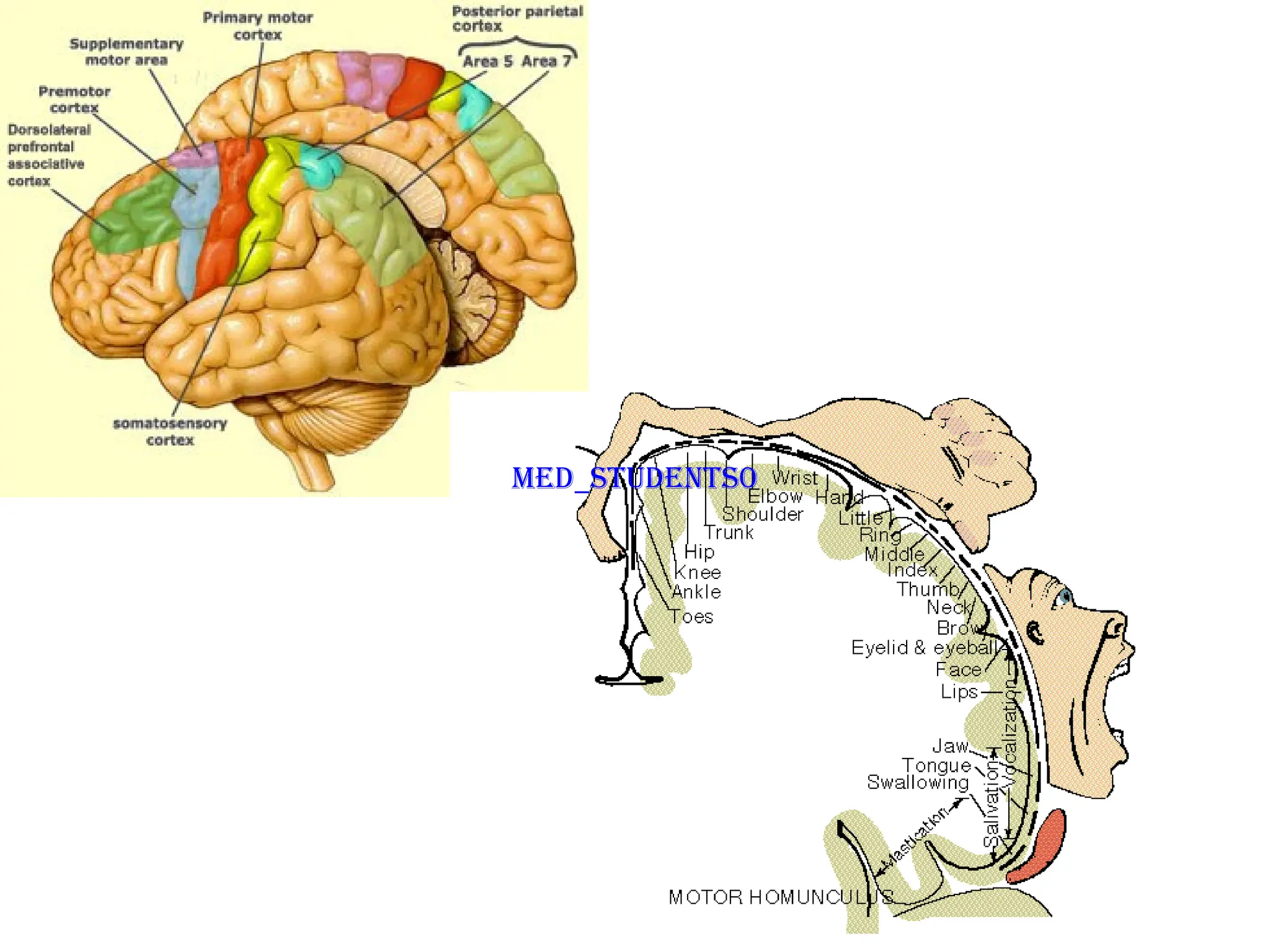
2) organized in a somatotopic manner with the feet at the upper medial
2) organized in a somatotopic manner with the feet at the upper medial
region of the gyrus and the face at the lower lateral region
region of the gyrus and the face at the lower lateral region
3) Area of representation is proportional with the complexity of function
3) Area of representation is proportional with the complexity of function
done by the muscle. So, muscles of hands and tongue occupies 50% of this
done by the muscle. So, muscles of hands and tongue occupies 50% of this
area
area
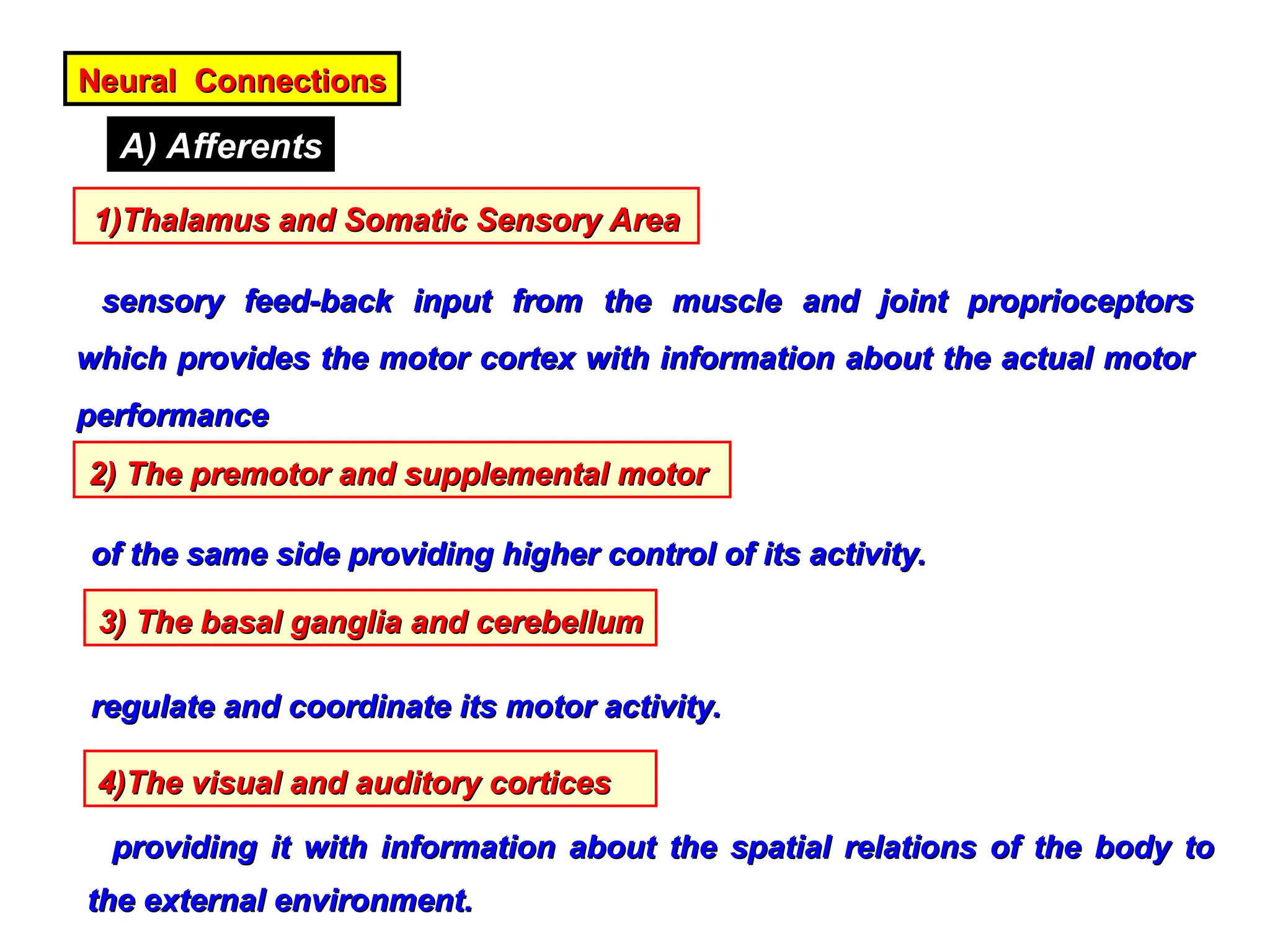
Neural Connections
Neural Connections
A)Afferents
1)Thalamus and Somatic Sensory Area
1)Thalamus and Somatic Sensory Area
sensory feed-back input from the muscle and joint proprioceptors
sensory feed-back input from the muscle and joint proprioceptors
which provides the motor cortex with information about the actual motor
which provides the motor cortex with information about the actual motor
performance
performance
2) The premotor and supplemental motor
2) The premotor and supplemental motor
of the same side providing higher control of its activity.
of the same side providing higher control of its activity.
3) The basal ganglia and cerebellum
3) The basal ganglia and cerebellum
regulate and coordinate its motor activity.
regulate and coordinate its motor activity.
4)The visual and auditory cortices
4)The visual and auditory cortices
providing it with information about the spatial relations of the body to
providing it with information about the spatial relations of the body to
the external environment.
the external environment.
27.
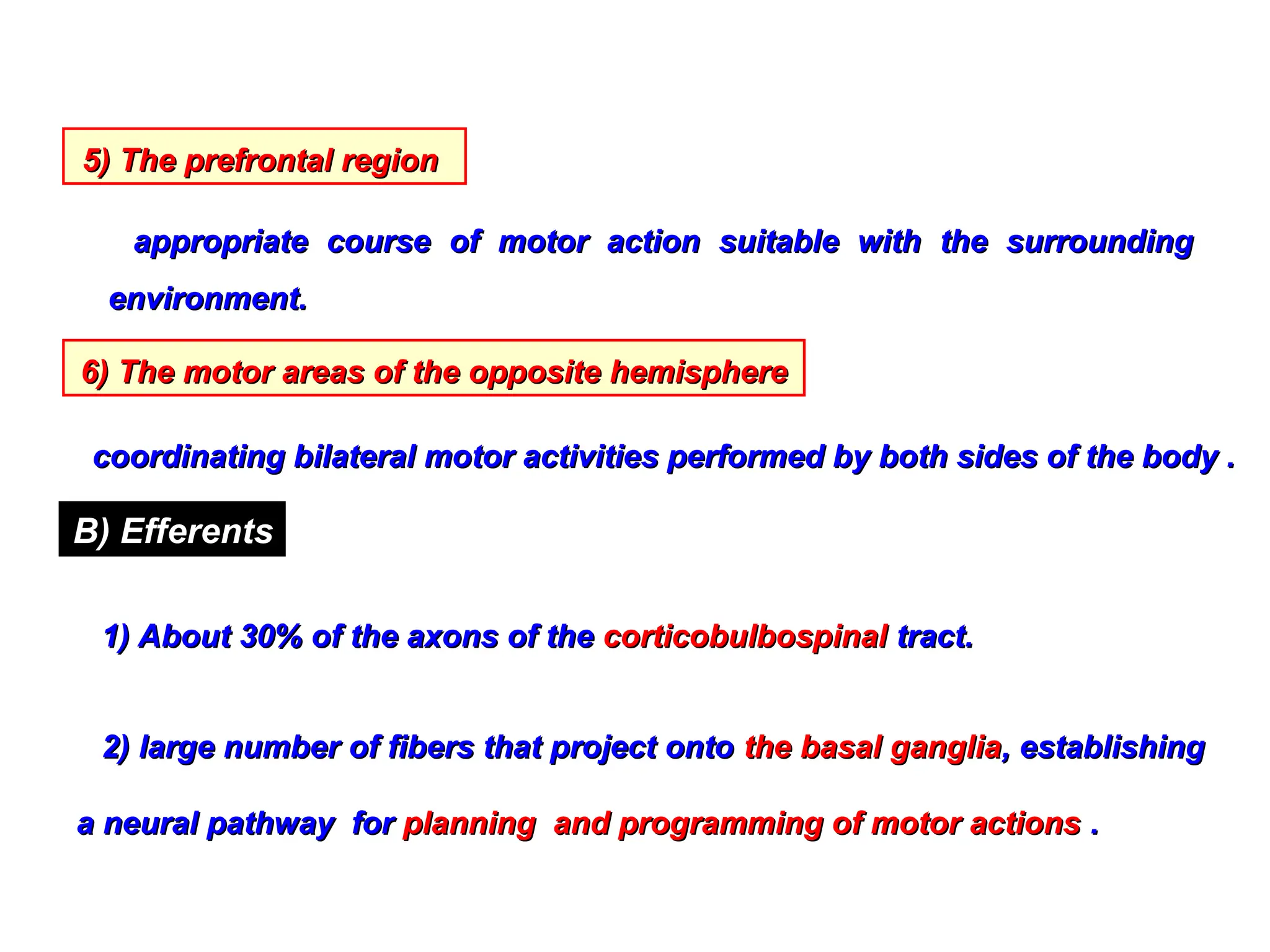
5) The prefrontalregion
5) The prefrontal region
appropriate course of motor action suitable with the surrounding
appropriate course of motor action suitable with the surrounding
environment.
environment.
6) The motor areas of the opposite hemisphere
6) The motor areas of the opposite hemisphere
coordinating bilateral motor activities performed by both sides of the body .
coordinating bilateral motor activities performed by both sides of the body .
B) Efferents
1) About 30% of the axons of the
1) About 30% of the axons of the corticobulbospinal
corticobulbospinal tract.
tract.
2) large number of fibers that project onto
2) large number of fibers that project onto the basal ganglia
the basal ganglia, establishing
, establishing
a neural pathway for
a neural pathway for planning and programming of motor actions
planning and programming of motor actions .
.
28.
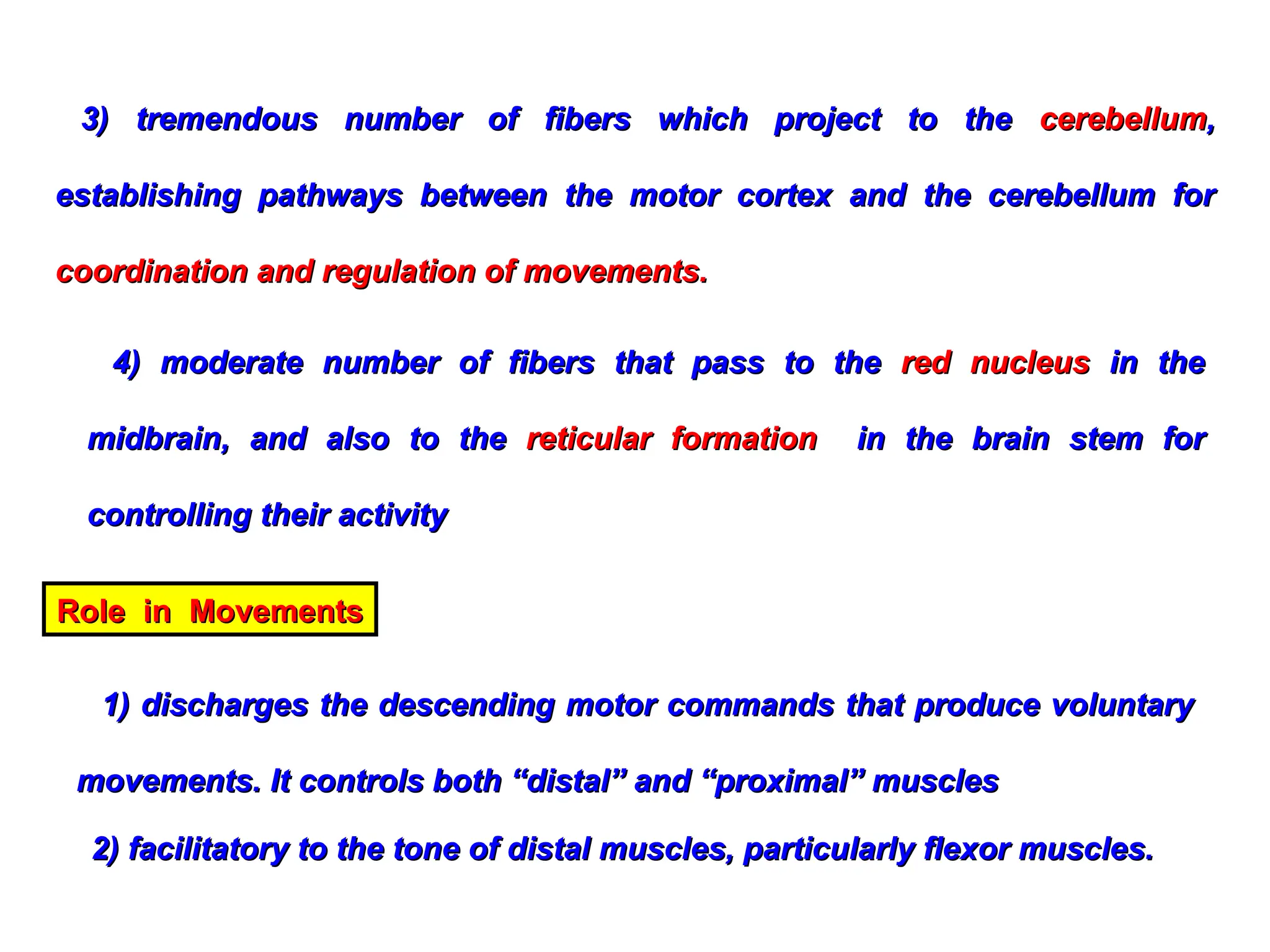
4) moderate numberof fibers that pass to the
4) moderate number of fibers that pass to the red nucleus
red nucleus in the
in the
midbrain, and also to the
midbrain, and also to the reticular formation
reticular formation in the brain stem for
in the brain stem for
controlling their activity
controlling their activity
3) tremendous number of fibers which project to the
3) tremendous number of fibers which project to the cerebellum
cerebellum,
,
establishing pathways between the motor cortex and the cerebellum for
establishing pathways between the motor cortex and the cerebellum for
coordination and regulation of movements.
coordination and regulation of movements.
Role in Movements
Role in Movements
1) discharges the descending motor commands that produce voluntary
1) discharges the descending motor commands that produce voluntary
movements. It controls both “distal” and “proximal” muscles
movements. It controls both “distal” and “proximal” muscles
2) facilitatory to the tone of distal muscles, particularly flexor muscles.
2) facilitatory to the tone of distal muscles, particularly flexor muscles.
29.
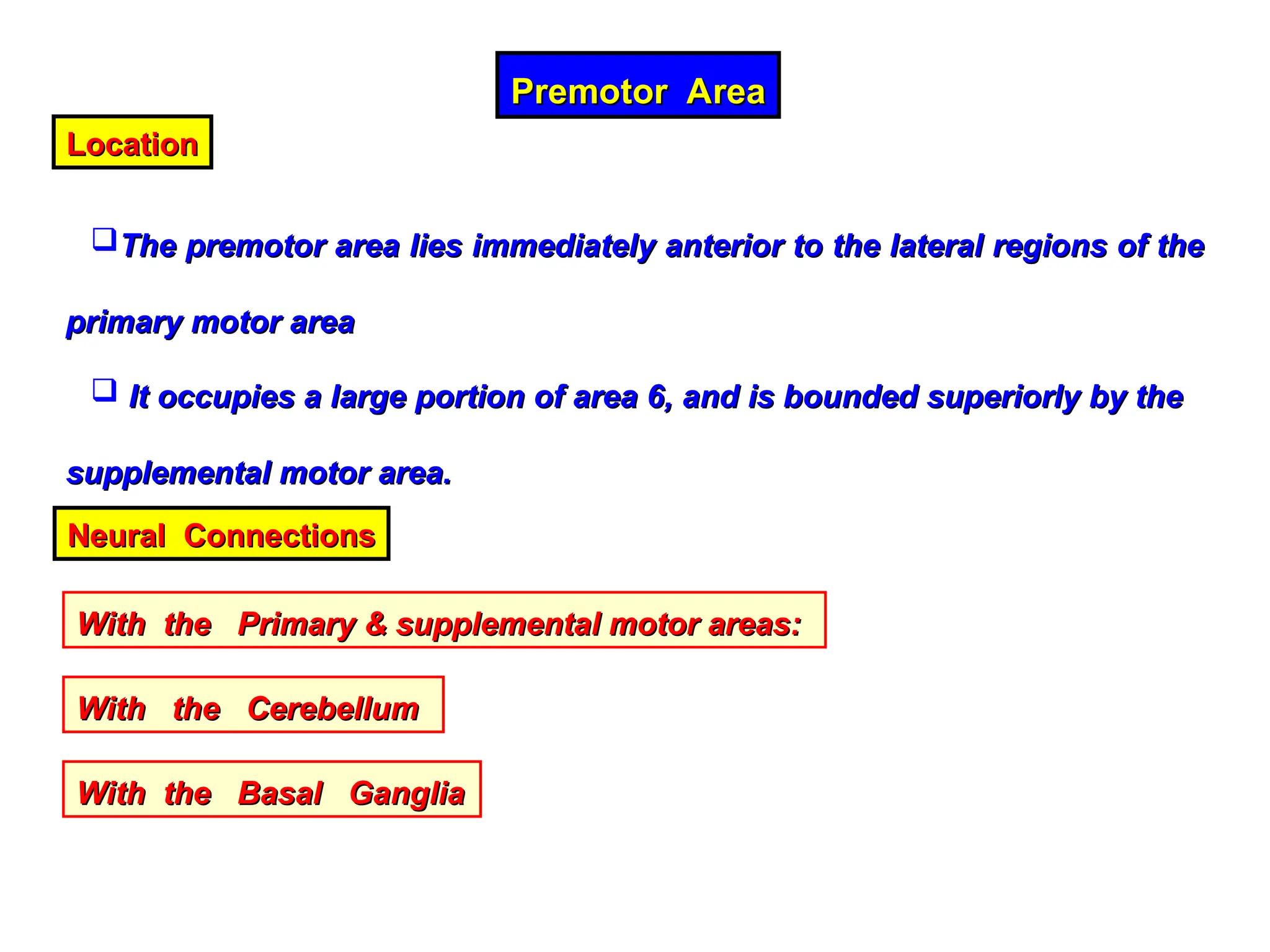
Premotor Area
Premotor Area
Location
Location
Thepremotor area lies immediately anterior to the lateral regions of the
The premotor area lies immediately anterior to the lateral regions of the
primary motor area
primary motor area
It occupies a large portion of area 6, and is bounded superiorly by the
It occupies a large portion of area 6, and is bounded superiorly by the
supplemental motor area.
supplemental motor area.
Neural Connections
Neural Connections
With the Primary & supplemental motor areas:
With the Primary & supplemental motor areas:
With the Cerebellum
With the Cerebellum
With the Basal Ganglia
With the Basal Ganglia
30.
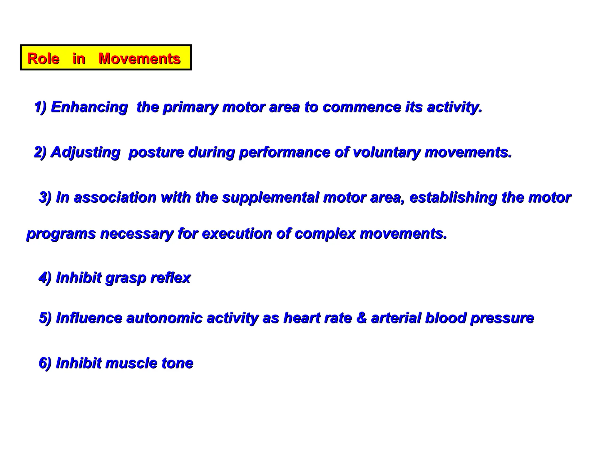
Role in Movements
Rolein Movements
1) Enhancing the primary motor area to commence its activity.
1) Enhancing the primary motor area to commence its activity.
2) Adjusting posture during performance of voluntary movements.
2) Adjusting posture during performance of voluntary movements.
3) In association with the supplemental motor area, establishing the motor
3) In association with the supplemental motor area, establishing the motor
programs necessary for execution of complex movements.
programs necessary for execution of complex movements.
4) Inhibit grasp reflex
4) Inhibit grasp reflex
5) Influence autonomic activity as heart rate & arterial blood pressure
5) Influence autonomic activity as heart rate & arterial blood pressure
6) Inhibit muscle tone
6) Inhibit muscle tone
31.
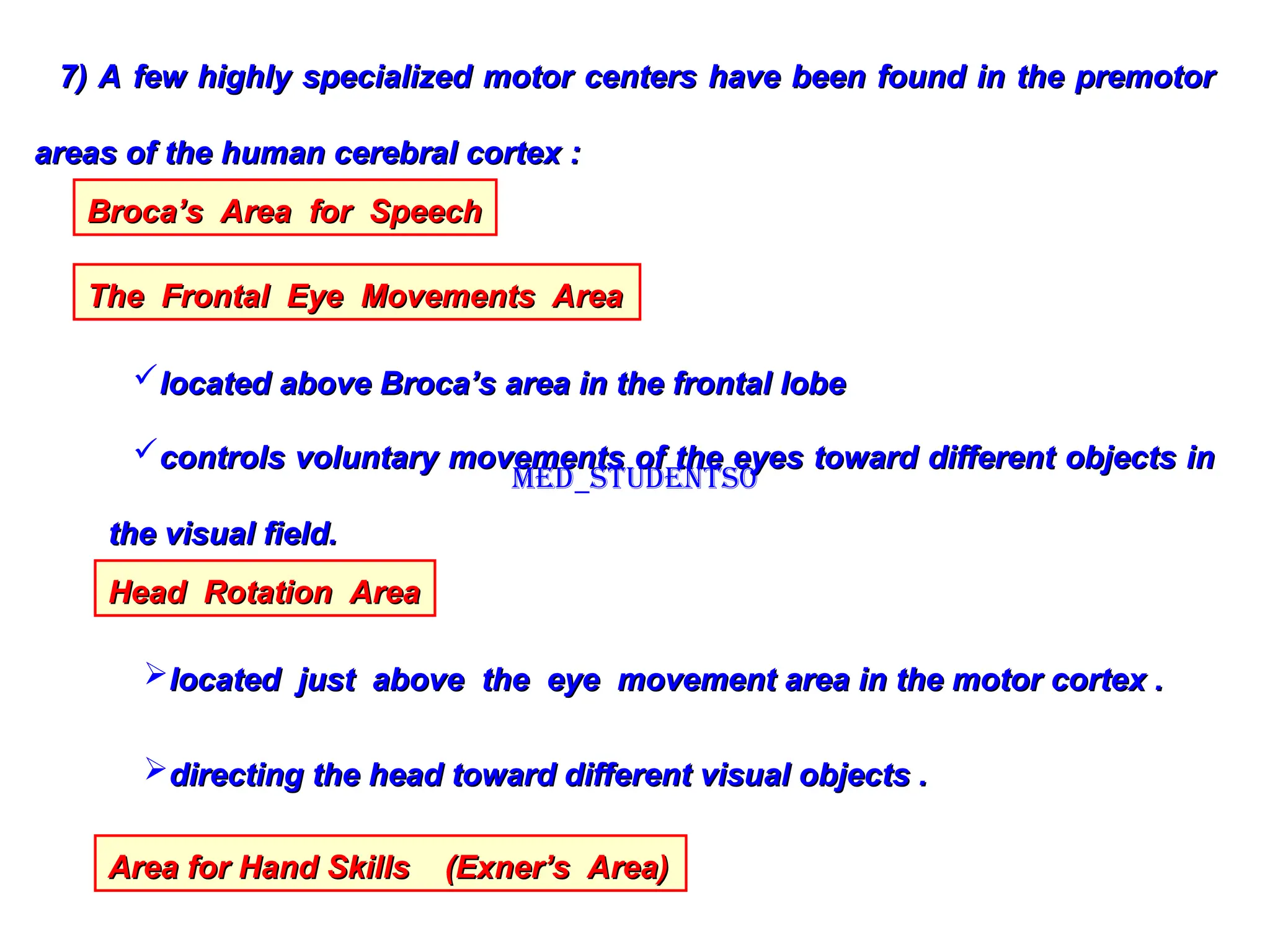
7) A fewhighly specialized motor centers have been found in the premotor
7) A few highly specialized motor centers have been found in the premotor
areas of the human cerebral cortex :
areas of the human cerebral cortex :
Broca’s Area for Speech
Broca’s Area for Speech
The Frontal Eye Movements Area
The Frontal Eye Movements Area
located above Broca’s area in the frontal lobe
located above Broca’s area in the frontal lobe
controls voluntary movements of the eyes toward different objects in
controls voluntary movements of the eyes toward different objects in
the visual field.
the visual field.
Head Rotation Area
Head Rotation Area
located just above the eye movement area in the motor cortex .
located just above the eye movement area in the motor cortex .
directing the head toward different visual objects .
directing the head toward different visual objects .
Area for Hand Skills (Exner’s Area)
Area for Hand Skills (Exner’s Area)
Med_students0