2. [1–4] and produce a steep voltage-dependent block at membrane po-
tentials (Vm) near the equilibrium potential (EK). The rectified cur-
rent–voltage (I–Vm) curve of the Kir channel shifts along the voltage
axis in parallel with shifts in EK as extracellular or intracellular K+
con-
centration changes (as in Fig. 3B). The extent of channel block correlates
with the driving force, (Vm − EK), for K+
conduction. The steep voltage
dependence of the block near EK could not be explained by the
Woodhull view [5], which assumed independent movements between
the conducting ions and the blocker and attributed the voltage depen-
dence to the fractional electrical field traversed by the positively
charged blocker. Previous blockage studies using various internal cation
blockers on Kir channels revealed that the measured voltage depen-
dence could be larger than unity, and that blockers with different va-
lences could have similar apparent voltage dependence [6,7].
The steep voltage dependence has been attributed to the multi-ionic
nature in early theoretical models [8,9]. A single-file multi-ion long pore
is a prerequisite for flux coupling. Movement and occupancy during the
binding process of the blockers are coupled with the conducting K+
ions
in the long pore [10–13]. X-ray crystal structural studies of the Kir chan-
nels have identified narrow long multi-ion cytoplasmic pores, which ex-
tend the single-file region to a 60 Å-long K+
conduction pathway
[14–17] (Fig. 1). The cytoplasmic pore provides electronegative lining
and weak binding ion sites for partial dehydrated K+
ions and can there-
by accommodate several K+
ions and facilitate flux-coupling effects.
However, even when the movement of multiple K+
ions within the cy-
toplasmic pore is taken into consideration, it seems to be difficult to rec-
oncile the strong voltage-dependent block with the fact that the
transmembrane potential drops only a small fraction along the cyto-
plasmic pore [18,19], because the transmembrane potential gradient is
mostly concentrated at the selectivity filter.
Our recent study on the mechanism of the “driving force”-depen-
dent block by intracellular Ba2+
on the cloned Kir2.1 channel presented
novel findings which may provide proper experimental and theoretical
explanations of inward rectification [11]. First, the increase in the appar-
ent affinity of Ba2+
blockage near EK results from the steep increase in
the flux-coupled encounter frequency between Ba2+
and the high-
affinity binding site, located near T141 at the internal entrance of the se-
lectivity filter (TIGYG, residues 142 to 146). Because the direction and
the magnitude of the unidirectional K+
flux changes dramatically at
voltages increase above EK, the encounter frequency and the apparent
binding rate experience a steep increase at a voltage range of (Vm −
EK) ~+10 to +40 mV. When the driving force (Vm − EK) is greater
than +40 mV, the apparent binding rates are limited by the intrinsic ac-
tivation barrier of the Ba2+
binding reaction to the high-affinity binding
site because the flux-dependent encounter frequency exceeds the acti-
vation rate.
Second, the slow unbinding rates of the internal Ba2+
block in the
Kir2.1 channel increase monotonically with voltage. These rates depend
mildly on voltage across the positive tested voltages, indicating that the
unbinding rates of Ba2+
are determined primarily by the intrinsic chem-
ical affinity of the high-affinity binding site. The positive voltage depen-
dence implies that Ba2+
ions may traverse through the selectivity filter
and dissociate outward to the extracellular solution [11]. The unbinding
process is little affected by changes in the direction of net K+
flux.
More interestingly, the driving force-dependence block has been
demonstrated even in the presence of a concentration gradient alone
by altering the extracellular K+
concentration and fixing the intracellu-
lar K+
concentration at a membrane voltage of 0 mV (see Fig. 8 in ref.
[11]), when a voltage difference across the membrane is absent. Under
this condition, the thermodynamic driving force is the chemical poten-
tial difference across the membrane. The relationship between the
unblocked current and the driving force is also demonstrated by the
strong rectification feature. These results suggest that the electrical po-
tential gradient across the flux-coupling region in the channel pore is
not essential for the steep change in the apparent blocking affinity
near the equilibrium point. Flux-coupled block can occur when K+
flux is driven either by concentration differences or by voltage differ-
ences across the membrane in the single-file multi-ion cytoplasmic
pore in Kir channels.
In this paper, we present a novel model, which combines comput-
er kinetic simulation and concepts from non-equilibrium thermody-
namics, to elucidate the mechanisms of inward rectification. The
simulations are based on experimental data from our recent study
of the internal Ba2+
block in the Kir2.1 channel [11]. The discrete
binding kinetics of the Ba2+
block have made it possible to dissect
the blocking events. The processes of the internal Ba2+
blockage on
Kir channels are described by sequential steps including association,
“driving force”- or flux-dependent encounter, binding, and dissocia-
tion. We apply the fluctuation theorem in a non-equilibrium small
system [20] to explain the flux-coupled encounter frequency by
interpreting the flux ratio of the unidirectional efflux to the influx
as the likelihood for a transfer event to occur down or against the
electrochemical potential gradient [21,22]. The theoretical and sim-
ulation studies here enforces that the “steep voltage-dependence”
in the block on Kir channels actually results from the driving force-
dependent block. Under this view, the inward rectification curve,
the molecular structural model, and the physiological experimental
data are consistent with one another.
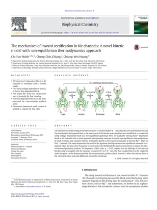
Fig. 1. The schematic model of the intracellular Ba2+
block in the Kir channel. The blocking process involves association, flux-coupled encounter, and chemical binding. The Ba2+
ion then
unbinds and dissociates to the external or internal solution. The backbone channel structure is a Kir2.1 channel model shown with Rasmol from the SWISS-MODEL Repository based on the
crystallography template structure of a Kir2.2 channel (PDB: 3sph) [14].
2 C.-P. Hsieh et al. / Biophysical Chemistry 212 (2016) 1–8
3. 2. Modeling methods and theories
2.1. The schematic model for intracellular Ba2+
block on the Kir channel
The backbone channel structure of a Kir2.1 channel model is shown
with Rasmol from the SWISS-MODEL Repository based on the crystal-
lography template structure of a Kir2.2 channel (PDB: 3sph) [14]
(Fig. 1). For an intracellular Ba2+
ion to move and bind onto the deep
high-affinity binding site near the T141 residue at the internal entrance
of the selectivity filter, three sequential steps must occur: (1) associa-
tion: Ba2+
ion diffusion from the intracellular solution to the cytoplas-
mic pore entrance, (2) translocation: “driving force”- or “K+
flux”-
coupled movement of the Ba2+
ion along the single-file multi-ion cyto-
plasmic pore to encounter the high-affinity binding site, and (3) bind-
ing: Ba2+
ion overcoming the activation energy barrier and binding on
the high-affinity binding site formed by the coordinating ligands. For
convenience of description, the states of the channel are denoted as
the following: “Open”, “Access”, “Encounter”, and “Block”. The kinetic
rate constants for transition between the states are denoted in Fig. 1.
The spatial arrangement of the deeper blocker binding site, accom-
panied by the shallower low-affinity binding sites that are always occu-
pied by K+
ions along the single-file cytoplasmic pore, makes it a
suitable structural environment for flux-dependent block. When the
driving force predominantly favors inward K+
current, there is always
at least one K+
ion binding to the shallower binding sites, which pre-
cludes the intracellular blocker from encountering the deeper blocker
binding site, resulting in no block. When the driving force favors out-
ward K+
flux, flux coupling effects facilitate the blocker ion to move
into the cytoplasmic pore, to occupy the inner vacancy, to encounter
the blocker binding sites, and to produce blockage.
2.2. Experimental data and parameters for kinetic modeling and
simulations
The experimental data of blocking kinetics by intracellular Ba2+
in
Kir2.1 channels were obtained from the previous study of Hsieh et al.
(2015) using inside-out patch-clamp recordings in Xenopus oocyte ex-
pression systems [11]. Experimental details and interpretation of the
data were described with details in the study. The apparent binding
and unbinding rate constants, kon and koff, and the voltage dependence
parameter used in the simulations are taken from kinetic studies of
E224G mutant Kir2.1 channels because the internal Ba2+
block in
E224G mutant channels exhibits a one-to-one relationship between
Ba2+
and a high-affinity binding site. This relationship makes kinetic
analysis feasible and, at the same time, preserves the feature of inward
rectification.
2.3. Computer simulation of channel blocking kinetics and model of inward
rectification
Computer simulations were performed with IonChannelLab (http://
www.jadesantiago.com/Electrophysiology/IonChannelLab/) [23], the
software that is used to build kinetic models and perform simulations
for channel currents using voltage-clamp recordings. The simulation is
based on Markov chain models to calculate the transition rates between
discrete conformational states. The Q-Matrix method was used. The
protocol of the voltage stimulus and ionic conditions in simulated
voltage-clamp recordings can be assigned in this software. The time
course of the amount of each channel state in the kinetic scheme
model can be simulated with appropriately assigned kinetic rate param-
eters. Each channel state can be set as permeant (open or unblocked) or
non-permeant (closed or blocked), and the macroscopic currents in
voltage-clamp recordings can be estimated in the simulations. The ion
currents in the voltage-clamp simulation were assumed to have an
ohmic linear relationship in the simulations in this paper for simplicity.
Although the actual current–voltage relationship of the unblocked open
wild-type and E224G Kir2.1 channels is not linear, the linear ohmic as-
sumption does not affect the results of the kinetic simulation except
for the current size. Single channel conductance was fixed at 38 pS, a
value in the E224G Kir2.1 channel in 100 mM symmetrical K+
at
−100 mV, as measured from single-channel recordings [24]. The total
number of channels was assigned as 1000. The construction of the ki-
netic scheme model will be discussed and be presented in Scheme 1.
Table 1 listed the parameters in kinetic simulations of the intracellular
Ba2+
block of Kir channels.
2.4. Interpretation of the Ussing flux ratio from the fluctuation theorem for
small non-equilibrium systems
The net K+
flux (Jnet) is the difference of the unidirectional outward
flux (the efflux, Jio) from the unidirectional inward flux (the influx, Joi):
Jnet = Jio − Joi. The ratio of efflux to influx has been estimated from the
Ussing flux ratio [25]:
Jio
Joi
¼
Kþ
 Ã
i
KþÂ Ã
o
exp
zK FVm
RT
¼ exp
zK F Vm−EKð Þ
RT
: ð1Þ
As Hodgkin and Keynes proposed in 1955 [26], for a multi-ion,
single-file long pore, the flux ratio equation is raised to the nth power:
Jio
Joi
¼
Kþ
 Ã
i
Kþ
 Ã
o
exp
zK FVm
RT
( )n
¼ exp
nzK F Vm−EKð Þ
RT
¼ exp
nΔ~μ
RT
;
ð2Þ
where n is the number of ions coupling in the pore. The electrochemical
potential difference across the membrane isΔ~μ= zKF(Vm − EK). zK is the
valence of K+
. F is the Faraday's constant. RT has the usual thermody-
namics meanings. Microscopically, the flux ratio can be interpreted
from the fluctuation theorem [20] as the probability ratio of the forward
ion transport step (down the electrochemical gradient, positive entropy
production) to the back transport step (against the electrochemical gra-
dient, negative entropy production) in a small non-equilibrium system
[21,22]. When there are n K+
ions lined up along a single-file pore, the
flux ratio increases to the nth power because a tracer ion is transferred
from one side to the other and requires n more transport cycles in the
Table 1
Parameters in simulations of the intracellular Ba2+
block of Kir channels.
Ionic condition 100 mM [K+
]out,
100 mM [K+
]in
20 mM [K+
]out,
100 mM [K+
]in
Various [K+
]out,
100 mM [K+
]in,
EK (mV) 0 mV −40 mV Various
Vm range in simulation −60 mV to +60 mV −100 mV to +20 mV at 0 mV
α1 (s−1
M−1
) 2.79 × 10⁴
exp(2.62 (Vm − EK)/25) 2.79 × 10⁴
([K+
]in/[K+
]out)2.62
β1 (s−1
) ~k2 ~k2
k1 (s−1
M−1
) 9.39 × 10⁵
exp(0.11 Vm/25) 9.39 × 10⁵
k2 (s−1
) 0.10 exp(−0.21 Vm/25) 0.10
k3 (s−1
) 0.71 exp(0.78 Vm/25) 0.71
All rate constants and parameters were from recordings of the inside-out patch clamp on the cloned Kir2.1 channels by Hsieh et al., 2015 (Figs. 3 and 4 in ref. [11]).
3C.-P. Hsieh et al. / Biophysical Chemistry 212 (2016) 1–8
4. forward direction than in the reverse direction. For example, when
(Vm − EK) = +25 mV at room temperature, the likelihood of a forward
transfer event is 2.72 if n = 1 and is 20.1 if n = 3.
3. Results
The schematic models of the sequential processes during the block-
age of internal Ba2+
on the Kir channels were shown in Fig. 1. The bio-
physical rationale of the model construction and the computer kinetic
simulation results were presented in this section.
3.1. The biophysical rationale of the “driving force”-dependent block in Kir
channels
The “driving force”-dependent block in Kir channels was shown to
result from the steep increase in the flux-coupled encounter frequency
when the driving force for K+
ion conduction, (Vm − EK), changes from
negative to positive across EK [11]. To illustrate this concept, consider
the association step required for the Ba2+
ions to access the cytoplasmic
pore entrance from the intracellular solution (from “Open” to “Access”
state). The fraction of the “Access” to “Open” state in equilibrium is
the following:
Access½ Š
Open½ Š
¼
Ba2þ
h i
Ka
; ð3Þ
where Ka is the dissociation constant of the association step. Ka = β/α
when Ka is expressed as the ratio of the binding and unbinding rate con-
stants (Fig. 1).
As soon as the Ba2+
ion associates with the entrance of the single-
file pore, its movement will be coupled by K+
ions lined up in the nar-
row long permeation pathway. The Ba2+
ion must move in concert
with the column of K+
ions to traverse the long cytoplasmic pore and
encounter the deep high-affinity binding site. Because there are only
weak ion binding sites along the cytoplasmic pore, the movement of
the Ba2+
ion is almost freely coupled by K+
ion flux. The flux ratio of
the unidirectional efflux to unidirectional influx in a multi-ion pore is
expressed as in Eq. (2), based on the interpretation from the fluctuation
theorem in a small non-equilibrium system [20–22]. If nf K+
ions have
to be transported outward to let the Ba2+
ion move from the “Access”
state to the “Encounter” state, the ratio of the rate (probability) of the
forward transport (down the driving force for K+
flux) to that of the
backward transport (against the driving force for K+
flux) would be ap-
proximately:
f 1
f 2
¼ exp
nf Δ~μ
RT
¼ exp
nf zK F Vm−EKð Þ
RT
: ð4Þ
The novel interpretation here is to apply the non-equilibrium fluctu-
ation theorem to describe the likelihood of the forward flux-coupled
Ba2+
translocation. Because the ratio in Eq. (4) is for time-dependent
flux, it would be inappropriate to view the ratio as the result of the
Boltzmann distribution for states in equilibrium.
To derive the apparent dissociation constant, consider the species
ratio at a steady state,
Encounter½ Š
Access½ Š
¼
f 1
f 2
¼ exp
nf zK F Vm−EKð Þ
RT
: ð5Þ
Combining Eqs. (3) and (5) gives:
Encounter½ Š
Open½ Š
¼
Ba2þ
h i
Ka
exp
nf zK F Vm−EKð Þ
RT
¼
Ba2þ
h i
Ka exp
−nf zK F Vm−EKð Þ
RT
:
ð6Þ
Hence, the apparent dissociation constant for the “Open” state to the
“Encounter” complex is
KOE ¼ Ka exp
−nf zK F Vm−EKð Þ
RT
: ð7Þ
In the experimental study of the internal Ba2+
block of the Kir2.1
channel, the steep decrease in the apparent dissociation constant at
test voltages from EK to (EK + 40 mV) was due to the steep increase
of the apparent binding rate constant at these voltage ranges, while
the unbinding rate was not affected by the driving force [11]. This find-
ing is reasonable because the apparent binding rate constant is limited
by the encounter frequency at the flux-coupled translocation step at
voltages near EK. Hence, we will assign the steep exponential increase
to the apparent binding rate constant in the kinetic simulation.
3.2. Kinetic model construction of the internal Ba2+
block on the Kir
channel
We performed a kinetic simulation for internal Ba2+
block in the Kir
channels using the software IonChannelLab. In our kinetic scheme
model, the association (from “Open” to “Access” state in Fig.1) and the
flux-coupled encounter (from “Access” to “Encounter” state) processes
were merged into one step because the experimental data for these pro-
cesses could not be separated and the measured apparent association
rate constants were the combined results of the sequential events.
Therefore, only three states were assigned in the kinetic scheme as the
following: “Open” state (unblocked), “Encounter” state (encounter but
not yet binding), and “Block” state (blocked). The “Block” state was
not conductive to K+
ions; the “Open” state and “Encounter” state was
conductive to K+
ions. The rate constants were denoted in Scheme 1:
ðScheme1Þ
The rate constants and the parameters used in the simulation are
listed in Table 1. The meanings of the rate constants are the following:
α1 =α0 expð
nf Δ~μ
RT Þ =α0 expð
nf zK
FðVmÀEKÞ
RT Þis the apparent binding rate
constant of the process from the “Open” to “Encounter” state, which is
significantly affected by the flux coupling effect in the single-file
multi-ion cytoplasmic pore, as mentioned in the aforementioned dis-
cussion. α1 is dependent on the driving force, Δ~μ = zKF(Vm − EK). α0
is the rate constant when Vm = EK.
k1 = k1(0 mV) expðzBδFVm
RT Þis the binding rate constant from the “En-
counter” state to the “Block” state. zB is the valence of the Ba2+
. δ is the
equivalent electrical distance needed to overcome the activation barrier
of the binding reaction. k1(0 mV) is the rate constant when Vm = 0 mV.
k2 = k2(0 mV) expðzBδFVm
RT Þ is the unbinding rate constant
required for Ba2+
to dissociate in the intracellular solution. k3 =
k3(0 mV) expðzBδFVm
RT Þ is the unbinding rate constant for Ba2+
to dissoci-
ate in the extracellular solution. Unbinding rate and voltage dependence
are determined by the intrinsic energy barrier for unbinding. As an ex-
perimental study [11] has shown, the mild positive voltage dependence
in koff over −60 mV to +100 mV suggests that Ba2+
ions may traverse
through the selectivity filter and dissociate outward in the extracellular
solution. At −120 mV to −160 mV, the Ba2+
ion primarily exits the in-
tracellular solution from the high-affinity binding site [11]. In the simu-
lation, we let β1 values, the rate of release of Ba2+
from the “Encounter”
state, equal the corresponding k2 values, so that the “Encounter” state
would not accumulate during the dissociation process.
4 C.-P. Hsieh et al. / Biophysical Chemistry 212 (2016) 1–8
5. 3.3. The computer simulation of the macroscopic currents in voltage-clamp
recordings in different voltage and ionic conditions
3.3.1. The simulation of the internal Ba2+
block with symmetrical
100 mM K+
The simulation results of internal Ba2+
block on the Kir channel
under the ionic condition of symmetrical 100 mM K+
(EK = 0 mV)
showed that inward rectification occurred in a dose-dependent fashion,
as internal Ba2+
were 1 μM, 10 μM, 100 μM, and 1 mM (Fig. 2). The volt-
age was held at −100 mV in the stimulus protocol, stepped to −60 mV
to +60 mV for 900 ms every 10 mV, and recovered to −100 mV for
12 s. The outward currents were blocked by internal 10 μM Ba2+
, and
the inward currents were relatively unaffected. At high blocker concen-
tration with 1 mM internal Ba2+
, the inward currents at −10 mV and
−20 mV were blocked to a significant extent (Fig. 2D and F). Block by
high concentration of spermine in the Kir2.1 channels at −10 mV and
−20 mV in symmetrical K+
concentration has been shown in single-
channel recordings [27]. This finding is still compatible with the flux-
dependent block model because at voltages negative, but near the equi-
librium potential, there is considerable unidirectional efflux, although
the net flux is inward. Therefore, when internal blocker concentration
is high, efflux-coupled block can still take place. From the perspective
Fig. 2. Simulated voltage-clamp currents with symmetrical 100 mM K+
concentration in (A) 1 μM, (B) 10 μM, (C) 100 μM, and (D) 1 mM internal Ba2+
. The voltage was stepped to a 900-
ms test pulse from −60 mV to +60 mV every 10 mV. (E) The long current tails reflected the slow unbinding rate (τ ~ 4 s) during recovery when the voltage was stepped back to −100 mV
for 12 s. (F) The normalized I–Vm curves showed inward rectification in a dose-dependent fashion in the block by internal Ba2+
.
5C.-P. Hsieh et al. / Biophysical Chemistry 212 (2016) 1–8
6. of non-equilibrium thermodynamics, reverse events can occur against
the thermodynamic driving force near the equilibrium point at
nanoscales in small systems; thus, backward transport against the elec-
trochemical gradient is inevitable [11,20,22]. This fact is reminiscent of
the “threshold” in the I–V curve of the semiconductor diode [28].
Our previous study presented the novel finding that the unbinding
process was slow when the voltage was stepped back to −100 mV
(τ = 4 s) from the steady-state Ba2+
block in Kir2.1 channel at positive
test voltages. Because the recovery rate is the same in the presence or
absence of internal Ba2+
(Fig. 3C in ref. [11]), the binding rate at
−100 mV could be neglected in the simulation. Here, we replayed the
long recovery tail in kinetic simulation during the dissociation process
at −100 mV (Fig. 2E). At first glance, the exceptionally slow unbinding
rate may seem to be inconsistent with the fact that the steady inward
currents were scarcely affected by internal Ba2+
. However, in our
model, inward rectification and slow unbinding rate were consistently
associated with each other because, as soon as the Ba2+
ion dissociated
from the high-affinity binding site at negative voltages, the inward K+
ions flowed immediately and occupied the inner K+
binding sites in
the single-file cytoplasmic pore. This flow steadily precluded intracellu-
lar Ba2+
ions from moving into the pore and encountering the deep
high-affinity binding site.
3.3.2. The computer simulation of the internal Ba2+
block at EK = −40 mV
We performed a kinetic simulation of the ionic condition with
20 mM external K+
and 100 mM internal K+
. EK = −40 mV. In the sim-
ulation, the driving force-dependent parameter, α1, which is the appar-
ent binding rate constant of the flux-dependent encounter process from
the “Open” to “Encounter” state, would shift −40 mV compared to the
conditions observed in symmetrical 100 mM K+
solutions. Other rate
constants remained the same in this simulation. The results of the sim-
ulation when the voltage was increased from −100 mV to +20 mV in
10 μM internal Ba2+
are shown in Fig. 3A. Inward rectification was
shown in the simulated currents and in the I–Vm curve. The simulated
I–Vm curves of the two ionic conditions, [K+
]out = 20 mM/[K+
]in =
100 mM and [K+
]out = 100 mM/[K+
]in = 100 mM, in the presence of
10 μM internal Ba2+
were compared in Fig. 3B. The I–Vm curve shifted
to the same extent as the shift in EK.
3.3.3. Simulation of the flux-dependent block by the concentration gradient
in the absence of a transmembrane electrical potential
According to our model, the steep increase in the apparent blockage
affinity from EK to (EK + 40 mV) is due to the steep increase in the flux-
dependent encounter frequency. We were curious whether the
similar flux-coupled block could be observed in the absence of a
transmembrane potential when the K+
flux was driven by the concen-
tration difference across the membrane. To examine this novel proposi-
tion, we conducted internal Ba2+
block experiments at 0 mV by
changing external K+
concentrations while keeping internal K+
con-
centration at 100 mM in the patch-clamp study (see Fig. 8 in ref. [11]).
The driving force-dependent block was observed under this condition,
even when there was no transmembrane electrical potential.
Intuitively, the flux ratio of the unidirectional efflux to influx at 0 mV
would be the concentration ratio, [K+
]in/[K+
]out. In a multi-ion pore, the
ratio would be ([K+
]in/[K+
]out)n
, where n is the flux ratio exponent.
When viewed from the perspective of the fluctuation theorem in
small non-equilibrium systems, the flux ratio driven by a concentration
gradient would be the following [22]:
Jio
Joi
¼ exp
−nΔμ
RT
; ð8Þ
where Δμ is the chemical potential difference across the membrane. By
definition,
Δμ ¼ μin−μout ¼ RTln
Kþ
 Ã
in
Kþ
 Ã
out
!
: ð9Þ
Substituting Eq. (9) into Eq. (8) also obtains,
Jio
Joi
¼
Kþ
 Ã
in
Kþ
 Ã
out
!n
: ð10Þ
Hence, we assign the flux-dependent rate constant, α1, in the simulation
as:
α1 ¼ α0 exp
nf Δμ
RT
¼ α0
Kþ
 Ã
in
Kþ
 Ã
out
!nf
: ð11Þ
Other rate constants in this simulation are the values at 0 mV
(Table 1).
If we express Eq. (9) in the form as:
Δμ ¼ RT ln
KþÂ Ã
in
Kþ
 Ã
out
!
¼ zK F −EKð Þ: ð12Þ
Under this view, the driving force for K+
conduction by a concentration
gradient alone at Vm = 0 mV is proportional to (-EK). When the driving
force by the concentration gradient is 1 RT, it would be similar to the
Fig. 3. (A) Simulated voltage-clamp currents with 20 mM external K+
and 100 mM internal K+
(EK = −40 mV) in 10 μM internal Ba2+
. The voltage was stepped to a 900-ms test pulse
from −100 mV to +20 mV every 10 mV. (B) The normalized I–Vm curve of the simulation in (A) and the I–Vm curve simulated with symmetrical 100 mM K+
in 10 μM internal Ba2+
were
compared.
6 C.-P. Hsieh et al. / Biophysical Chemistry 212 (2016) 1–8
7. driving force when (Vm − EK) = +25 mV. For example, if Δμ=1 RT,
[K+
]out should be 37 mM, [K+
]in = 100 mM, and EK = −25 mV. Thus,
the driving force, (Vm − EK), for K+
flux at Vm = 0 mV is +25 mV.
We simulate the internal Ba2+
block when K+
flux is driven by con-
centration differences alone (Vm = 0 mV) across the membrane under
various ionic conditions listed in Table 2. The internal K+
concentration
was kept at 100 mM; thus, the effect of competition between the Ba2+
and K+
during the binding process should be similar. The external K+
concentrations were designed to make Δμ be −2.0 RT, −1.5 RT, −1.0
RT, …, to +2.0 RT. In the simulation of voltage-clamp recordings, the
voltage was stepped to 0 mV under various ionic K+
conditions from a
holding voltage of −100 mV. Fig. 4A shows the simulated K+
currents
in 10 μM internal Ba2+
. The currents were blocked in a manner that sug-
gests inward rectification, although all the simulation experiments were
performed at 0 mV. The relationship between the currents and the driv-
ing force (Δμ) was plotted in Fig. 4B, which resembled the I–Vm curves
of Kir channels shown previously. The driving force-dependent effect
of every +1 RT of Δμ from the equilibrium point is similar to that caused
by every +25 mV increase in the I–Vm curves when the horizontal axis
is the membrane voltage. The simulated currents that are driven by con-
centration differences or by transmembrane potentials at a comparable
driving force of +2 RT or +50 mV are shown in Fig. 4C. The black trace
was the current at 0 mV when [K+
]out = 14 mM and [K+
]in = 100 mM
(EK = −50 mV). The blue trace was the current at +50 mV under sym-
metrical [K+
] = 100 mM (EK = 0 mV). The blocking rates were similar.
The steady-state unblocked current was larger at +50 mV under sym-
metrical K+
because the unbinding rate at positive voltage would be
larger in this case.
4. Discussion
We have demonstrated that the “driving force”- or flux-dependent
block in inward rectifier K+
channels can be explained by the concen-
tration gradient alone. In addition, the block can be explained by the
more general condition when the driving force is the electrochemical
potential difference, Δ~μ = zKF(Vm − EK), across the membrane, both
in the real patch-clamp experiments [11] and in the kinetic simulation
study presented here. The single-file multi-ion cytoplasmic pore in Kir
channels is the essential structural element for flux coupling. Inward
rectification caused by pore block is a result of the evolutionary
Table 2
[K+
] in simulations, Δμ at 0 mV, and EK.
[K+
]in (mM) [K+
]out (mM) Δμ (RT) EK (mV)
100 739 −2.0 +50
100 448 −1.5 +37.5
100 272 −1.0 +25
100 165 −0.5 +12.5
100 100 0.0 0
100 61 +0.5 −12.5
100 37 +1.0 −25
100 22 +1.5 −37.5
100 14 +2.0 −50
Fig. 4. (A) Superimposed simulated currents in 10 μM internal Ba2+
at 0 mV driven by various K+
concentration gradients across the membrane. The voltage was held at −100 mV and
stepped to 0 mV at various K+
concentrations. The ionic conditions and the corresponding Δμ are listed in Table 2. (B) The simulated unblocked currents in 10 μM internal Ba2+
were
plotted with the chemical potential difference (Δμ) across the membrane at 0 mV. (C) The simulated blockage by 10 μM internal Ba2+
when the K+
current was driven by +2.0 RT of
chemical potential difference (Vm = 0 mV, [K+
]out = 14 mM, [K+
]in = 100 mM) (black trace) was compared to blockage at Vm = +50 mV in symmetrical 100 mM K+
(blue trace)
when the electrochemical potential difference was also +2.0 RT. (For interpretation of the references to color in this figure legend, the reader is referred to the web version of this article.)
7C.-P. Hsieh et al. / Biophysical Chemistry 212 (2016) 1–8
8. molecular design of a deep binding site for the blocker accompanied
with low-affinity ion binding sites for the blocker ion and K+
ions
along the cytoplasmic pore. Although we have discussed the flux-
coupling block based on the single-file multi-ion nature of the pore, it
should be noted that the cytoplasmic pores of Kir channels may not be
strictly single-filed along the whole conduction pathway. Molecular dy-
namics simulation studies have shown that the wider central pore of the
cytoplasmic domain of Kir2.1 channels could accommodate 5–10 K+
ions in 1 M KCl [18]. However, one partially hydrated K+
ion near the
cytoplasmic entrance were always present during the molecular dy-
namics simulation [18] and also in the crystal structure studies [15,
16]. Functional studies using patch-clamp recordings and site-directed
mutagenesis on cloned Kir2.1 channels with the homology structure
model suggested that three single-file ion binding sites may present
near residues M301 and A306 at the cytoplasmic domain and residue
D172 in the water cavity [29]. This is consistent with the nf values
~2.6 which is determined from our previous Ba2+
block study [11]
and is used in the simulation study here. The blockage of internal
Ba2+
from the cytoplasmic solution to the high-affinity binding site
near T141 may be coupled with the outward movement of 2 to 3 K+
ions.
The major contribution of this paper is to properly explain the bio-
physical mechanism underlying the steep voltage dependence of blocks
near the equilibrium potential in inward rectifier K+
channels. Conven-
tional explanations have attributed the steep voltage-dependent block
near EK in Kir channels to be the result of voltage drop caused by multi-
ple K+
ions across the local electrical potential gradient [13,19]. Howev-
er, this view is not consistent with the fact that the transmembrane
potential drops mostly across the selectivity filter 35 Å away from the
cytoplasmic domain [18]. The model we propose here may provide a so-
lution to this conundrum and reconcile the controversy. In our model,
we applied the fluctuation theorem in non-equilibrium thermodynam-
ics to describe the flux ratio [22]. We attribute this idea to a novel
driving force-dependent apparent association binding rate constant,
α1 = α0 expð
nf zK
FðVmÀEKÞ
RT Þ. The driving force, (Vm − EK), is a non-
equilibrium thermodynamic tendency for ion flux, which is determined
by the concentrations and electrical potentials of the intracellular and
extracellular bulk solutions. The driving force determines the direction
of the net K+
flux and the flux ratio of the efflux to the influx. The
single-file K+
ion array interacts and moves in concert through the en-
tire permeation pathway, from the innermost cytoplasmic entrance to
the outermost exit of the selectivity filter. Therefore, the blocker in the
cytoplasmic pore can be influenced by the multi-ion flux coupling effect,
regardless of whether there is a drop in local electrical potential or not.
For this reason, the local electrical potential gradient along the cytoplas-
mic pore is not necessary for “the steep voltage-dependent block” near
EK. Our novel experimental and simulation studies performed at 0 mV,
when the K+
flux is driven by concentration gradient alone, further sup-
port the idea that flux-dependent block and steep inward rectification
can be demonstrated even when there is no transmembrane electrical
potential (Fig. 4 and [11]).
Thermodynamic driving forces, fluctuations in small systems, and
flux coupling are the concepts of non-equilibrium thermodynamics. In
a bulk solution in physiological conditions, every ion is randomly
pushed by the surrounding water molecules and jiggles without a spe-
cific direction, with a correlation time between two collisions of
~10−13
s. The mean random walk step is only 0.5 Å. This means that
an ion would change its direction every 10−13
s after it moves in less
than one atomic radius. For comparison, the average distance between
two K+
ions is 25 Å in a 100 mM K+
solution. Hence, each ion just
moves independently, and the thermodynamic driving force does not
exert a directional force on the ion in the bulk solution or in a very
wide pore. However, in the nanoscale single-file multi-ion pore, the
driving force can exert a directional impact on the movement of the
ions in the pore and thereby make the Kir channel act as a membrane
diode at the molecular level. We hope that this simulation study based
on a structural model and physiological experimental data would en-
hance the understanding of the mechanisms of inward rectification.
Acknowledgments
This study was supported by a grant from the Far Eastern Memorial
Hospital (FEMH-2015-D-050).
References
[1] A.N. Lopatin, E.N. Makhina, C.G. Nichols, Potassium channel block by cytoplasmic
polyamines as the mechanism of intrinsic rectification, Nature 372 (1994) 366–369.
[2] H. Matsuda, M. Hayashi, M. Okada, Voltage-dependent block by internal spermine of
the murine inwardly rectifying K+
channel, Kir2.1, with asymmetrical K+
concen-
trations, J. Physiol. 588 (2010) 4673–4681.
[3] H. Matsuda, A. Saigusa, H. Irisawa, Ohmic conductance through the inwardly recti-
fying K channel and blocking by internal Mg2+
, Nature 325 (1987) 156–159.
[4] C.A. Vandenberg, Inward rectification of a potassium channel in cardiac ventricular
cells depends on internal magnesium ions, Proc. Natl. Acad. Sci. U. S. A. 84 (1987)
2560–2564.
[5] A.M. Woodhull, Ionic blockage of sodium channels in nerve, J. Gen. Physiol. 61
(1973) 687–708.
[6] D. Guo, Z. Lu, Interaction mechanisms between polyamines and IRK1 inward recti-
fier K+
channels, J. Gen. Physiol. 122 (2003) 485–500.
[7] M. Spassova, Z. Lu, Coupled ion movement underlies rectification in an inward-
rectifier K+
channel, J. Gen. Physiol. 112 (1998) 211–221.
[8] B. Hille, Ion Channels of Excitable Membranes, Sinauer, Sunderland, MA, 2001.
[9] B. Hille, W. Schwarz, Potassium channels as multi-ion single-file pores, J. Gen. Phys-
iol. 72 (1978) 409–442.
[10] V.A. Baronas, H.T. Kurata, Inward rectifiers and their regulation by endogenous poly-
amines, Front. Physiol. 5 (2014) 325.
[11] C.P. Hsieh, C.C. Kuo, C.W. Huang, Driving force-dependent block by internal Ba2+
on
the Kir2.1 channel: Mechanistic insight into inward rectification, Biophys. Chem.
202 (2015) 40–57.
[12] C.W. Huang, C.C. Kuo, Flow- and voltage-dependent blocking effect of ethosuximide
on the inward rectifier K+
(Kir2.1) channel, Pflugers Arch. 467 (2015) 1733–1746.
[13] Z. Lu, Mechanism of rectification in inward-rectifier K+
channels, Annu. Rev. Physiol.
66 (2004) 103–129.
[14] S.B. Hansen, X. Tao, R. MacKinnon, Structural basis of PIP2 activation of the classical
inward rectifier K+
channel Kir2.2, Nature 477 (2011) 495–498.
[15] M. Nishida, M. Cadene, B.T. Chait, R. MacKinnon, Crystal structure of a Kir3.1-pro-
karyotic Kir channel chimera, EMBO J. 26 (2007) 4005–4015.
[16] S. Pegan, C. Arrabit, P.A. Slesinger, S. Choe, Andersen's syndrome mutation effects on
the structure and assembly of the cytoplasmic domains of Kir2.1, Biochemistry 45
(2006) 8599–8606.
[17] X. Tao, J.L. Avalos, J. Chen, R. MacKinnon, Crystal structure of the eukaryotic strong
inward-rectifier K+
channel Kir2.2 at 3.1 Å resolution, Science 326 (2009)
1668–1674.
[18] J.L. Robertson, L.G. Palmer, B. Roux, Multi-ion distributions in the cytoplasmic do-
main of inward rectifier potassium channels, Biophys. J. 103 (2012) 434–443.
[19] H.G. Shin, Y.P. Xu, Z. Lu, Evidence for sequential ion-binding loci along the inner pore
of the IRK1 inward-rectifier K+
channel, J. Gen. Physiol. 126 (2005) 123–135.
[20] C. Bustamante, J. Liphardt, F. Ritort, The nonequilibrium thermodynamics of small
systems, Phys. Today 58 (2005) 43–48.
[21] D.A. Beard, H. Qian, Relationship between thermodynamic driving force and one-
way fluxes in reversible processes, PLoS One 2 (2007) e144.
[22] C.P. Hsieh, Interpretation of the Ussing flux ratio from the fluctuation theorem,
Biophys. Chem. 139 (2009) 57–62.
[23] J.A. Santiago-Castillo, M. Covarrubias, J.E. Sanchez-Rodriguez, P. Perez-Cornejo, J.
Arreola, Simulating complex ion channel kinetics with IonChannelLab, Channels
(Austin) 4 (2010) 422–428.
[24] H.K. Chang, S.H. Yeh, R.C. Shieh, Charges in the cytoplasmic pore control intrinsic in-
ward rectification and single-channel properties in Kir1.1 and Kir2.1 channels, J.
Membr. Biol. 215 (2007) 181–193.
[25] H.H. Ussing, The distinction by means of tracers between active transport and diffu-
sion. The transfer of iodide across the isolated frog skin, Acta Physiol. Scand. 19
(1949) 43–56.
[26] A.L. Hodgkin, R.D. Keynes, The potassium permeability of a giant nerve fibre, J.
Physiol. 128 (1955) 61–88.
[27] L.H. Xie, S.A. John, J.N. Weiss, Spermine block of the strong inward rectifier potassi-
um channel Kir2.1: dual roles of surface charge screening and pore block, J. Gen.
Physiol. 120 (2002) 53–66.
[28] W. Ehrenberg, Maxwell's demon, Sci. Am. 217 (1967) 103–110.
[29] H.K. Chang, L.J. Marton, K.K. Liang, R.C. Shieh, K+
binding in the G-loop and water
cavity facilitates Ba2+
movement in the Kir2.1 channel, Biochim. Biophys. Acta
1788 (2009) 500–506.
8 C.-P. Hsieh et al. / Biophysical Chemistry 212 (2016) 1–8

![[1–4] and produce a steep voltage-dependent block at membrane po-
tentials (Vm) near the equilibrium potential (EK). The rectified cur-
rent–voltage (I–Vm) curve of the Kir channel shifts along the voltage
axis in parallel with shifts in EK as extracellular or intracellular K+
con-
centration changes (as in Fig. 3B). The extent of channel block correlates
with the driving force, (Vm − EK), for K+
conduction. The steep voltage
dependence of the block near EK could not be explained by the
Woodhull view [5], which assumed independent movements between
the conducting ions and the blocker and attributed the voltage depen-
dence to the fractional electrical field traversed by the positively
charged blocker. Previous blockage studies using various internal cation
blockers on Kir channels revealed that the measured voltage depen-
dence could be larger than unity, and that blockers with different va-
lences could have similar apparent voltage dependence [6,7].
The steep voltage dependence has been attributed to the multi-ionic
nature in early theoretical models [8,9]. A single-file multi-ion long pore
is a prerequisite for flux coupling. Movement and occupancy during the
binding process of the blockers are coupled with the conducting K+
ions
in the long pore [10–13]. X-ray crystal structural studies of the Kir chan-
nels have identified narrow long multi-ion cytoplasmic pores, which ex-
tend the single-file region to a 60 Å-long K+
conduction pathway
[14–17] (Fig. 1). The cytoplasmic pore provides electronegative lining
and weak binding ion sites for partial dehydrated K+
ions and can there-
by accommodate several K+
ions and facilitate flux-coupling effects.
However, even when the movement of multiple K+
ions within the cy-
toplasmic pore is taken into consideration, it seems to be difficult to rec-
oncile the strong voltage-dependent block with the fact that the
transmembrane potential drops only a small fraction along the cyto-
plasmic pore [18,19], because the transmembrane potential gradient is
mostly concentrated at the selectivity filter.
Our recent study on the mechanism of the “driving force”-depen-
dent block by intracellular Ba2+
on the cloned Kir2.1 channel presented
novel findings which may provide proper experimental and theoretical
explanations of inward rectification [11]. First, the increase in the appar-
ent affinity of Ba2+
blockage near EK results from the steep increase in
the flux-coupled encounter frequency between Ba2+
and the high-
affinity binding site, located near T141 at the internal entrance of the se-
lectivity filter (TIGYG, residues 142 to 146). Because the direction and
the magnitude of the unidirectional K+
flux changes dramatically at
voltages increase above EK, the encounter frequency and the apparent
binding rate experience a steep increase at a voltage range of (Vm −
EK) ~+10 to +40 mV. When the driving force (Vm − EK) is greater
than +40 mV, the apparent binding rates are limited by the intrinsic ac-
tivation barrier of the Ba2+
binding reaction to the high-affinity binding
site because the flux-dependent encounter frequency exceeds the acti-
vation rate.
Second, the slow unbinding rates of the internal Ba2+
block in the
Kir2.1 channel increase monotonically with voltage. These rates depend
mildly on voltage across the positive tested voltages, indicating that the
unbinding rates of Ba2+
are determined primarily by the intrinsic chem-
ical affinity of the high-affinity binding site. The positive voltage depen-
dence implies that Ba2+
ions may traverse through the selectivity filter
and dissociate outward to the extracellular solution [11]. The unbinding
process is little affected by changes in the direction of net K+
flux.
More interestingly, the driving force-dependence block has been
demonstrated even in the presence of a concentration gradient alone
by altering the extracellular K+
concentration and fixing the intracellu-
lar K+
concentration at a membrane voltage of 0 mV (see Fig. 8 in ref.
[11]), when a voltage difference across the membrane is absent. Under
this condition, the thermodynamic driving force is the chemical poten-
tial difference across the membrane. The relationship between the
unblocked current and the driving force is also demonstrated by the
strong rectification feature. These results suggest that the electrical po-
tential gradient across the flux-coupling region in the channel pore is
not essential for the steep change in the apparent blocking affinity
near the equilibrium point. Flux-coupled block can occur when K+
flux is driven either by concentration differences or by voltage differ-
ences across the membrane in the single-file multi-ion cytoplasmic
pore in Kir channels.
In this paper, we present a novel model, which combines comput-
er kinetic simulation and concepts from non-equilibrium thermody-
namics, to elucidate the mechanisms of inward rectification. The
simulations are based on experimental data from our recent study
of the internal Ba2+
block in the Kir2.1 channel [11]. The discrete
binding kinetics of the Ba2+
block have made it possible to dissect
the blocking events. The processes of the internal Ba2+
blockage on
Kir channels are described by sequential steps including association,
“driving force”- or flux-dependent encounter, binding, and dissocia-
tion. We apply the fluctuation theorem in a non-equilibrium small
system [20] to explain the flux-coupled encounter frequency by
interpreting the flux ratio of the unidirectional efflux to the influx
as the likelihood for a transfer event to occur down or against the
electrochemical potential gradient [21,22]. The theoretical and sim-
ulation studies here enforces that the “steep voltage-dependence”
in the block on Kir channels actually results from the driving force-
dependent block. Under this view, the inward rectification curve,
the molecular structural model, and the physiological experimental
data are consistent with one another.
Fig. 1. The schematic model of the intracellular Ba2+
block in the Kir channel. The blocking process involves association, flux-coupled encounter, and chemical binding. The Ba2+
ion then
unbinds and dissociates to the external or internal solution. The backbone channel structure is a Kir2.1 channel model shown with Rasmol from the SWISS-MODEL Repository based on the
crystallography template structure of a Kir2.2 channel (PDB: 3sph) [14].
2 C.-P. Hsieh et al. / Biophysical Chemistry 212 (2016) 1–8](data:image/gif;base64,R0lGODlhAQABAIAAAAAAAP///yH5BAEAAAAALAAAAAABAAEAAAIBRAA7)