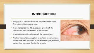
PTERYGIUM(SURFER’S EYES)
PRESENTED BY : ALAO NICHOLAS
: EJEDAWE PROMISE
OUTLINE
• INTRODUCTION
• EPIDEMIOLOGY
• FEATURES
• MORPHOLOGY (FEATURES & PARTS)
• AETIOLOGY & RISK FACTORS
• CLINICAL FEATURES
• TYPES
• INVESTIGATIONS
• TREATMENT
• PROGNOSIS
• COMPLICATION
INTRODUCTION
• Pterygium is derived from the ancient Greek word,
Pterygion, which means wing.
• It is a noncancerous fibrovascular growth of the
conjunctiva and can extend to the cornea.
• it is a degenerative disease of the conjunctiva.
• Another name for pterygium is ‘surfer’s eye’s because
surfers are well exposed to the elements (sun,wind,salt,
water) that can give rise to the growth.
EPIDEMIOLOGY
• Worldwide about 10-12% of the population is affected
• In Nigeria, the prevalence rates of pterygium in the general population
varies from 4 to 9%.
• Males are more often affected than females with a ratio of 2:1
• It is common in adults from the ages 20-40yrs and less common in children
<12yrs.
FEATURES
• Shape : Triangular/wing-like
• -It is raised
• -Color : white or pink
• -Location: within the interpalpebral fissure. It can be found both in the nasal
/temporal region or both( here it is called ,double headed pyerygium). But
it is more commonly found in the nasal region.
PARTS
• Parts of the pterygium includes;
• Apex
• Head
• Body
• Base
• Stocker’s line(deposition of iron seen in the cornea epithelium at the apex
of the pterygium)
RISK FACTORS
• Prolonged exposure to UV radiation
• Certain occupations like farming and sailors
• Age(20-40yrs high incidence)
• Genetics
• Vitamin A deficiency
• Wind
• Dust
PATHOPHYSIOLOGY
• The aetiopathohenesis of pterygium is unclear, however, UV radiation plays
a significant role in the development and progression.
• Prolonged exposure to UV radiation leads to mutations in the conjunctival
cells leading to an abnormal cell growth, it also stimulates fibroblast leading
to excessive collagen deposition.
• UV radiation also generates reactive oxygen species, damaging cellular
components.
TYPES
• 1) Progressive
• -high vascularity
• -thick
• -progressively encroaches towards the centre of the cornea
• 2) Regressive (atrophic)
• -thin
• -little vascularity
CLINICAL FEATURES
• Ranges from mild to severe
• -conjuctivitis
• -redness of eyes
• -dry eye
• -watery eye
• -grittiness/foreign body sensation
• -itching
• -eye pain/ocular ache
• -blur/double vision
• -Discomfort with contact lens
DIAGNOSIS
• -History taking
• -physical examination and other investigations including
• -visual acuity test
• -opthalmic examination using slit lamp biomicroscopy
• -corneal topography
• Corneal surface is examined to knw about it’s shape,curvature and thickness-pachymetry
• For this,one can use slit lamp,Optical coherence tomography (OCT)
TREATMENT
• This depends on severity
• A) Conservative management
• -giving lubricating eyedrops
• -using sunglasses for UV protection
• -avoidance of irritants ,wearing protective eyewears,hats e.t.c
TREATMENT CONT’D
• B) Surgical management
• -pterygion excision
• -A hole can be left behind which is filled using conjunctival autograft or amniotic tissue graft.
•
• Also after surgery, medications are given
•
• -antibiotics: prevent infec tions ,like fluoroquinolones (ofloxacin,levofloxacin),macrolides
(azithromycin)
• -steroid eyedrops : prevent pterygium from growing back
PROGNOSIS
• Good with prompt treatment and proper management.
COMPLICATIONS
• -Recurrence after surgery ,may occur due to risk factor exposure, non-
compliance to medications.
• -astigmatism
• -vision loss
• -corneal scarring

PTERYGIUM.pdf..,.........................

  • 1.
    PTERYGIUM(SURFER’S EYES) PRESENTED BY: ALAO NICHOLAS : EJEDAWE PROMISE
  • 2.
    OUTLINE • INTRODUCTION • EPIDEMIOLOGY •FEATURES • MORPHOLOGY (FEATURES & PARTS) • AETIOLOGY & RISK FACTORS • CLINICAL FEATURES • TYPES • INVESTIGATIONS • TREATMENT • PROGNOSIS • COMPLICATION
  • 3.
    INTRODUCTION • Pterygium isderived from the ancient Greek word, Pterygion, which means wing. • It is a noncancerous fibrovascular growth of the conjunctiva and can extend to the cornea. • it is a degenerative disease of the conjunctiva. • Another name for pterygium is ‘surfer’s eye’s because surfers are well exposed to the elements (sun,wind,salt, water) that can give rise to the growth.
  • 4.
    EPIDEMIOLOGY • Worldwide about10-12% of the population is affected • In Nigeria, the prevalence rates of pterygium in the general population varies from 4 to 9%. • Males are more often affected than females with a ratio of 2:1 • It is common in adults from the ages 20-40yrs and less common in children <12yrs.
  • 5.
    FEATURES • Shape :Triangular/wing-like • -It is raised • -Color : white or pink • -Location: within the interpalpebral fissure. It can be found both in the nasal /temporal region or both( here it is called ,double headed pyerygium). But it is more commonly found in the nasal region.
  • 6.
    PARTS • Parts ofthe pterygium includes; • Apex • Head • Body • Base • Stocker’s line(deposition of iron seen in the cornea epithelium at the apex of the pterygium)
  • 7.
    RISK FACTORS • Prolongedexposure to UV radiation • Certain occupations like farming and sailors • Age(20-40yrs high incidence) • Genetics • Vitamin A deficiency • Wind • Dust
  • 8.
    PATHOPHYSIOLOGY • The aetiopathohenesisof pterygium is unclear, however, UV radiation plays a significant role in the development and progression. • Prolonged exposure to UV radiation leads to mutations in the conjunctival cells leading to an abnormal cell growth, it also stimulates fibroblast leading to excessive collagen deposition. • UV radiation also generates reactive oxygen species, damaging cellular components.
  • 9.
    TYPES • 1) Progressive •-high vascularity • -thick • -progressively encroaches towards the centre of the cornea • 2) Regressive (atrophic) • -thin • -little vascularity
  • 10.
    CLINICAL FEATURES • Rangesfrom mild to severe • -conjuctivitis • -redness of eyes • -dry eye • -watery eye • -grittiness/foreign body sensation • -itching • -eye pain/ocular ache • -blur/double vision • -Discomfort with contact lens
  • 11.
    DIAGNOSIS • -History taking •-physical examination and other investigations including • -visual acuity test • -opthalmic examination using slit lamp biomicroscopy • -corneal topography • Corneal surface is examined to knw about it’s shape,curvature and thickness-pachymetry • For this,one can use slit lamp,Optical coherence tomography (OCT)
  • 12.
    TREATMENT • This dependson severity • A) Conservative management • -giving lubricating eyedrops • -using sunglasses for UV protection • -avoidance of irritants ,wearing protective eyewears,hats e.t.c
  • 13.
    TREATMENT CONT’D • B)Surgical management • -pterygion excision • -A hole can be left behind which is filled using conjunctival autograft or amniotic tissue graft. • • Also after surgery, medications are given • • -antibiotics: prevent infec tions ,like fluoroquinolones (ofloxacin,levofloxacin),macrolides (azithromycin) • -steroid eyedrops : prevent pterygium from growing back
  • 14.
    PROGNOSIS • Good withprompt treatment and proper management.
  • 15.
    COMPLICATIONS • -Recurrence aftersurgery ,may occur due to risk factor exposure, non- compliance to medications. • -astigmatism • -vision loss • -corneal scarring