2. BILE DUCT INJURY (I)
• Any injury to the bile duct during
cholecystectomy is a dreaded complication.
• Major bile duct injuries may require biliary-
enteric reconstruction
• Many patients, their consultans, and their
lawyers believe these treatments result in a
lifetime of disability (Moraca R.J et al : Arch Surg 2003,
137:889-894)
3. BILE DUCT INJURY (2)
• The occurrence of an accidental bile duct
injury strikes the patient and surgeons with
great force, as neither is prepared for this
complication
• Often the surgeons is not immediately aware
of disaster, and a delayed diagnosis adds
further difficulty to the potentially disturbed
relationship between doctor and patient.
(Gouma DJ and Obertrop H : BJS 2002,89,385-386)
4. Complications of Laparoscopic Cholecystectomy :
A National Survey of 4,292 Hospitals and an
Analysis of 77,604 Cases
• 1.750 respondents
• 1.2% laparotomy for treatment of complications
• 0.6% mean rate of bile duct injury (exclusive of cystic duct),
that will be lowered after performing > 100 LC
• 50% of bile duct injury was recognized postoperatively,
required anastomotic repair
• 33 pts died, 18 of them due to operative injury
• 0.14% bowel injuries
• 0.25% vascular injuries
Deziel D J et al Chicago Illinois - Am J of Surg 165 January 1993
Most lethal complications
5. • Since 35 years ago, bile duct reconstructions were
performed in every imaginable way : end-to-end
repair, hepatico gastrotomy, hepatico-duodenostomy
(HD), loop hepatico-jejunostomy, and hepatico-
jejunostomy Roux-en-Y (HJ)
• Analysis of the results showed that HD and HJ
produced the lowest rates of recurrent stricture
formation, and these two have been the accepted
operations eversince
(Moraca R.J et al : Arch Surg 2003, 137:889-894)
BILE DUCT INJURY (3)
10. CHD DRAINS FREELY IN TO THE
PERITONEAL CAVITY
Lahey Clinic, Burlington,MA 1994
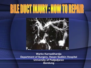
11. Common varians of bile duct anatomy
Lahey Clinic, Burlington, MA.1994
12. MANNER OF CONFLUENCE RIGHT
SECTORAL DUCTS
Blumgart LH. Surg Clin N Am. 1994.74.4
13. CLINICAL PRESENTATION
• Many injuries are unrecognizes at the time of
the initial operation, and their presentation
will vary
• Those with associated bile leak will present
early and often acutely ill from bile peritonitis
or subhepatic abscess
14. BILE LEAK IS RECOGNIZED
EARLIER
Presentation:
• Acutely ill
• Gut failure
Warko karnadihardja- 2004
15. CLINICAL PRESENTAION
• Those with an injury but not leak, usually
develop jaundice sometime after discharge
from hospital, depending of the nature of the
injury
• Some injuries evolve slowly or cause partial
obstruction
• Stricture may involve principally the right or
left hepatic duct or one of the right sectorial
hepatic ducts
16. TIPS & TRICKS TO DIAGNOSE
BILE DUCT INJURY
History of unexplained fevers, pain, abnormal
liver function test results, or pruritus
Should prompt an investigation
17. MANAGEMENT OF BILE DUCT INJURY (1)
• IMPORTANCE
– Preoperative investigation
– Patient Preparation
• BEFORE OPERATION
• The surgeon must define completely the extent
of injury and treat co existing conditions that
will increase operative morbidity and reduce
the likelihood of a successful repair
18. MANAGEMENT OF BILE DUCT INJURY (2)
• Preoperative imaging
– Is there subhepatic abscess or
collection?
– Is there ongoing bile leakage ?
– What is the level of biliary injury ?
– Are there associated vascular injuries /
– Is there evidence of lobar atrophy ?
19. TYPES OF IMAGING INVESTIGATION (1)
• Doppler Ultrasonography : May reveal the
level of:
– ductul injury and an associated vascular
injury or fluid collection
– Inadequate to define the extent of stricture
– Of little value if bile ducts are
decompressed
20. TYPES OF IMAGING INVESTIGATION (2)
• Cholangiography
– PTC is superior to ERCP
– MRCP : Noninvasive, provides striking images of
biliary tree
• Arteriography and Splenoportography
– If any suspection of vascular injury or portal
hypertension
• Isotopic scanning
– Functional assessment of incomplete stricture or
strictures of a sectoral hepatic duct (Bismuth
types)
21. TYPES OF IMAGING INVESTIGATION (3)
• Contrast-enchanced CT
– Probably the best initial study
– May define level of injury, fluid collection
or ascites
– May suggest the possibility of vascular
damage
– Reveal lobar atrophy
22. IMAGING OF BILE DUCT INJURY
Radiologist Society of North America :Radiology 1998
PTC MRCP: Surgical Clip After Multiple Attempts
to Repair (MD-CT)
23. ATROPHY OF THE LEFT HEPATIC LOBE
WITH DILATED AND CROWDED
INTRAHEPATIC DUCTS
Jarnagin WR and Blumgart LH; Arch Surg 134,1999
24. RIGHT LOBE ATROPHY AND
COMPENSATORY LEFT LOBE
HYPERTROPHY
Blumgart,LH,Surg Clin North Am. 1994,vol 74 no.4
25. OPENING THE UMBILICAL FISSURE BY
DIVIDING THE BRIDGE OF LIVER TISSUE
THAT CONNECTS SEGMENT III AND IV
Blumgart, LH, Surgery of the Liver and Biliary tract, 1994
26. EXPOSING THE HILAR PLATE
Blumgart. LH, Surg Clin North Am,1994. vol 74 no.4
27. MOBILIZATION OF HILAR PLATE FOR
HIGH BILIARY STRICTURES
Extension of bile duct
opening to permit wide
biliary enteric
anastomosis
Blumgart LH: Surg Clin N Am.1984 vo.74 1994
Lahey Clinic, Burlington, MA.1994
28. CREATING A SEPTA BETWEEN MULTIPLE BILE
DUCTS TO FORM A COMMON CHANNEL TO BE
ANASTOMOSED TO SINGLE OPENING OF THE
JEJUNUM
Lahey Clinic, Burlington, MA.1994
29. ANTERIOR AND POSTERIOR ROW OF
SUTURES
Blumgart LH, Surg of the Liver & Biliary tract, 1994
30. • Biliary function to be normal at more than 4 years
after biliary-enteric reconstruction for bile duct
injury
• When surgically feasable, we prefer HD to HJ
• 9 years study: February 1.1993-Januari 1. 2002
Depart of General, Vascular and Thoracic Surgery, Virginia Mason Medical
Center, Seatle, Wash
Arch Surg, vol 137, Aug.2002
31. OPERATIVE TECHNIQUE (1)
• A generous incision-full mobilization of the inferior
surface of the liver identify the site of bile duct injury
• Avoid dissection that might devascularize the
remaining bile duct, that is of the hepatic arterial and
portal venous systems
• Sharp debridement was used for damaged or
devitalized bile duct wall to the level of normal
mucosa
• Identify each patients unique anatomy for the right
and left hepatic ducts and their relationship to the
bifurcation by : Surgical Instrumentation,
cholangiography or choledochoscopy
Virginia Mason Medical Center, Seattle, Wash
32. OPERATIVE TECHNIQUE (2)
• Biliary enteric anastomosis were performed using
magnification for a mucosa-to- mucosa anastomosis
with the use of single layer of multiple, fine,
interrupted, absorbable sutures for a watertight
closure
• Temporary transanastomotic stents were various
used including
– Percutaneous transhepatic
– Percutaneous trans-enteric
– Internal small silicone stents anchored to mucosa
– Or no stent
Virginia Mason Medical Center, Seatle. Wash.2002
33. TEMPORARY
TRANSANASTOMOTIC STENTS
Blumgart LH : Surg N Am; 1994, vol. 74 no. 4
A. Percutaneous trans-enteric
B. Percutaneous transhepatic
C. U tube
D. Internal small silicone stent
anchored to mucosa
34. OPERATIVE TECHNIQUE (2)
• For Hepaticoduodenostomy
– Wide Kocherization of the duodenum to create a
tension free anastomosis end to side was
accomplished
• Roux-en-Y Jejunal Limbs
– Were made intentionally short so that
postoperatively endoscopic inspection of the
anastomotic site could be attempted when
indicated
– Hepaticojejunostomy was done end-to-side
35. ROUX-EN-Y HEPATICO JEJUNOSTOMY
WITH EXTENDED ACCESS LOOP
Blumgart LH,Surgery of the Liver and Biliary Tract, 1994
“Burried Subcutaneous
Stoma”, marked by clip
Open skin stoma
Warko Karnadihardja-BDG
36. OPERATIVE TECHNIQUE (3)
• A generous incision-full mobilization of the inferior
surface of the liver identify the site of bile duct injury
• Avoid dissection that might devascularize the
remaining bile duct, that is of the hepatic arterial and
portal venous systems
• Sharp debridement was used for damaged or
devitalized bile duct wall to the level of normal
mucosa
• Identify each patients unique anatomy for the right
and left hepatic ducts and their relationship to the
bifurcation by : Surgical Instrumentation,
cholangiography or choledochoscopy
Virginia Mason Medical Center, Seattle, Wash 2002
37. Kegunaan kombinasi Kent & sweetheart retractors
Tersedia hampir di
semua Rumah Sakit
di Bandung
(peralatan standar)
38. OPERATIVE TECHNIQUE (4)
• Closed suction drains were placed below and near
biliary-enteric anastomosis
• All transanatomotic stents were removed
postoperatively within 3 weeks after cholangiography
demonstrated patent anastomoses
• Internal anastomotic stents are allowed to pass
spontaneously
• No long-term stenting
• Patients with HJ were treated with long-term
prophylactic medication to avoid peptic ulceration
Virginia Mason Medical Center, Seattle, Wash 2002
39. COROSION CAST OF ADULT LIVER
Van Damme and Bonte J : Vascular Anatomy of in
Abdominal Surg. Thieme 1990
BLOOD SUPPLY TO CBD
Surgical Clin N. Am,1994
40. GOOD VASCULARIZATION OF THE
PROXIMAL JEJUNUM
Vascularization of the duodenojejunal angle
Van Damme J P and Bonte J : Vascular Anatomy of in
Abdominal Surgery Thieme, 1990
42. “ More careful and accurate communication between
doctor and patient, before and after primary surgery
as well as before and after surgery, may help to
prevent disappointing results”
“ Studies not only to have focused on outcome in terms
of laboratory and imaging results, rather than in
terms of general well-being or quality of life”
British Journal of Surg 2002.89
Department of Surgery Academic Medical Centre Amsterdam
43. • “ A stricture of the biliary tree can be one of the most
challenges that a surgeon can face”
• “If unrecognized or managed improperly, life-threatening
complications, such as biliary cirrhosis, portal hypertension
and cholangitis can develop”
• “Management with pre-op cholangiography to delineate the
anatomy and placement of percutaneous biliary catheters,
followed by surgical reconstruction with a Roux-en-Y
hepaticojejunostomy, is associated with a successful
outcome in up to 98% of patients”
John Hopkins Hospital, Baltimore, Maryland. Ann Surg, September 2000
44. • “ The initial management of patients with proximal
bile duct injuries will depend on the type of injury
and time of recognition”
• “ If the injury is recognized immediately, surgeons
must consider their ability to repair it immediately”
• “ If the surgeon is unable to effect a reasonable
repair and competent help is not available, then
the patient should undergo adequate drainage and
be referred to a more experienced surgeons
Arch Surg vol 134, July 1999
45. • “ Good results are obtained with a Roux-en-y
hepatico jejunostomy after complex injuries’
• “The use at of transanastomotic stents has to be
selective according to the individual characteristics
of each patient and the experience of each
surgeon”
• We recommend their use when unhealthy ie:
ischemic, scarred and small ducts < 4 mm are
found”
Mercado MA et al depart Surg, INCMNSZ, Mexico City
A dilema not answered ?
Arch Surg vol 137, July 2002
46. Damage control surgery for
uncontroled bleeding of hepatic
rupture, bile leakage and sepsis
51. Bile leakage after laparoscopic cholecystectomy
and after laparotomy repair, stenting CBD with
small stent 7 F
Continuing SIRS and Sepsis
52. Replacing drainage for source control
with bigger CBD stent 10 F, before
definitive surgery
53. CONCLUSIONS
• Bile duct injury during cholecystectomy, either
laparoscopic or open, is a complex and a dreaded
complication
• The proximal bile duct is at greater risk for injury in
laparoscopic surgery and may require biliary-enteric
reconstruction
• Many patients, their consultants and their lawyers
believe these treatments result in a lifetime of
disability
• Among the surgical strategies for repair, hepatico
jejunostomy yields the most favorable results, as far
as we consider the good principles of surgery, such
as, should be tension free, good vascularization,
healty duct and the widest diameter of bile duct
available