This presentation outlines the basic management steps for a gastric outlet obstruction, including a basic introduction of gastric outlet obstruction, some statistics and finally the management.
Contents
1. Introduction
2. Causes
3.Etiology (Benign and Malignant)
4. Pathophysiology
5. Clinical features
6. Physical examinations
7. Metabolic effects
8. Investigations
9. Management (conservative and surgical along with indications)
10.
Summary
3.
INTRODUCTION
• Gastric outletobstruction (GOO, pyloric obstruction) is not a
single entity
• Clinical and pathophysiological consequence
of any disease process that produces a mechanical impediment
to gastric emptying
4.
CAUSES
• Two well-definedgroups of causes
BENIGN AND MALIGNANT
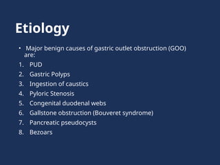
• Benign causes include pyloric stenosis secondary to peptic
ulceration
• While, Malignant causes include Gastric cancer.
• Previously, peptic ulcer diseases were more common.
• Now, with the decrease in the incidence of peptic ulceration and
the advent of potent medical treatments, gastric outlet
obstruction should be treated as malignant unless proven
otherwise.
5.
• Only 37%have benign disease, with the rest having malignant
cause.
• The term ‘pyloric stenosis’ is a misnomer as the stenosis is
seldom at the pylorus.
• Commonly, when the condition is due to underlying peptic ulcer
disease the stenosis is found in the first part of duodenum(most
common site for peptic ulcer)
• True pyloric stenosis however, can occur as a result of fibrosis
around a pyloric channel ulcer.
6.
Diagnostic and treatmentdilemma
• Exclude functional non-mechanical causes of obstruction,
such as diabetic gastroparesis
• Once mechanical obstruction is established, differentiate
between benign and malignant ( definitive treatment varies)
• Diagnosis and treatment is Urgent, because delay further
compromises patient’s nutritional status
Delay also further compromises edematous tissue and
complicates surgical intervention
7.
Frequency
• The incidenceoccurs in less than 5% in patients. With peptic
ulcer disease being the leading benign cause
• Peripancreatic malignancy, the most common malignant
etiology- 15-20%.
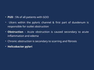
• PUD :5% of all patients with GOO
• Ulcers within the pyloric channel & first part of duodenum is
responsible for outlet obstruction
• Obstruction – Acute obstruction is caused secondary to acute
inflammation and edema
• Chronic obstruction is secondary to scarring and fibrosis
• Helicobacter pylori
10.
• Pediatric agegroup- Congenital Pyloric stenosis
• Occurs in 4 per 1000 births
• Boys˃ Girls (4 : 1)
• More common in first-born children
• It is familial.
• PYLORIC STENOSIS occurs between 3rd
and 6th
week of age
of an infant, which is the time taken for gradual
hypertrophy of the circular smooth muscle of the pylorus to
cause complete obstruction.
• Visible gastric peristalsis is seen.
11.
• Pancreatic canceris the most common malignancy causing GOO
• Outlet obstruction may occur in 10-20%
Other tumors include
1. Ampullary cancer
2. Duodenal cancer
3. Cholangiocarcinoma
4. Gastric cancer
5. Metastases to the gastric outlet by other primary tumors
12.
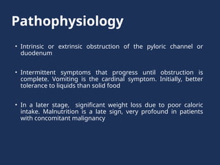
Pathophysiology
• Intrinsic orextrinsic obstruction of the pyloric channel or
duodenum
• Intermittent symptoms that progress until obstruction is
complete. Vomiting is the cardinal symptom. Initially, better
tolerance to liquids than solid food
• In a later stage, significant weight loss due to poor caloric
intake. Malnutrition is a late sign, very profound in patients
with concomitant malignancy
13.
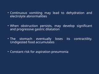
• Continuous vomitingmay lead to dehydration and
electrolyte abnormalities
• When obstruction persists, may develop significant
and progressive gastric dilatation
• The stomach eventually loses its contractility.
Undigested food accumulates
• Constant risk for aspiration pneumonia
14.
Clinical features
Gastric outletobstruction from a duodenal ulcer or
incomplete obstruction typically present with symptoms
of the following:
1. Gastric retention, including early satiety, bloating or epigastric
fullness, indigestion, anorexia, nausea, vomiting, epigastric
pain, and weight loss
2. Pain is severe, persistent, in the epigastric region, and also
with feeling of fullness
3. Nausea and vomiting are the cardinal symptoms
15.
4.Vomiting – Non-bilious,and it characteristically
contains undigested food particles
5.Loss of periodicity.
6.Early stages: vomiting, intermittent and usually
occurs within 1 hour of a meal
7.Very often it is possible to recognize foodstuff taken
several days previously
8. Patient loses weight, appears unwell and dehydrated
16.
9.Frequently malnourished anddehydrated and have a
metabolic insufficiency
10. Weight loss , most significant with malignant
disease
11.Abdominal pain is not frequent and usually relates to
the underlying cause, such as PUD or Pancreatic Cancer.
17.
Physical examination
• Chronicdehydration and Malnutrition
On examination :
1. Distended abdomen and a succussion splash may be
audible on shaking the patient’s abdomen
Positive succussion splash is done with 4 hours empty
stomach, by placing a stethoscope over the epigastric
region and shaking the patient adequately.
18.
2.A dilated stomachmay be appreciated as a tympanic
mass in the epigastric area and/or left upper quadrant
3.Visible gastric peristalsis (VGP) may be elicited by asking
the patient to drink a cup of water.
4.Auscultopercussion test shows dilated stomach.
( This test is done by placing a stethoscope over epigastric
region. Skin is scratched from left side downwards, at
several points away from the epigastrium using finger and
these points are joined. Normally the greater curvature of
the stomach lies above the level of umbilicus, while in GOO
it lies below the level of umbilicus.)
19.
5.Goldstein saline loadtest: half and hour after
installation of 750ml of saline, if volume remained
and if more than 250ml is present, suggests
obstruction.
20.
Metabolic effects
• Dehydrationand electrolyte abnormalities- Increase in BUN
and creatinine are late features of dehydration
• Prolonged vomiting causes loss of hydrochloric acid &
produces an increase of bicarbonate in the plasma to
compensate for the lost chloride, hypokalemic hypochloremic
metabolic alkalosis
• Alkalosis shifts the intracellular potassium to the extracellular
compartment, and the serum potassium is increased
factitiously
21.
• With continuedvomiting, the renal excretion of
potassium increases in order to preserve sodium
• The adrenocortical response to hypovolemia intensifies
the exchange of potassium for sodium at the distal
tubule, with subsequent aggravation of the hypokalemia
Paradoxically acidic
urine
• Initially,the urine has a low chloride and high bicarbonate
content, reflecting the primary metabolic abnormality
• This bicarbonate is excreted along with sodium and so, with
time, the patient becomes progressively hyponatremic and more
profoundly dehydrated.
• Because of the dehydration, a phase of sodium retention follows
and potassium and hydrogen are excreted in preference.
• This results in the urine becoming paradoxically acidic
• Alkalosis leads to a lowering of the circulating ionised calcium,
and gastric tetany can occur.
24.
• Clinical featuresof Paradoxical aciduria
1. Irritability, confused status, dehydration
2. Often convulsions can occur.
3. Features of alkalosis like rapid breathing (Cheyne-stokes
breathing and tetany)
• Investigations
1. Serum electrolytes
2. Arterial blood gas analysis
3. Serum calcium level estimation
• Treatment : Double strength normal saline with IV
potassium under ECG monitoring. Plus IV magnesium.
25.
Investigations
1.Barium meal study:
Absenceof duodenal cap.
Dilated stomach where greater curvature is below the level of iliac
crest.
Mottled stomach
Barium does not pass into duodenum.
2.Gastroscopy to rule out stomach carcinoma and to visualize the
stenosed area.
3.Electrolyte study for the correction of electrolyte imbalance.
4.ECG to check for hypokalemia.
26.
Management
1. Correcting themetabolic and electrolyte abnormality by IV
fluids.
2. Rehydrated with intravenous isotonic saline with potassium
supplementation or double strength slaine, calcium,
potassium, magnesium.
3. Replacing the sodium chloride and water allows the kidney to
correct the acid–base abnormality
4. Following rehydration it may become obvious that the patient is
also anemic.
5. Blood transfusion if given if there is anemia.
27.
6. TPN support.
7.STOMACH WASH: The stomach should be emptied using a Wide-
bore gastric tube/Eswald’s tube. Pass an orogastric tube and lavage
the stomach until it is completely emptied.
8. It reduces the edema of the stomach wall and improves gastric
emptying time by increasing the gastric muscle tone.
9. Then endoscopy and contrast radiology
10.Biopsy of the area around the pylorus is essential to exclude
malignancy
11.The patient should also have an anti-secretory agent, initially given
intravenously to ensure absorption
28.
• Early cases: settle with conservative treatment, presumably as
oedema around the ulcer diminishes as the ulcer is healed
• Endoscopic treatment with balloon dilatation useful in early
cases
(Dilating the duodenal stenosis may result in perforation, and
the dilatation may have to be performed several times and may
not be successful in the long term)
29.
Surgical management
• Highlyselective vagotomy(HSV) with gastrojejunostomy is
present recommendation even though it is technically difficult.
• HSV is better than Truncal vagotomy as it maintains the nerve
supply of the chronically obstructed antrum and so may
eventually reduce the chronic emptying problems.
• Vagotomy, antrectomy (acid secreting area) with Billroth I
anastomosis along with feeding jejunostomy for nutrition is
the other option.
30.
Indications for Surgery
•Gastric outlet obstruction due to benign ulcer disease may be
treated medically if results of imaging studies or endoscopy
determine - acute inflammation and edema are the principle
causes (as opposed to scarring and fibrosis, which may be
fixed)
• If medical therapy fails, then surgical therapy
• Typically, if resolution or improvement is not seen within 48-
72 hours, surgical intervention is necessary
31.
• The choiceof surgical procedure depends upon the patient's
particular circumstances
• In cases of malignant obstruction, weigh the extent of surgical
intervention for the relief of obstruction against the malignancy's
type and extent, as well as the patient's anticipated long-term
prognosis
• As a guiding principle, undertake major tumor resections in the
absence of metastatic disease
32.
• In patientswith largely metastatic disease, determine the
degree of surgical intervention for palliation in the light of
patient’s realistic prognosis and personal wishes.
33.
Summary
Gastric outlet obstructionis most commonly associated with
longstanding peptic ulcer disease and gastric cancer
• The metabolic abnormality of hypochloraemic alkalosis is
usually only seen with peptic ulcer disease and should be
treated with isotonic saline with potassium supplementation.
• Endoscopic biopsy is essential to determine whether the
cause of the problem is malignancy
34.
• Endoscopic dilatationof the gastric outlet may be
effective in the less severe cases of benign stenosis
• Operation is normally required, with a drainage
procedure being performed for benign disease and
appropriate resectional surgery if malignant.
35.
Other causes ofGastric outlet
obstruction
• Adult pyloric stenosis
This is a rare condition and its relationship to childhood condition is
unclear, although some patients have a long history of problems with
gastric emptying. It commonly treated by pyloroplasty that
pyloromyotomy.
• Pyloric mucosal diaphragm
The origin of this rare condition is unknown. It usually does not
become apparent until mid life. When found, simple excision of
mucosal diaphragm is all that is required.