1. Objective
Introduction
Expression vectors
Results and Discussion
Conclusions
Future Research
Acknowledgements
References
• I would like to thank Dr. Hall for giving me the opportunity to work in his lab
and continued guidance, Ashley Meyers for her continued support and
guidance throughout all the experiments as well as all the members of the
Hall lab for their support.
Development Of A Single Step Immunoassay Using A Single Domain VHH Circularly Permutated GFP Fusion Protein
To Detect Toxin B from Clostridium difficile
Prashant Tanwar, Ashley Meyers1, J. C. Hall1
1School of Environmental Sciences, University of Guelph, Guelph, ON, N1G 2W1, Canada
• To develop a single step immunoassay for the detection TcdB using a single
domain variable heavy-chain antibody (VHH) cpGFP (VHH-cpGFP).
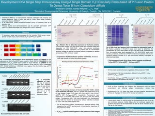
Fig. 1 Schematic representation of the expression vectors. A) pAM203 for the
expression of the VHH39-sfGFP fusion protein in E.coli HB2151. B) pAM204 for the
expression of VHH39-cpGFP fusion protein in E.coli HB2151. C) pAM205 for the
expression of VHH39/167-cpGFP fusion protein in E.coli HB2151. sfGFP, superfolder
GFP; G4SGF, (G4S)3 and KGSG4S, linker regions; His6, 6x Histidine tag; cpGFP,
circularly permutated GFP.
1500 bp
10000 bp
4000 bp
1500 bp
1000 bp
A) pAM203
4453 bp
1122 bp
10000 bp
4000 bp
1000 bp
B) pAM204
4453 bp
1167 bp
10000 bp
4000 bp
1500 bp
5000 bp
C) pAM205
1572 bp
4453 bp
Successful transformation of E. coli cells.
Fig 3. Western Blot to detect the expression of the fusion proteins.
Crude protein extracts from the soluble lysate and insoluble lysate were
separated on 12% SDS-PAGE, under non-reducing conditions,
transferred to PVDF membrane and probed with an anti-PentaHis
antibody followed by a goat anti-mouse antibody conjugated to alkaline
phosphatase. MW, molecular weight.
• Expression of all three fusion proteins confirmed. (All future
work was carried out using the soluble lysate).
50
75
100
150
MW
(kDa)
37
25
20
10
Lysate 1 (soluble) Lysate 2 (insoluble)
Fig 5. SDS-PAGE and western blot to compare the expression levels of
the fusion proteins. A Bradford assay was used to determine the
concentration of TSP in each lysate. 50 ug of TSP from crude protein extracts
from the soluble lysate were separated on 12% SDS-PAGE, under non-
reducing conditions, transferred to PVDF membrane and probed with an anti-
PentaHis antibody followed by a goat anti-mouse antibody conjugated to
alkaline phosphatase.
• The expression levels of the three fusion proteins are different:
VHH39-sfGFP > VHH39-cpGFP > VHH39/167-cpGFP.
• All three fusion proteins fluoresce regardless of the presence of TcdB.
• The expression of all three molecules is different. VHH39-sfGFP > VHH39-
cpGFP > VHH39/167-cpGFP.
• Nonetheless, VHH39/167-cpGFP fluorescence is higher meaning the construct
comes together in the presence of Toxin B.
• Purify all three molecules and measure the fluorescence with the same
concentration and differing antigen concentration, across time.
• Development of a VHH fusion that does not come together spontaneously
perhaps by changing the linkers of the circular permutations.
50 ug TSP
50
75
100
150
MW
(kDa)
37
25
20
10
50
75
100
150
MW
(kDa)
37
25
20
10
50 ug TSP
• Clostridium difficile is a Gram-positive anaerobic pathogen that produces two
primary exotoxins, Toxin A (TcdA) and toxin B (TcdB), which cause C.difficile-
associated disease (CDAD)1,2
• In its native form, Green Fluorescent Protein (GFP) is commonly used as a
detection tool
• Previous studies have demonstrated the use of a circularly permutated GFP
(cpGFP) to detect the antigen ICP using an scFv-cpGFP antibody3
0%
5%
10%
15%
20%
25%
30%
35%
40%
0 100 200 300 400 500
PercentageIncreaseInFluorescenceWhenTcdB
IsAdded
Time (minutes)
Percentage Increase In Fluorescence in the Presence of TcdB
203
204
205
Molecules
Fig 4. The percentage increase in fluorescence after TcdB is added
to the molecules over time. TcdB (0.5 ug/well) was mixed with 6.25 ug
of Total Soluble Protein (TSP) from 203 (VHH39-sfGFP), 204 (VHH39-
cpGFP), 205 (VHH39/167-cpGFP), respectively. Fluorescence was
measured in a microtiter plate reader over 8 hours with and without
antigen. Fluorescence with TcdB was subtracted from fluorescence
without TcdB then divided by fluorescence without TcdB then multiplied
by 100%. Data was plotted against time.
• For all three fusion proteins, fluorescence is observed without TcdB
suggesting that cpGFP spontaneously comes together regardless of
the presence of TcdB.
• VHH39/167-cpGFP comes together in the presence of the antigen.
1. Hussack G et al. 2011. Neutralization of Clostridium difficile Toxin A with Single-domain Antibodies Targeting the
Cell Receptor Binding Domain. J. Biol. Chem 286: 8961-8976.
2. Rupnik M et al. 2009. Clostridium difficile infection: new developments in epidemiology and pathogenesis. Nature
Reviews Microbiology 7: 526-536.
3. Kojima M et al. 2011. Activation of circularly permutated beta-lactamase tethered to antibody domains by specific
small molecules. Bioconjugate chemistry 22, 633-41.
Fig. 2 Enzymatic
Digestion of DNA.
500 ng of plasmid
DNA was digested
using 20 units of both
Nco I and Not I
restriction enzymes
then run on a 1%
agarose gel. C
represents colony.
A
B
C VHH 1673` cpGFP5` cpGFP
Lac
promoter
PelB leader ColE1 ori
NcoI
VHH 39
NotI
His6
G4SGF (G4S)3 KGSG4S
3` cpGFP5` cpGFP
Lac
promoter
PelB leader ColE1 ori
NcoI
VHH 39
NotI
His6
G4SGF (G4S)3
sfGFP
Lac
promoter
PelB leader ColE1 ori
NcoI
VHH 39
NotI
His6
G4SGF