SlideShare a Scribd company logo
1
Contrast Filled Rectal Balloons in Post-Prostatectomy
Proton Therapy Alignment
Rachel Rendall, B.S. R.T.(T), Lauren Curran, B.S. R.T.(T), John Han-Chih
Chang, MD, Mingcheng Gao, PhD, Daniel Spring, B.S. R.T.(R)(T),
Charles Yoo, B.S., MBA, Mark Pankuch, PhD, William Hartsell, MD
CDH Proton Center, a ProCure Center – Warrenville, IL, USA
Sagittal, axial and coronal view of ERB in XIO for treatment planning. Green outlines TV,
brown outlines the rectum, red outlines the ERB.
Axial, Coronal, Sagittal, and 3D views of planning CT with Re-CT overlay. Position of ERB is
confirmed to be reproducible throughout treatment.
Orthogonal films taken during treatment showing
proper alignment of ERB within contour and
bony anatomy.
Graph 1: Difference in amount of table shifts per fraction.
Graphs 2 and 3: Evaluation of rectum receiving dose. Red line is deviation tolerance.
Introduction
The use of prostate gland fiducial markers in prostate alignment is a
commonly used practice in photon therapy and proton therapy. These
fiducials assist in image fusion, anatomy contouring, creation of
target volumes (TV), and is used for daily target alignment for image
guidance. In the absence of the prostate gland, the localization of
treatment targets is more challenging. Some prostatectomy surgeries
leave surgical clips which can be contoured as a fiducial, however
some surgeries leave no clips. These patients need an alternative way
to localize the prostate bed that can also be used as an alignment
device. Proper immobilization using endorectal balloons (ERB) is
essential to reduce margins, which reduce the dose to normal
structures. ERBs serve as a setup reference device by distending the
rectal wall out of the treatment volume, decreasing the amount of
high dose to the rectum volume.
Methods and Materials
In patients with gold or carbon prostate gland fiducials, the fiducial is
contoured in dosimetry planning with a margin of 1mm to aid in daily
setup. An ERB is contoured with no margin due to the size differential
between the balloon and fiducial. After all contours are finished, the
dosimetrist and physician must evaluate the TV, ERB and rectum
contours. The rectum contour must be outside of the ERB contour
and the TV contour must not overlap the ERB contour.
Results
Discussion and Conclusion
Contrast filled ERBs have shown equivalent results compared to
patients with fiducial markers. Using ERBs is now an alternative
procedure to allow patients without fiducial markers or surgical clips
to be treated at CDH Proton Center, a ProCure Center. ERBs have
shown to be well-tolerated, reliable, reproducible, and an effective
tool for alignment of post-prostatectomy patients as well as efficient
device for treatment planning, so that proper dose sparing of normal
tissue can occur in post-prostatectomy patients.
The entire ERB contour is forced to a
density of 1.0, using the relative
stopping power ratio of the contrast
mix inserted in the ERB to the water
tissue equivalent based on Hounsfield
units. Beam arrangement for ERB
patients are picked to give minimal
dose to the bladder and rectum. The
ideal beamline position has been
determined to be opposing right and
left laterals. After the range and
modulation are determined, the plan is
then checked for deviations regarding
dose limitations and tolerances of
surrounding tissue. If deviations are
present, the dosimetrist will continue
to optimize the plan by editing
apertures and compensators. Once the
plan is determined optimal for the
patient, it is then ready to be exported
to milling, quality assurance and the
treatment room.
Preparation of the ERB is very
important because it is used
as an immobilization device as
well as an alignment device. A
syringe is filled with a pre-
determined amount of
contrast and attached to the
ERB. During the simulation process, it is evaluated whether barium
contrast or a 50-50 Cystografin-water will be used during treatment.
When the patient is positioned on the treatment table, orthogonal x-
rays are taken and the patient is aligned using the anterior ERB
border with the planned ERB contour. Bony anatomy is used to align
the patient superiorly and right to left. A verification film is taken after
table shifts to confirm proper alignment.
Axial and sagittal views of isodose
lines for planned treatment. Red
shaded area is TV, purple outline is
ERB, brown outline is rectum, inner
yellow line is conedown shape.
Evaluation of the dosage to the tumor volume and areas requiring
protection from the proton particle beam compared to the patients
with fiducials show a similar rectal dose to the 50% volume and 70%
volume tolerance limit.
The number of table shifts in prostate patients with fiducial markers
averages two per fraction compared to ERB patients’ average of one
per fraction.
In this study, the ERB patient’s setup parameters and outcomes were
compared to 10 patients who were aligned with fiducial markers daily.
ERB patients did not have any additional acute complications
compared to those with fiducial markers. The highest reported
urinary frequency toxicity was grade one. ERBs average the same
size of localized offset corrections as fiducials, but reduce the
average number of table shifts per treatment by half.

More Related Content

Similar to Poster ERB Final

IGRT APP.pdf
IGRT APP.pdfIGRT APP.pdf
IGRT APP.pdf
Kanhu Charan
 
EBRT of Ca Cervix
EBRT of Ca CervixEBRT of Ca Cervix
EBRT of Ca Cervix
Shyamsunder Kumawat
 
IMRT IN CANCER CERVIX
IMRT IN CANCER CERVIXIMRT IN CANCER CERVIX
IMRT IN CANCER CERVIX
Dr.T.Sujit :-)
 
Effects of rapid palatal expansion on the sagittal and vertical dimensions of...
Effects of rapid palatal expansion on the sagittal and vertical dimensions of...Effects of rapid palatal expansion on the sagittal and vertical dimensions of...
Effects of rapid palatal expansion on the sagittal and vertical dimensions of...
EdwardHAngle
 
Cervix External Beam Radiotherapy techniques
Cervix External Beam Radiotherapy techniquesCervix External Beam Radiotherapy techniques
Cervix External Beam Radiotherapy techniques
Animesh Agrawal
 
Volumetric-Based Analysis of In-Vivo and Ex-Vivo Quantita-tive MR Diffusion P...
Volumetric-Based Analysis of In-Vivo and Ex-Vivo Quantita-tive MR Diffusion P...Volumetric-Based Analysis of In-Vivo and Ex-Vivo Quantita-tive MR Diffusion P...
Volumetric-Based Analysis of In-Vivo and Ex-Vivo Quantita-tive MR Diffusion P...
JohnJulie1
 
Volumetric-Based Analysis of In-Vivo and Ex-Vivo Quantita-tive MR Diffusion P...
Volumetric-Based Analysis of In-Vivo and Ex-Vivo Quantita-tive MR Diffusion P...Volumetric-Based Analysis of In-Vivo and Ex-Vivo Quantita-tive MR Diffusion P...
Volumetric-Based Analysis of In-Vivo and Ex-Vivo Quantita-tive MR Diffusion P...
JapaneseJournalofGas
 
Electronic portal imaging by rose wekesa
Electronic portal imaging by rose wekesaElectronic portal imaging by rose wekesa
Electronic portal imaging by rose wekesa
Kesho Conference
 
Erb tendourology section
Erb tendourology sectionErb tendourology section
Erb tendourology section
Ahmed Eliwa
 
STUDY OF eTEP FOR VENTRAL HERNIA REPAIR.pptx
STUDY OF eTEP FOR VENTRAL HERNIA REPAIR.pptxSTUDY OF eTEP FOR VENTRAL HERNIA REPAIR.pptx
STUDY OF eTEP FOR VENTRAL HERNIA REPAIR.pptx
AnandaHegde1
 
Sarcoma brachytherapy updates
Sarcoma brachytherapy updatesSarcoma brachytherapy updates
Sarcoma brachytherapy updates
Ashutosh Mukherji
 
Efficacy Of Imrt Through Dmlc And Inverse Planning
Efficacy Of Imrt Through Dmlc And Inverse PlanningEfficacy Of Imrt Through Dmlc And Inverse Planning
Efficacy Of Imrt Through Dmlc And Inverse Planningfondas vakalis
 
MRI procedure of pelvis and hip suman duwal
MRI procedure of pelvis and hip suman duwalMRI procedure of pelvis and hip suman duwal
MRI procedure of pelvis and hip suman duwal
suman duwal
 
EBRT IN CARCINOMA CERVIX
EBRT IN CARCINOMA CERVIXEBRT IN CARCINOMA CERVIX
EBRT IN CARCINOMA CERVIX
Isha Jaiswal
 
root resoption.pptx
root resoption.pptxroot resoption.pptx
root resoption.pptx
SadhuAbhijeet
 
Radical Prostate Radiotherapy
Radical Prostate RadiotherapyRadical Prostate Radiotherapy
Radical Prostate Radiotherapy
Catherine Holborn
 
Role of SBRT in lung cancer
Role of SBRT in lung cancerRole of SBRT in lung cancer
Role of SBRT in lung cancer
DrAyush Garg
 
SBRT Contouring Guidelines
SBRT  Contouring  GuidelinesSBRT  Contouring  Guidelines
SBRT Contouring Guidelines
Dr Rushi Panchal
 
Intra Operative Radiotherapy
Intra Operative RadiotherapyIntra Operative Radiotherapy
Intra Operative Radiotherapy
Sasikumar Sambasivam
 

Similar to Poster ERB Final (20)

IGRT APP.pdf
IGRT APP.pdfIGRT APP.pdf
IGRT APP.pdf
 
EBRT of Ca Cervix
EBRT of Ca CervixEBRT of Ca Cervix
EBRT of Ca Cervix
 
IMRT IN CANCER CERVIX
IMRT IN CANCER CERVIXIMRT IN CANCER CERVIX
IMRT IN CANCER CERVIX
 
ICRU 38 nayana
ICRU 38 nayanaICRU 38 nayana
ICRU 38 nayana
 
Effects of rapid palatal expansion on the sagittal and vertical dimensions of...
Effects of rapid palatal expansion on the sagittal and vertical dimensions of...Effects of rapid palatal expansion on the sagittal and vertical dimensions of...
Effects of rapid palatal expansion on the sagittal and vertical dimensions of...
 
Cervix External Beam Radiotherapy techniques
Cervix External Beam Radiotherapy techniquesCervix External Beam Radiotherapy techniques
Cervix External Beam Radiotherapy techniques
 
Volumetric-Based Analysis of In-Vivo and Ex-Vivo Quantita-tive MR Diffusion P...
Volumetric-Based Analysis of In-Vivo and Ex-Vivo Quantita-tive MR Diffusion P...Volumetric-Based Analysis of In-Vivo and Ex-Vivo Quantita-tive MR Diffusion P...
Volumetric-Based Analysis of In-Vivo and Ex-Vivo Quantita-tive MR Diffusion P...
 
Volumetric-Based Analysis of In-Vivo and Ex-Vivo Quantita-tive MR Diffusion P...
Volumetric-Based Analysis of In-Vivo and Ex-Vivo Quantita-tive MR Diffusion P...Volumetric-Based Analysis of In-Vivo and Ex-Vivo Quantita-tive MR Diffusion P...
Volumetric-Based Analysis of In-Vivo and Ex-Vivo Quantita-tive MR Diffusion P...
 
Electronic portal imaging by rose wekesa
Electronic portal imaging by rose wekesaElectronic portal imaging by rose wekesa
Electronic portal imaging by rose wekesa
 
Erb tendourology section
Erb tendourology sectionErb tendourology section
Erb tendourology section
 
STUDY OF eTEP FOR VENTRAL HERNIA REPAIR.pptx
STUDY OF eTEP FOR VENTRAL HERNIA REPAIR.pptxSTUDY OF eTEP FOR VENTRAL HERNIA REPAIR.pptx
STUDY OF eTEP FOR VENTRAL HERNIA REPAIR.pptx
 
Sarcoma brachytherapy updates
Sarcoma brachytherapy updatesSarcoma brachytherapy updates
Sarcoma brachytherapy updates
 
Efficacy Of Imrt Through Dmlc And Inverse Planning
Efficacy Of Imrt Through Dmlc And Inverse PlanningEfficacy Of Imrt Through Dmlc And Inverse Planning
Efficacy Of Imrt Through Dmlc And Inverse Planning
 
MRI procedure of pelvis and hip suman duwal
MRI procedure of pelvis and hip suman duwalMRI procedure of pelvis and hip suman duwal
MRI procedure of pelvis and hip suman duwal
 
EBRT IN CARCINOMA CERVIX
EBRT IN CARCINOMA CERVIXEBRT IN CARCINOMA CERVIX
EBRT IN CARCINOMA CERVIX
 
root resoption.pptx
root resoption.pptxroot resoption.pptx
root resoption.pptx
 
Radical Prostate Radiotherapy
Radical Prostate RadiotherapyRadical Prostate Radiotherapy
Radical Prostate Radiotherapy
 
Role of SBRT in lung cancer
Role of SBRT in lung cancerRole of SBRT in lung cancer
Role of SBRT in lung cancer
 
SBRT Contouring Guidelines
SBRT  Contouring  GuidelinesSBRT  Contouring  Guidelines
SBRT Contouring Guidelines
 
Intra Operative Radiotherapy
Intra Operative RadiotherapyIntra Operative Radiotherapy
Intra Operative Radiotherapy
 

Poster ERB Final

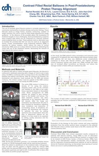
  • 1. 1 Contrast Filled Rectal Balloons in Post-Prostatectomy Proton Therapy Alignment Rachel Rendall, B.S. R.T.(T), Lauren Curran, B.S. R.T.(T), John Han-Chih Chang, MD, Mingcheng Gao, PhD, Daniel Spring, B.S. R.T.(R)(T), Charles Yoo, B.S., MBA, Mark Pankuch, PhD, William Hartsell, MD CDH Proton Center, a ProCure Center – Warrenville, IL, USA Sagittal, axial and coronal view of ERB in XIO for treatment planning. Green outlines TV, brown outlines the rectum, red outlines the ERB. Axial, Coronal, Sagittal, and 3D views of planning CT with Re-CT overlay. Position of ERB is confirmed to be reproducible throughout treatment. Orthogonal films taken during treatment showing proper alignment of ERB within contour and bony anatomy. Graph 1: Difference in amount of table shifts per fraction. Graphs 2 and 3: Evaluation of rectum receiving dose. Red line is deviation tolerance. Introduction The use of prostate gland fiducial markers in prostate alignment is a commonly used practice in photon therapy and proton therapy. These fiducials assist in image fusion, anatomy contouring, creation of target volumes (TV), and is used for daily target alignment for image guidance. In the absence of the prostate gland, the localization of treatment targets is more challenging. Some prostatectomy surgeries leave surgical clips which can be contoured as a fiducial, however some surgeries leave no clips. These patients need an alternative way to localize the prostate bed that can also be used as an alignment device. Proper immobilization using endorectal balloons (ERB) is essential to reduce margins, which reduce the dose to normal structures. ERBs serve as a setup reference device by distending the rectal wall out of the treatment volume, decreasing the amount of high dose to the rectum volume. Methods and Materials In patients with gold or carbon prostate gland fiducials, the fiducial is contoured in dosimetry planning with a margin of 1mm to aid in daily setup. An ERB is contoured with no margin due to the size differential between the balloon and fiducial. After all contours are finished, the dosimetrist and physician must evaluate the TV, ERB and rectum contours. The rectum contour must be outside of the ERB contour and the TV contour must not overlap the ERB contour. Results Discussion and Conclusion Contrast filled ERBs have shown equivalent results compared to patients with fiducial markers. Using ERBs is now an alternative procedure to allow patients without fiducial markers or surgical clips to be treated at CDH Proton Center, a ProCure Center. ERBs have shown to be well-tolerated, reliable, reproducible, and an effective tool for alignment of post-prostatectomy patients as well as efficient device for treatment planning, so that proper dose sparing of normal tissue can occur in post-prostatectomy patients. The entire ERB contour is forced to a density of 1.0, using the relative stopping power ratio of the contrast mix inserted in the ERB to the water tissue equivalent based on Hounsfield units. Beam arrangement for ERB patients are picked to give minimal dose to the bladder and rectum. The ideal beamline position has been determined to be opposing right and left laterals. After the range and modulation are determined, the plan is then checked for deviations regarding dose limitations and tolerances of surrounding tissue. If deviations are present, the dosimetrist will continue to optimize the plan by editing apertures and compensators. Once the plan is determined optimal for the patient, it is then ready to be exported to milling, quality assurance and the treatment room. Preparation of the ERB is very important because it is used as an immobilization device as well as an alignment device. A syringe is filled with a pre- determined amount of contrast and attached to the ERB. During the simulation process, it is evaluated whether barium contrast or a 50-50 Cystografin-water will be used during treatment. When the patient is positioned on the treatment table, orthogonal x- rays are taken and the patient is aligned using the anterior ERB border with the planned ERB contour. Bony anatomy is used to align the patient superiorly and right to left. A verification film is taken after table shifts to confirm proper alignment. Axial and sagittal views of isodose lines for planned treatment. Red shaded area is TV, purple outline is ERB, brown outline is rectum, inner yellow line is conedown shape. Evaluation of the dosage to the tumor volume and areas requiring protection from the proton particle beam compared to the patients with fiducials show a similar rectal dose to the 50% volume and 70% volume tolerance limit. The number of table shifts in prostate patients with fiducial markers averages two per fraction compared to ERB patients’ average of one per fraction. In this study, the ERB patient’s setup parameters and outcomes were compared to 10 patients who were aligned with fiducial markers daily. ERB patients did not have any additional acute complications compared to those with fiducial markers. The highest reported urinary frequency toxicity was grade one. ERBs average the same size of localized offset corrections as fiducials, but reduce the average number of table shifts per treatment by half.