SlideShare a Scribd company logo
1 of 85
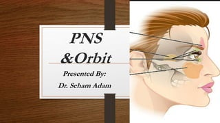
PNS
&Orbit
Presented By:
Dr. Seham Adam
SINUS ANATOMY:
The para-nasal sinuses consists of usually four
paired air-filled spaces. They have several
functions of which reducing the weight of the head
is the most important. Other functions are air
humidification and aiding in voice resonance.
There are named for the facial bones in which they
are located:
Frontal sinus
Maxillary sinus .
Sphenoid sinus .
Ethmoid sinus .
The maxillary ostium opens superioly into the infundibulum, which is
the channel between the inferomedial aspect of the orbit laterally and
uncinated process medially.
The ostium , infundibulum and middle meatus is important clinically
and is known as the ostiomeatal complex .
Ostiomeatal complex :
 The ostiomeatal complex (or unit), is a common channel that
links the frontal sinus, anterior and middle ethmoid sinus and
the maxillary sinus to the middle meatus that allows air
flow and mucociliary drainage.
The posterior ethmoidal Sinus empty to the superior meatus.
Sphenoidal Sinus opens to the sphenoethmoidal recess.
IMAGING MODALITY OF SINUSES:
• Plain Radiography :
• evaluate normal sinuses (Transradiant Because Contain Air )
• Showing mucosal thickening ,air- fluid level , bone
destruction and fractures.
• Computed Tomography :is the gold standard imaging
investigation in sinus disease.
• Magnetic resonance imaging (MRI):
usually used the primary investigation.
Ct sinus scan
1
1
1
1
2
Mucosal Thickening
 Thickened mucosa can be recognized providing there is some air in the
sinus by noting the soft tissue interface between the air in the sinus and the
bony wall.
• The mucosal thickening may be smooth in outline or it may be polypoid .
• Allergy and infection both cause mucosal thickening and it is often
impossible to say radiological which condition is responsible.
• Such changes are often seen in asymptomatic people.
Opaque Sinus:
The sinus becomes opaque when all the air is replaced.
CAUSES OF AN OPAQUE SINUS:
• Infection.
• Allergy
• Mucocele.
• Antrochoanal polyp.
• Truma with heamorrhage
• Carcinoma of sinus or nasal cavity.
Sinusitis
 inflammation of the paranasal sinus mucosa.
 can occur in any of the paranasal sinuses.
 Involvement: maxillary > ethmoidal, frontal >
sphenoidal sinus.
 Types:
• Infectious sinusitis( Acute ,Subacute and Chronic).
• Noninfectious (allergic) .
• Dental infection and sinusitis (20% maxillary antra ).
Radiographic Features:
• Acute sinusitis is usually diagnosed and treated
clinically, but CT is recommended if symptoms persist.
• Both CT and MRI can elegantly demonstrate mucosal
thickening and fluid levels as well as displaying the
bony walls of the sinuses.
• Opacified sinus may be partial or complete
• Mucosal thickening
• Air-fluid levels( diagnostic for acute sinusitis )
• Chronic sinusitis: mucosal thickening and hyperostosis
of bone.
Acute Sinusitis
plain radiography
Normal sinuses Acute sinusitis
Acute Sinusitis:
PNS CT
Normal sinuses Acute sinusitis
Chronic Sinusitis:
plain radiography
Normal sinuses Chronic sinuses
Chronic sinusitis
PNS Ct
Normal sinuses Chronic sinusitis
Fungal Sinusitis :
• Predisposing factors: diabetes, prolonged antibiotic or steroid therapy,
immune-compromised patient
Radiographic Features :
• Bony destruction and rapid extension into adjacent anatomic spaces
• Indistinguishable from tumor: biopsy required
• Main role of CT/MRI is to determine extent of disease
• Aspergillosis may appear hyperdense on CT .
Mucocele:
• A largely clinically silent mucus-containing expansil lesion of the
paranasal sinuses with secondary obstruction of the main ostium of the
affected paranasal sinus.
•The lesion is lined by ciliated columnar respiratory epithelium
•It becomes symptomatic only when it extends into surrounding tissue
planes secondary to remodeling of the surrounding bone and erosion.
•Most common sinus involvement is frontal (67%), followed
by ethmoidal (20%), maxillary (10%), and sphenoidal (3%).
CT scan of the head shows
diffuse opacification of the
frontal and ethmoidal sinuses
with remodeling of the bony
walls with cortical erosion
(arrow) of the superior part of
the right lamina papyracea.
(a) T1-weighted axial image reveals high T1 signal (arrow) at right frontal
sinus. The left frontal sinus is not pneumatized. (b) FLAIR-weighted axial
image reveals high T2 signal (arrow) at right frontal sinus. (c) Postcontrast
T1-weighted imaging does not reveal any abnormal postcontrast
enhancement (arrow) of the sinus lesion
Sino-nasal polyposis:
• Refers to the presence of multiple benign polyps in
the nasal cavity and paransal sinuses , expansion and
opacification of PNS with thinning septa . It causes a
specific pattern of chronic sinusitis
Sino-nasal polyp:
Antro- choanal polyp : are solitary sino-nasal polyp that arise within
the maxillary sinus. They pass through and enlarge the sinus
ostium and posterior nasal cavity to the nasopharynx .
 This results in unilateral opacification of the maxillary antrum and the
frontal and anterior ethmoidal sinuses due to obstruction of their
drainage pathways.
Polyps are common intranasal mass lesions, resulting
from trapped fluid in the deeper layers of the mucosa of the
nasal cavity. These can occur in any age group and can extend
into the adjacent paranasal sinuses as well asposteriorly into
the pharynx.
Sino-nasal polyp:
Coronal plane , sagittal and Axial
reconstructed contrast enhance CT images
there is an extensive fluid-attenuated lesion
filling the right maxillary sinus, nasal cavity ,
and nasopharynx (black arrows), without
disruption of adjacent structures.
On CT, polyps are of low (fluid)
attenuation. However, polyps with
more inspissated secretions can be
of higher density. Such polyps
may be difficult to differentiate
from other lesions by CT alone
Sagittal T2-weighted with extensive T2prolongation (white arrow).
and contrast-enhanced axial T1-weighted fat-suppressed
images demonstrate a predominantly peripherally enhancing focus (white arrows)
PNS Tumor:
• Types SCC( 90% ), Maxillary sinus( 80%).
• In all opaque sinuses, particularly the antra , special
attention should be paid to the bony margins on CT, because
if these are destroyed a diagnosis of carcinoma needs
exclusion.
• CT is superior at visualizing bone destruction, but MRI
is preferred for demonstrating tumour invasion by
showing the extent of any soft tissue mass beyond the
sinus cavity.
• Both CT and MRI have an important role in treatment
planning and in assessing response to treatment.
On CT scan, unilateral sinonasal soft tissue mass with patchy
postcontrast enhancement, destruction of the bony wall of sinus,
and aggressive regional spread is seen.
Moderately enhancing aggressive soft
tissue mass at right maxillary sinus with
destruction of medial and posterior wall of
right maxilla extends into masticator space
(arrow), involving the pterygopalatine
fossae (arrow), pterygoid muscles (arrow).
(A-C)Low T1 and intermediate T2 signal mass exhibits patchy post-
contrast enhancement. Local infiltration of right temporalis muscle is
more apparent. (D) Invasion of the hard palate (arrow), floor of maxillary
sinus (arrow) with extension into buccal space (arrow), and into the soft
tissue of the cheek (arrow) is seen.
Nasopharyngeal Carcinoma
Sinus trauma :
A fracture of the sinus wall may
result in hemorrhage (high density)
and opacification of the sinus.
Computed tomography
demonstrated a depressed fracture of
the anterior wall of the right
maxillary sinus . Mild mucosal
thickening with air-fluid level in the
maxillary sinus that suggests blood.
The Orbit
Orbital Anatomy
Anterior view of the orbit
demonstrating
extra ocular muscles, superior
orbital fissure and content of
the intraconal space
Orbital Anatomy
Lateral view of the orbit demonstrating:
oculomotor nerve (CN III) with its inferior and
superior division
Imaging modalities:
• Computed tomography and MRI clearly demonstrate the
anatomy of the orbits.
• Imaging is indicated in all patients with exophthalmos because
it is important to distinguish between masses arising within the
orbit, masses arising outside the orbit and thyroid eye disease.
• With an intraorbital mass, its relationship to the optic nerve can
be determined.
1.Retr orbital fat
2.Medial rectus
3.Lens
4.Lateral rectus
5.Optic nerve
1
2
3
4 5
Ct Scan Of The Orbit
Retinoblastoma:
Malignant tumor that arises from neuro-ectodermal cells of retina.
Clinical:
• leukocoria (white mass behind pupil).
• Age: <3 years
• 30% bilateral, 30% multifocal within one eye
• 10% of patients have a familial history of retinoblastoma
.
Retinoblastoma:
In an infant with a soft tissue, partially calcified mass within the optic
globe.
Radiographic Features:
• Intraocular mass
• Calcifications are common (90%);
• Associated with other malignancies most common
(osteosarcoma).
On orbital CT, a soft tissue mass is typically
noted. Associated calcifications are present in
70% of cases and strongly suggest the
diagnosis
Contrast-enhanced
orbital CT shows a
partially calcified soft
tissue lesion along the
posterior aspect of the
right optic globe
(arrow
MRI demonstrates slight T1 hyperintensity of
the tumor with low T2 signal and some degree
of contrast enhancement.
Optic Nerve Glioma :
Most common cause of diffuse optic nerve enlargement,
especially in childhood.
Clinical findings include loss of vision, proptosis (bulky
tumors).
In neurofibromatosis (NF-1) the disease may be bilateral.
Lesions can involve any portion of the nerve from the orbit to
the optic tract; it can be bilateral
Radiographic Features:
•fusiform enlargement of optic nerve.
• Contrast enhancement variable.
• Calcifications rare.
MRI is the imaging modality of choice.
Optic Nerve Meningioma
Optic nerve meningiomas are benign tumors arising from the
arachnoid cap cells of the optic nerve sheath.
Age: 4th decade (80% female); younger patients typically
have NF.
Progressive loss of vision interfere with blood supply to the
optic nerve.
Radiographic Features:
Mass surrounding the optic nerve,
• Calcification (common)
• Intense contrast enhancement “tram track sign”
Orbital infection:
 The orbital septum represents a barrier to infectious spread from
anterior to posterior structures.
Common causes of orbital infection include spread from infected sinus
and trauma.
 Orbital infectious processes divided into preseptal and postseptal
Preseptal “periorbital” cellulitis can be due to several causes including
trauma, dental disease, and adjacent soft tissue inflammatory disease
Postseptal “orbital” cellulitis is almost always seen in the associated
setting of paranasal sinus (ethmoidal or frontal) disease.
• Postseptal infection (true orbital cellulitis): Sub periosteal
infiltrate or abscess ,Stranding of retro bulbar fat ,Lateral
displacement of enlarged medial rectus muscle , Proptosis.
• Sub-periosteal abscess is the extreme end of the spectrum of orbital
cellulitis, usually presenting as fluid collection between the lateral
rectus and the lamina papyracea.
There is extensive right periorbital and intraorbital soft tissue swelling.
A rim-enhancing fluid collection is present along the right lamina
papyracea (arrow). The adjacent right ethmoid sinuses are extensively
opacified (arrowheads).
Thyroid Orbitopathy :
is a thyroid-associated process that results in mucopolysaccharide deposition
within the extraocular muscles resulting in early enlargement of the muscle,
with relative sparing of the tendon.
•Mucopolysaccharide deposition may result in relative low attenuation
centers of the muscles involved.
•Muscular involvement typically follows the temporal pattern of IMSLO,
where the inferior rectus is most frequently affected , followed by the medial,
superior, and lateral recti, and least frequently, the oblique muscles.
Thyroid Orbito-pathy:
Radiographic Features:
• Exophthalmos .
• Muscle involvement.
• Inferior (most common) Medial Superior Lateral.
• Spares tendon insertions.
• Often bilateral, symmetrical.
• patient presenting with painless proptosis involving both orbits.
Thyroid Ophthalmopathy
Ct . scan through the orbits showing enlargement of
the extraocular muscles, particularly the inferior and
medial rectus.
Orbital pseudotumor:
• Idiopathic inflammatory condition ,
The exact etiology is not known but an association with many
inflammatory/autoimmune diseases is reported.
• Infiltrating, intraconal, retro-orbital, non-granulomatous mass,
usually unilateral and involving extraocular musculature, including
tendinous portion in patient with painful proptosis.
Orbital pseudotumor:
Intense postcontrast enhancement, based on evaluated fat suppressed
postgadolinium T1-weighted MRI.
It is possible to have only muscular involvement; however, the
tendinous portion is always involved. The lacrimal gland, uvea,
sclera, optic nerve sheath, and bony orbits can be variably involved.
Coronal noncontrast CT shows an infiltrating
intraconal retrobulbar mass (arrow) involving the
superior and lateral rectus muscle.
a) T1-weighted sagittal MRI reveals infiltrating retrobulbar low T1 signal mass
(arrow) involving the superior rectus muscle. (b) T2-weighted coronal MRI
reveals patchily high T2 signal intraconal mass (arrow) involving expanded
superior rectus and lateral rectus muscles. (c) Postcontrast fat-suppressed T1-
weighted image reveals infiltrative intensely enhancing intra-conal retro-orbital
mass(arrow) involving the superior and lateral rectus muscles.
Blow out fracture
A direct blow to the eye raises the intraorbital pressure and can
result in a fracture of the orbital floor, which is the weakest part
of the orbit.
The break in the orbital floor allows herniation of orbital
contents into the antrum, which may result in diplopia.
Orbital blowout fractures occur when there is a fracture of one
of the walls of orbit but the orbital rim remains intact. This is
typically caused by a direct blow to the central orbit from a fist
or ball.
Imaging is best performed by CT with coronal reconstructions,
which show a crescentic soft tissue mass in the roof of the antrum
this should not be confused with mucosal thickening.
A fracture of the orbital floor may also be visible
The arrow refers to the
herniated orbital contents
Frontal Air-fluid level in the right maxillary
antra associated with a "tear drop" arising
from the orbital floor consistent with
prolapsed orbital content
Inferior and medial blow out fractures. Note distortion
of inferior rectus and inferior herniation of orbital fat
PNS , Orbit and   Neck .pptx

More Related Content

Similar to PNS , Orbit and Neck .pptx

Imaging of paranasal sinuses
Imaging of paranasal sinusesImaging of paranasal sinuses
Imaging of paranasal sinusesMilan Silwal
 
Sinonasal polyposis
Sinonasal polyposisSinonasal polyposis
Sinonasal polyposisShaista Amir
 
Radiological approach to PNS (Paranasal sinus) Masses
Radiological approach to PNS (Paranasal sinus) MassesRadiological approach to PNS (Paranasal sinus) Masses
Radiological approach to PNS (Paranasal sinus) MassesDr Anuj Aggarwal
 
Presentation1.pptx, radiological imaging of the nasopharyngeal diseases.
Presentation1.pptx, radiological imaging of the nasopharyngeal diseases.Presentation1.pptx, radiological imaging of the nasopharyngeal diseases.
Presentation1.pptx, radiological imaging of the nasopharyngeal diseases.Abdellah Nazeer
 
Complications of rhinosinusitis
Complications of rhinosinusitisComplications of rhinosinusitis
Complications of rhinosinusitisMohammed Nishad N
 
Imaging of paranasal sinuses
Imaging of paranasal sinusesImaging of paranasal sinuses
Imaging of paranasal sinusesArchana Koshy
 
Skull base tumors &amp; perineural spread radiology ppt
Skull base tumors &amp; perineural spread radiology pptSkull base tumors &amp; perineural spread radiology ppt
Skull base tumors &amp; perineural spread radiology pptDr pradeep Kumar
 
MASTOIDECTOMY (BY DR.RICHARD & DR.BUKUKU)
MASTOIDECTOMY (BY DR.RICHARD & DR.BUKUKU)MASTOIDECTOMY (BY DR.RICHARD & DR.BUKUKU)
MASTOIDECTOMY (BY DR.RICHARD & DR.BUKUKU)RitchieShija
 
MASTOIDECTOMY PRESENTATION
MASTOIDECTOMY  PRESENTATIONMASTOIDECTOMY  PRESENTATION
MASTOIDECTOMY PRESENTATIONRitchieShija
 
Carcinoma neuroendocrino del setto un rarissimo tumore nasale
Carcinoma neuroendocrino del setto un rarissimo tumore nasaleCarcinoma neuroendocrino del setto un rarissimo tumore nasale
Carcinoma neuroendocrino del setto un rarissimo tumore nasaleMerqurio
 
Carcinoma neuroendocrino del setto un rarissimo tumore nasale
Carcinoma neuroendocrino del setto un rarissimo tumore nasaleCarcinoma neuroendocrino del setto un rarissimo tumore nasale
Carcinoma neuroendocrino del setto un rarissimo tumore nasaleMerqurio
 
SEMINAR_ON_MAXILLARY_SINUS.ppt
SEMINAR_ON_MAXILLARY_SINUS.pptSEMINAR_ON_MAXILLARY_SINUS.ppt
SEMINAR_ON_MAXILLARY_SINUS.pptRehana116725
 
Tumours Of The Cp Afinal Power Pressed
Tumours Of The Cp Afinal Power PressedTumours Of The Cp Afinal Power Pressed
Tumours Of The Cp Afinal Power PressedLiew Boon Seng
 
4086742..................................
4086742..................................4086742..................................
4086742..................................srinivaspennam88
 

Similar to PNS , Orbit and Neck .pptx (20)

Imaging of paranasal sinuses
Imaging of paranasal sinusesImaging of paranasal sinuses
Imaging of paranasal sinuses
 
Sinonasal polyposis
Sinonasal polyposisSinonasal polyposis
Sinonasal polyposis
 
Radiological approach to PNS (Paranasal sinus) Masses
Radiological approach to PNS (Paranasal sinus) MassesRadiological approach to PNS (Paranasal sinus) Masses
Radiological approach to PNS (Paranasal sinus) Masses
 
Imaging in ent
Imaging in entImaging in ent
Imaging in ent
 
Maxillary Sinus
Maxillary SinusMaxillary Sinus
Maxillary Sinus
 
Presentation1.pptx, radiological imaging of the nasopharyngeal diseases.
Presentation1.pptx, radiological imaging of the nasopharyngeal diseases.Presentation1.pptx, radiological imaging of the nasopharyngeal diseases.
Presentation1.pptx, radiological imaging of the nasopharyngeal diseases.
 
Temporal & infra temporal region
Temporal & infra temporal regionTemporal & infra temporal region
Temporal & infra temporal region
 
Complications of rhinosinusitis
Complications of rhinosinusitisComplications of rhinosinusitis
Complications of rhinosinusitis
 
Imaging of paranasal sinuses
Imaging of paranasal sinusesImaging of paranasal sinuses
Imaging of paranasal sinuses
 
Skull base tumors &amp; perineural spread radiology ppt
Skull base tumors &amp; perineural spread radiology pptSkull base tumors &amp; perineural spread radiology ppt
Skull base tumors &amp; perineural spread radiology ppt
 
Functional endoscopic sinus surgery
Functional endoscopic sinus surgeryFunctional endoscopic sinus surgery
Functional endoscopic sinus surgery
 
MASTOIDECTOMY PPT
MASTOIDECTOMY PPTMASTOIDECTOMY PPT
MASTOIDECTOMY PPT
 
MASTOIDECTOMY (BY DR.RICHARD & DR.BUKUKU)
MASTOIDECTOMY (BY DR.RICHARD & DR.BUKUKU)MASTOIDECTOMY (BY DR.RICHARD & DR.BUKUKU)
MASTOIDECTOMY (BY DR.RICHARD & DR.BUKUKU)
 
MASTOIDECTOMY PRESENTATION
MASTOIDECTOMY  PRESENTATIONMASTOIDECTOMY  PRESENTATION
MASTOIDECTOMY PRESENTATION
 
Carcinoma neuroendocrino del setto un rarissimo tumore nasale
Carcinoma neuroendocrino del setto un rarissimo tumore nasaleCarcinoma neuroendocrino del setto un rarissimo tumore nasale
Carcinoma neuroendocrino del setto un rarissimo tumore nasale
 
Carcinoma neuroendocrino del setto un rarissimo tumore nasale
Carcinoma neuroendocrino del setto un rarissimo tumore nasaleCarcinoma neuroendocrino del setto un rarissimo tumore nasale
Carcinoma neuroendocrino del setto un rarissimo tumore nasale
 
SEMINAR_ON_MAXILLARY_SINUS.ppt
SEMINAR_ON_MAXILLARY_SINUS.pptSEMINAR_ON_MAXILLARY_SINUS.ppt
SEMINAR_ON_MAXILLARY_SINUS.ppt
 
Tumours Of The Cp Afinal Power Pressed
Tumours Of The Cp Afinal Power PressedTumours Of The Cp Afinal Power Pressed
Tumours Of The Cp Afinal Power Pressed
 
4086742..................................
4086742..................................4086742..................................
4086742..................................
 
Sino-nasal malignancy
Sino-nasal malignancySino-nasal malignancy
Sino-nasal malignancy
 

Recently uploaded

💎VVIP Kolkata Call Girls Parganas🩱7001035870🩱Independent Girl ( Ac Rooms Avai...
💎VVIP Kolkata Call Girls Parganas🩱7001035870🩱Independent Girl ( Ac Rooms Avai...💎VVIP Kolkata Call Girls Parganas🩱7001035870🩱Independent Girl ( Ac Rooms Avai...
💎VVIP Kolkata Call Girls Parganas🩱7001035870🩱Independent Girl ( Ac Rooms Avai...Taniya Sharma
 
All Time Service Available Call Girls Marine Drive 📳 9820252231 For 18+ VIP C...
All Time Service Available Call Girls Marine Drive 📳 9820252231 For 18+ VIP C...All Time Service Available Call Girls Marine Drive 📳 9820252231 For 18+ VIP C...
All Time Service Available Call Girls Marine Drive 📳 9820252231 For 18+ VIP C...Arohi Goyal
 
Top Rated Bangalore Call Girls Mg Road ⟟ 8250192130 ⟟ Call Me For Genuine Sex...
Top Rated Bangalore Call Girls Mg Road ⟟ 8250192130 ⟟ Call Me For Genuine Sex...Top Rated Bangalore Call Girls Mg Road ⟟ 8250192130 ⟟ Call Me For Genuine Sex...
Top Rated Bangalore Call Girls Mg Road ⟟ 8250192130 ⟟ Call Me For Genuine Sex...narwatsonia7
 
Call Girls Bhubaneswar Just Call 9907093804 Top Class Call Girl Service Avail...
Call Girls Bhubaneswar Just Call 9907093804 Top Class Call Girl Service Avail...Call Girls Bhubaneswar Just Call 9907093804 Top Class Call Girl Service Avail...
Call Girls Bhubaneswar Just Call 9907093804 Top Class Call Girl Service Avail...Dipal Arora
 
Call Girl Number in Panvel Mumbai📲 9833363713 💞 Full Night Enjoy
Call Girl Number in Panvel Mumbai📲 9833363713 💞 Full Night EnjoyCall Girl Number in Panvel Mumbai📲 9833363713 💞 Full Night Enjoy
Call Girl Number in Panvel Mumbai📲 9833363713 💞 Full Night Enjoybabeytanya
 
Call Girls Service Surat Samaira ❤️🍑 8250192130 👄 Independent Escort Service ...
Call Girls Service Surat Samaira ❤️🍑 8250192130 👄 Independent Escort Service ...Call Girls Service Surat Samaira ❤️🍑 8250192130 👄 Independent Escort Service ...
Call Girls Service Surat Samaira ❤️🍑 8250192130 👄 Independent Escort Service ...CALL GIRLS
 
Bangalore Call Girls Hebbal Kempapura Number 7001035870 Meetin With Bangalor...
Bangalore Call Girls Hebbal Kempapura Number 7001035870  Meetin With Bangalor...Bangalore Call Girls Hebbal Kempapura Number 7001035870  Meetin With Bangalor...
Bangalore Call Girls Hebbal Kempapura Number 7001035870 Meetin With Bangalor...narwatsonia7
 
Lucknow Call girls - 8800925952 - 24x7 service with hotel room
Lucknow Call girls - 8800925952 - 24x7 service with hotel roomLucknow Call girls - 8800925952 - 24x7 service with hotel room
Lucknow Call girls - 8800925952 - 24x7 service with hotel roomdiscovermytutordmt
 
Bangalore Call Girls Nelamangala Number 7001035870 Meetin With Bangalore Esc...
Bangalore Call Girls Nelamangala Number 7001035870  Meetin With Bangalore Esc...Bangalore Call Girls Nelamangala Number 7001035870  Meetin With Bangalore Esc...
Bangalore Call Girls Nelamangala Number 7001035870 Meetin With Bangalore Esc...narwatsonia7
 
Call Girl Coimbatore Prisha☎️ 8250192130 Independent Escort Service Coimbatore
Call Girl Coimbatore Prisha☎️  8250192130 Independent Escort Service CoimbatoreCall Girl Coimbatore Prisha☎️  8250192130 Independent Escort Service Coimbatore
Call Girl Coimbatore Prisha☎️ 8250192130 Independent Escort Service Coimbatorenarwatsonia7
 
VIP Russian Call Girls in Varanasi Samaira 8250192130 Independent Escort Serv...
VIP Russian Call Girls in Varanasi Samaira 8250192130 Independent Escort Serv...VIP Russian Call Girls in Varanasi Samaira 8250192130 Independent Escort Serv...
VIP Russian Call Girls in Varanasi Samaira 8250192130 Independent Escort Serv...Neha Kaur
 
Top Rated Bangalore Call Girls Richmond Circle ⟟ 8250192130 ⟟ Call Me For Gen...
Top Rated Bangalore Call Girls Richmond Circle ⟟ 8250192130 ⟟ Call Me For Gen...Top Rated Bangalore Call Girls Richmond Circle ⟟ 8250192130 ⟟ Call Me For Gen...
Top Rated Bangalore Call Girls Richmond Circle ⟟ 8250192130 ⟟ Call Me For Gen...narwatsonia7
 
Night 7k to 12k Chennai City Center Call Girls 👉👉 7427069034⭐⭐ 100% Genuine E...
Night 7k to 12k Chennai City Center Call Girls 👉👉 7427069034⭐⭐ 100% Genuine E...Night 7k to 12k Chennai City Center Call Girls 👉👉 7427069034⭐⭐ 100% Genuine E...
Night 7k to 12k Chennai City Center Call Girls 👉👉 7427069034⭐⭐ 100% Genuine E...hotbabesbook
 
Call Girls Siliguri Just Call 9907093804 Top Class Call Girl Service Available
Call Girls Siliguri Just Call 9907093804 Top Class Call Girl Service AvailableCall Girls Siliguri Just Call 9907093804 Top Class Call Girl Service Available
Call Girls Siliguri Just Call 9907093804 Top Class Call Girl Service AvailableDipal Arora
 
Call Girls Colaba Mumbai ❤️ 9920874524 👈 Cash on Delivery
Call Girls Colaba Mumbai ❤️ 9920874524 👈 Cash on DeliveryCall Girls Colaba Mumbai ❤️ 9920874524 👈 Cash on Delivery
Call Girls Colaba Mumbai ❤️ 9920874524 👈 Cash on Deliverynehamumbai
 
Russian Escorts Girls Nehru Place ZINATHI 🔝9711199012 ☪ 24/7 Call Girls Delhi
Russian Escorts Girls  Nehru Place ZINATHI 🔝9711199012 ☪ 24/7 Call Girls DelhiRussian Escorts Girls  Nehru Place ZINATHI 🔝9711199012 ☪ 24/7 Call Girls Delhi
Russian Escorts Girls Nehru Place ZINATHI 🔝9711199012 ☪ 24/7 Call Girls DelhiAlinaDevecerski
 
Vip Call Girls Anna Salai Chennai 👉 8250192130 ❣️💯 Top Class Girls Available
Vip Call Girls Anna Salai Chennai 👉 8250192130 ❣️💯 Top Class Girls AvailableVip Call Girls Anna Salai Chennai 👉 8250192130 ❣️💯 Top Class Girls Available
Vip Call Girls Anna Salai Chennai 👉 8250192130 ❣️💯 Top Class Girls AvailableNehru place Escorts
 
Call Girls Darjeeling Just Call 9907093804 Top Class Call Girl Service Available
Call Girls Darjeeling Just Call 9907093804 Top Class Call Girl Service AvailableCall Girls Darjeeling Just Call 9907093804 Top Class Call Girl Service Available
Call Girls Darjeeling Just Call 9907093804 Top Class Call Girl Service AvailableDipal Arora
 

Recently uploaded (20)

💎VVIP Kolkata Call Girls Parganas🩱7001035870🩱Independent Girl ( Ac Rooms Avai...
💎VVIP Kolkata Call Girls Parganas🩱7001035870🩱Independent Girl ( Ac Rooms Avai...💎VVIP Kolkata Call Girls Parganas🩱7001035870🩱Independent Girl ( Ac Rooms Avai...
💎VVIP Kolkata Call Girls Parganas🩱7001035870🩱Independent Girl ( Ac Rooms Avai...
 
All Time Service Available Call Girls Marine Drive 📳 9820252231 For 18+ VIP C...
All Time Service Available Call Girls Marine Drive 📳 9820252231 For 18+ VIP C...All Time Service Available Call Girls Marine Drive 📳 9820252231 For 18+ VIP C...
All Time Service Available Call Girls Marine Drive 📳 9820252231 For 18+ VIP C...
 
Top Rated Bangalore Call Girls Mg Road ⟟ 8250192130 ⟟ Call Me For Genuine Sex...
Top Rated Bangalore Call Girls Mg Road ⟟ 8250192130 ⟟ Call Me For Genuine Sex...Top Rated Bangalore Call Girls Mg Road ⟟ 8250192130 ⟟ Call Me For Genuine Sex...
Top Rated Bangalore Call Girls Mg Road ⟟ 8250192130 ⟟ Call Me For Genuine Sex...
 
Call Girls Bhubaneswar Just Call 9907093804 Top Class Call Girl Service Avail...
Call Girls Bhubaneswar Just Call 9907093804 Top Class Call Girl Service Avail...Call Girls Bhubaneswar Just Call 9907093804 Top Class Call Girl Service Avail...
Call Girls Bhubaneswar Just Call 9907093804 Top Class Call Girl Service Avail...
 
Call Girl Number in Panvel Mumbai📲 9833363713 💞 Full Night Enjoy
Call Girl Number in Panvel Mumbai📲 9833363713 💞 Full Night EnjoyCall Girl Number in Panvel Mumbai📲 9833363713 💞 Full Night Enjoy
Call Girl Number in Panvel Mumbai📲 9833363713 💞 Full Night Enjoy
 
Call Girls Service Surat Samaira ❤️🍑 8250192130 👄 Independent Escort Service ...
Call Girls Service Surat Samaira ❤️🍑 8250192130 👄 Independent Escort Service ...Call Girls Service Surat Samaira ❤️🍑 8250192130 👄 Independent Escort Service ...
Call Girls Service Surat Samaira ❤️🍑 8250192130 👄 Independent Escort Service ...
 
Bangalore Call Girls Hebbal Kempapura Number 7001035870 Meetin With Bangalor...
Bangalore Call Girls Hebbal Kempapura Number 7001035870  Meetin With Bangalor...Bangalore Call Girls Hebbal Kempapura Number 7001035870  Meetin With Bangalor...
Bangalore Call Girls Hebbal Kempapura Number 7001035870 Meetin With Bangalor...
 
Lucknow Call girls - 8800925952 - 24x7 service with hotel room
Lucknow Call girls - 8800925952 - 24x7 service with hotel roomLucknow Call girls - 8800925952 - 24x7 service with hotel room
Lucknow Call girls - 8800925952 - 24x7 service with hotel room
 
Bangalore Call Girls Nelamangala Number 7001035870 Meetin With Bangalore Esc...
Bangalore Call Girls Nelamangala Number 7001035870  Meetin With Bangalore Esc...Bangalore Call Girls Nelamangala Number 7001035870  Meetin With Bangalore Esc...
Bangalore Call Girls Nelamangala Number 7001035870 Meetin With Bangalore Esc...
 
Call Girl Coimbatore Prisha☎️ 8250192130 Independent Escort Service Coimbatore
Call Girl Coimbatore Prisha☎️  8250192130 Independent Escort Service CoimbatoreCall Girl Coimbatore Prisha☎️  8250192130 Independent Escort Service Coimbatore
Call Girl Coimbatore Prisha☎️ 8250192130 Independent Escort Service Coimbatore
 
VIP Russian Call Girls in Varanasi Samaira 8250192130 Independent Escort Serv...
VIP Russian Call Girls in Varanasi Samaira 8250192130 Independent Escort Serv...VIP Russian Call Girls in Varanasi Samaira 8250192130 Independent Escort Serv...
VIP Russian Call Girls in Varanasi Samaira 8250192130 Independent Escort Serv...
 
Top Rated Bangalore Call Girls Richmond Circle ⟟ 8250192130 ⟟ Call Me For Gen...
Top Rated Bangalore Call Girls Richmond Circle ⟟ 8250192130 ⟟ Call Me For Gen...Top Rated Bangalore Call Girls Richmond Circle ⟟ 8250192130 ⟟ Call Me For Gen...
Top Rated Bangalore Call Girls Richmond Circle ⟟ 8250192130 ⟟ Call Me For Gen...
 
Escort Service Call Girls In Sarita Vihar,, 99530°56974 Delhi NCR
Escort Service Call Girls In Sarita Vihar,, 99530°56974 Delhi NCREscort Service Call Girls In Sarita Vihar,, 99530°56974 Delhi NCR
Escort Service Call Girls In Sarita Vihar,, 99530°56974 Delhi NCR
 
Night 7k to 12k Chennai City Center Call Girls 👉👉 7427069034⭐⭐ 100% Genuine E...
Night 7k to 12k Chennai City Center Call Girls 👉👉 7427069034⭐⭐ 100% Genuine E...Night 7k to 12k Chennai City Center Call Girls 👉👉 7427069034⭐⭐ 100% Genuine E...
Night 7k to 12k Chennai City Center Call Girls 👉👉 7427069034⭐⭐ 100% Genuine E...
 
Call Girls Siliguri Just Call 9907093804 Top Class Call Girl Service Available
Call Girls Siliguri Just Call 9907093804 Top Class Call Girl Service AvailableCall Girls Siliguri Just Call 9907093804 Top Class Call Girl Service Available
Call Girls Siliguri Just Call 9907093804 Top Class Call Girl Service Available
 
Call Girls Colaba Mumbai ❤️ 9920874524 👈 Cash on Delivery
Call Girls Colaba Mumbai ❤️ 9920874524 👈 Cash on DeliveryCall Girls Colaba Mumbai ❤️ 9920874524 👈 Cash on Delivery
Call Girls Colaba Mumbai ❤️ 9920874524 👈 Cash on Delivery
 
Russian Escorts Girls Nehru Place ZINATHI 🔝9711199012 ☪ 24/7 Call Girls Delhi
Russian Escorts Girls  Nehru Place ZINATHI 🔝9711199012 ☪ 24/7 Call Girls DelhiRussian Escorts Girls  Nehru Place ZINATHI 🔝9711199012 ☪ 24/7 Call Girls Delhi
Russian Escorts Girls Nehru Place ZINATHI 🔝9711199012 ☪ 24/7 Call Girls Delhi
 
Vip Call Girls Anna Salai Chennai 👉 8250192130 ❣️💯 Top Class Girls Available
Vip Call Girls Anna Salai Chennai 👉 8250192130 ❣️💯 Top Class Girls AvailableVip Call Girls Anna Salai Chennai 👉 8250192130 ❣️💯 Top Class Girls Available
Vip Call Girls Anna Salai Chennai 👉 8250192130 ❣️💯 Top Class Girls Available
 
Call Girls Darjeeling Just Call 9907093804 Top Class Call Girl Service Available
Call Girls Darjeeling Just Call 9907093804 Top Class Call Girl Service AvailableCall Girls Darjeeling Just Call 9907093804 Top Class Call Girl Service Available
Call Girls Darjeeling Just Call 9907093804 Top Class Call Girl Service Available
 
Russian Call Girls in Delhi Tanvi ➡️ 9711199012 💋📞 Independent Escort Service...
Russian Call Girls in Delhi Tanvi ➡️ 9711199012 💋📞 Independent Escort Service...Russian Call Girls in Delhi Tanvi ➡️ 9711199012 💋📞 Independent Escort Service...
Russian Call Girls in Delhi Tanvi ➡️ 9711199012 💋📞 Independent Escort Service...
 

PNS , Orbit and Neck .pptx

  • 2. SINUS ANATOMY: The para-nasal sinuses consists of usually four paired air-filled spaces. They have several functions of which reducing the weight of the head is the most important. Other functions are air humidification and aiding in voice resonance. There are named for the facial bones in which they are located: Frontal sinus Maxillary sinus . Sphenoid sinus . Ethmoid sinus .
  • 3.
  • 4.
  • 5. The maxillary ostium opens superioly into the infundibulum, which is the channel between the inferomedial aspect of the orbit laterally and uncinated process medially. The ostium , infundibulum and middle meatus is important clinically and is known as the ostiomeatal complex .
  • 6. Ostiomeatal complex :  The ostiomeatal complex (or unit), is a common channel that links the frontal sinus, anterior and middle ethmoid sinus and the maxillary sinus to the middle meatus that allows air flow and mucociliary drainage. The posterior ethmoidal Sinus empty to the superior meatus. Sphenoidal Sinus opens to the sphenoethmoidal recess.
  • 7.
  • 8. IMAGING MODALITY OF SINUSES: • Plain Radiography : • evaluate normal sinuses (Transradiant Because Contain Air ) • Showing mucosal thickening ,air- fluid level , bone destruction and fractures. • Computed Tomography :is the gold standard imaging investigation in sinus disease. • Magnetic resonance imaging (MRI): usually used the primary investigation.
  • 9.
  • 10.
  • 11.
  • 12.
  • 13.
  • 15. 1
  • 17.
  • 18.
  • 19. Mucosal Thickening  Thickened mucosa can be recognized providing there is some air in the sinus by noting the soft tissue interface between the air in the sinus and the bony wall. • The mucosal thickening may be smooth in outline or it may be polypoid . • Allergy and infection both cause mucosal thickening and it is often impossible to say radiological which condition is responsible. • Such changes are often seen in asymptomatic people.
  • 20. Opaque Sinus: The sinus becomes opaque when all the air is replaced. CAUSES OF AN OPAQUE SINUS: • Infection. • Allergy • Mucocele. • Antrochoanal polyp. • Truma with heamorrhage • Carcinoma of sinus or nasal cavity.
  • 21. Sinusitis  inflammation of the paranasal sinus mucosa.  can occur in any of the paranasal sinuses.  Involvement: maxillary > ethmoidal, frontal > sphenoidal sinus.
  • 22.  Types: • Infectious sinusitis( Acute ,Subacute and Chronic). • Noninfectious (allergic) . • Dental infection and sinusitis (20% maxillary antra ).
  • 23. Radiographic Features: • Acute sinusitis is usually diagnosed and treated clinically, but CT is recommended if symptoms persist. • Both CT and MRI can elegantly demonstrate mucosal thickening and fluid levels as well as displaying the bony walls of the sinuses. • Opacified sinus may be partial or complete • Mucosal thickening • Air-fluid levels( diagnostic for acute sinusitis ) • Chronic sinusitis: mucosal thickening and hyperostosis of bone.
  • 24.
  • 25. Acute Sinusitis plain radiography Normal sinuses Acute sinusitis
  • 26. Acute Sinusitis: PNS CT Normal sinuses Acute sinusitis
  • 28. Chronic sinusitis PNS Ct Normal sinuses Chronic sinusitis
  • 29. Fungal Sinusitis : • Predisposing factors: diabetes, prolonged antibiotic or steroid therapy, immune-compromised patient Radiographic Features : • Bony destruction and rapid extension into adjacent anatomic spaces • Indistinguishable from tumor: biopsy required • Main role of CT/MRI is to determine extent of disease • Aspergillosis may appear hyperdense on CT .
  • 30.
  • 31. Mucocele: • A largely clinically silent mucus-containing expansil lesion of the paranasal sinuses with secondary obstruction of the main ostium of the affected paranasal sinus. •The lesion is lined by ciliated columnar respiratory epithelium •It becomes symptomatic only when it extends into surrounding tissue planes secondary to remodeling of the surrounding bone and erosion. •Most common sinus involvement is frontal (67%), followed by ethmoidal (20%), maxillary (10%), and sphenoidal (3%).
  • 32. CT scan of the head shows diffuse opacification of the frontal and ethmoidal sinuses with remodeling of the bony walls with cortical erosion (arrow) of the superior part of the right lamina papyracea.
  • 33. (a) T1-weighted axial image reveals high T1 signal (arrow) at right frontal sinus. The left frontal sinus is not pneumatized. (b) FLAIR-weighted axial image reveals high T2 signal (arrow) at right frontal sinus. (c) Postcontrast T1-weighted imaging does not reveal any abnormal postcontrast enhancement (arrow) of the sinus lesion
  • 34. Sino-nasal polyposis: • Refers to the presence of multiple benign polyps in the nasal cavity and paransal sinuses , expansion and opacification of PNS with thinning septa . It causes a specific pattern of chronic sinusitis
  • 35.
  • 36. Sino-nasal polyp: Antro- choanal polyp : are solitary sino-nasal polyp that arise within the maxillary sinus. They pass through and enlarge the sinus ostium and posterior nasal cavity to the nasopharynx .  This results in unilateral opacification of the maxillary antrum and the frontal and anterior ethmoidal sinuses due to obstruction of their drainage pathways.
  • 37.
  • 38. Polyps are common intranasal mass lesions, resulting from trapped fluid in the deeper layers of the mucosa of the nasal cavity. These can occur in any age group and can extend into the adjacent paranasal sinuses as well asposteriorly into the pharynx. Sino-nasal polyp:
  • 39. Coronal plane , sagittal and Axial reconstructed contrast enhance CT images there is an extensive fluid-attenuated lesion filling the right maxillary sinus, nasal cavity , and nasopharynx (black arrows), without disruption of adjacent structures. On CT, polyps are of low (fluid) attenuation. However, polyps with more inspissated secretions can be of higher density. Such polyps may be difficult to differentiate from other lesions by CT alone
  • 40. Sagittal T2-weighted with extensive T2prolongation (white arrow). and contrast-enhanced axial T1-weighted fat-suppressed images demonstrate a predominantly peripherally enhancing focus (white arrows)
  • 41. PNS Tumor: • Types SCC( 90% ), Maxillary sinus( 80%). • In all opaque sinuses, particularly the antra , special attention should be paid to the bony margins on CT, because if these are destroyed a diagnosis of carcinoma needs exclusion.
  • 42. • CT is superior at visualizing bone destruction, but MRI is preferred for demonstrating tumour invasion by showing the extent of any soft tissue mass beyond the sinus cavity. • Both CT and MRI have an important role in treatment planning and in assessing response to treatment.
  • 43. On CT scan, unilateral sinonasal soft tissue mass with patchy postcontrast enhancement, destruction of the bony wall of sinus, and aggressive regional spread is seen. Moderately enhancing aggressive soft tissue mass at right maxillary sinus with destruction of medial and posterior wall of right maxilla extends into masticator space (arrow), involving the pterygopalatine fossae (arrow), pterygoid muscles (arrow).
  • 44. (A-C)Low T1 and intermediate T2 signal mass exhibits patchy post- contrast enhancement. Local infiltration of right temporalis muscle is more apparent. (D) Invasion of the hard palate (arrow), floor of maxillary sinus (arrow) with extension into buccal space (arrow), and into the soft tissue of the cheek (arrow) is seen.
  • 46. Sinus trauma : A fracture of the sinus wall may result in hemorrhage (high density) and opacification of the sinus.
  • 47. Computed tomography demonstrated a depressed fracture of the anterior wall of the right maxillary sinus . Mild mucosal thickening with air-fluid level in the maxillary sinus that suggests blood.
  • 48.
  • 49.
  • 51. Orbital Anatomy Anterior view of the orbit demonstrating extra ocular muscles, superior orbital fissure and content of the intraconal space
  • 52. Orbital Anatomy Lateral view of the orbit demonstrating: oculomotor nerve (CN III) with its inferior and superior division
  • 53.
  • 54. Imaging modalities: • Computed tomography and MRI clearly demonstrate the anatomy of the orbits. • Imaging is indicated in all patients with exophthalmos because it is important to distinguish between masses arising within the orbit, masses arising outside the orbit and thyroid eye disease. • With an intraorbital mass, its relationship to the optic nerve can be determined.
  • 55. 1.Retr orbital fat 2.Medial rectus 3.Lens 4.Lateral rectus 5.Optic nerve 1 2 3 4 5 Ct Scan Of The Orbit
  • 56.
  • 57. Retinoblastoma: Malignant tumor that arises from neuro-ectodermal cells of retina. Clinical: • leukocoria (white mass behind pupil). • Age: <3 years • 30% bilateral, 30% multifocal within one eye • 10% of patients have a familial history of retinoblastoma .
  • 58. Retinoblastoma: In an infant with a soft tissue, partially calcified mass within the optic globe. Radiographic Features: • Intraocular mass • Calcifications are common (90%); • Associated with other malignancies most common (osteosarcoma).
  • 59. On orbital CT, a soft tissue mass is typically noted. Associated calcifications are present in 70% of cases and strongly suggest the diagnosis Contrast-enhanced orbital CT shows a partially calcified soft tissue lesion along the posterior aspect of the right optic globe (arrow MRI demonstrates slight T1 hyperintensity of the tumor with low T2 signal and some degree of contrast enhancement.
  • 60. Optic Nerve Glioma : Most common cause of diffuse optic nerve enlargement, especially in childhood. Clinical findings include loss of vision, proptosis (bulky tumors). In neurofibromatosis (NF-1) the disease may be bilateral. Lesions can involve any portion of the nerve from the orbit to the optic tract; it can be bilateral
  • 61. Radiographic Features: •fusiform enlargement of optic nerve. • Contrast enhancement variable. • Calcifications rare. MRI is the imaging modality of choice.
  • 62.
  • 63.
  • 64. Optic Nerve Meningioma Optic nerve meningiomas are benign tumors arising from the arachnoid cap cells of the optic nerve sheath. Age: 4th decade (80% female); younger patients typically have NF. Progressive loss of vision interfere with blood supply to the optic nerve.
  • 65. Radiographic Features: Mass surrounding the optic nerve, • Calcification (common) • Intense contrast enhancement “tram track sign”
  • 66.
  • 67.
  • 68. Orbital infection:  The orbital septum represents a barrier to infectious spread from anterior to posterior structures. Common causes of orbital infection include spread from infected sinus and trauma.  Orbital infectious processes divided into preseptal and postseptal Preseptal “periorbital” cellulitis can be due to several causes including trauma, dental disease, and adjacent soft tissue inflammatory disease Postseptal “orbital” cellulitis is almost always seen in the associated setting of paranasal sinus (ethmoidal or frontal) disease.
  • 69. • Postseptal infection (true orbital cellulitis): Sub periosteal infiltrate or abscess ,Stranding of retro bulbar fat ,Lateral displacement of enlarged medial rectus muscle , Proptosis. • Sub-periosteal abscess is the extreme end of the spectrum of orbital cellulitis, usually presenting as fluid collection between the lateral rectus and the lamina papyracea.
  • 70. There is extensive right periorbital and intraorbital soft tissue swelling. A rim-enhancing fluid collection is present along the right lamina papyracea (arrow). The adjacent right ethmoid sinuses are extensively opacified (arrowheads).
  • 71. Thyroid Orbitopathy : is a thyroid-associated process that results in mucopolysaccharide deposition within the extraocular muscles resulting in early enlargement of the muscle, with relative sparing of the tendon. •Mucopolysaccharide deposition may result in relative low attenuation centers of the muscles involved. •Muscular involvement typically follows the temporal pattern of IMSLO, where the inferior rectus is most frequently affected , followed by the medial, superior, and lateral recti, and least frequently, the oblique muscles.
  • 72. Thyroid Orbito-pathy: Radiographic Features: • Exophthalmos . • Muscle involvement. • Inferior (most common) Medial Superior Lateral. • Spares tendon insertions. • Often bilateral, symmetrical. • patient presenting with painless proptosis involving both orbits.
  • 74. Ct . scan through the orbits showing enlargement of the extraocular muscles, particularly the inferior and medial rectus.
  • 75.
  • 76. Orbital pseudotumor: • Idiopathic inflammatory condition , The exact etiology is not known but an association with many inflammatory/autoimmune diseases is reported. • Infiltrating, intraconal, retro-orbital, non-granulomatous mass, usually unilateral and involving extraocular musculature, including tendinous portion in patient with painful proptosis.
  • 77. Orbital pseudotumor: Intense postcontrast enhancement, based on evaluated fat suppressed postgadolinium T1-weighted MRI. It is possible to have only muscular involvement; however, the tendinous portion is always involved. The lacrimal gland, uvea, sclera, optic nerve sheath, and bony orbits can be variably involved.
  • 78.
  • 79. Coronal noncontrast CT shows an infiltrating intraconal retrobulbar mass (arrow) involving the superior and lateral rectus muscle.
  • 80. a) T1-weighted sagittal MRI reveals infiltrating retrobulbar low T1 signal mass (arrow) involving the superior rectus muscle. (b) T2-weighted coronal MRI reveals patchily high T2 signal intraconal mass (arrow) involving expanded superior rectus and lateral rectus muscles. (c) Postcontrast fat-suppressed T1- weighted image reveals infiltrative intensely enhancing intra-conal retro-orbital mass(arrow) involving the superior and lateral rectus muscles.
  • 81. Blow out fracture A direct blow to the eye raises the intraorbital pressure and can result in a fracture of the orbital floor, which is the weakest part of the orbit. The break in the orbital floor allows herniation of orbital contents into the antrum, which may result in diplopia. Orbital blowout fractures occur when there is a fracture of one of the walls of orbit but the orbital rim remains intact. This is typically caused by a direct blow to the central orbit from a fist or ball.
  • 82. Imaging is best performed by CT with coronal reconstructions, which show a crescentic soft tissue mass in the roof of the antrum this should not be confused with mucosal thickening. A fracture of the orbital floor may also be visible
  • 83. The arrow refers to the herniated orbital contents Frontal Air-fluid level in the right maxillary antra associated with a "tear drop" arising from the orbital floor consistent with prolapsed orbital content
  • 84. Inferior and medial blow out fractures. Note distortion of inferior rectus and inferior herniation of orbital fat