Persistent Right Umbilical Vein Diagnosis by Sonography
•
0 likes•321 views

This document discusses a case study of a fetus found to have a persistent extrahepatic right umbilical vein (PRUV) during a routine 18-week ultrasound scan. A PRUV is a vascular anomaly where the right umbilical vein persists instead of regressing as it normally would. In this case, further ultrasound exams and fetal echocardiograms found the PRUV to be an isolated finding with no other anomalies present. The document then provides background information on normal umbilical vein development, the different types of PRUV, prevalence rates found in previous studies, and risk of associated birth defects depending on the type of PRUV.

Recommended
More Related Content
Similar to Persistent Right Umbilical Vein Diagnosis by Sonography
Similar to Persistent Right Umbilical Vein Diagnosis by Sonography (20)
More from Võ Tá Sơn
More from Võ Tá Sơn (20)
Recently uploaded
Recently uploaded (20)
Persistent Right Umbilical Vein Diagnosis by Sonography
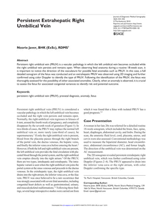
- 1. Journal of Diagnostic Medical Sonography 26(5) 253–256 © The Author(s) 2010 Reprints and permission: http://www. sagepub.com/journalsPermissions.nav DOI: 10.1177/8756479310381130 http://jdm.sagepub.com Persistent Extrahepatic Right Umbilical Vein Noorie Javer, BHK (ExSc), RDMS1 Abstract Persistent right umbilical vein (PRUV) is a vascular pathology in which the left umbilical vein becomes occluded while the right umbilical vein persists and remains open. When observing fetal anatomy during a routine 18-week scan, it is important to notice the direction of the vasculature for possible fetal anomalies such as PRUV. In this case, a full detailed sonogram of the fetus was conducted and an extrahepatic PRUV was observed using 2D imaging and further confirmed using color Doppler to identify the type of PRUV. Following the identification of the PRUV, the fetus was thoroughly assessed for the possibility of other associated anomalies. Clearly, when an anomaly is observed, it is crucial to assess the fetus for associated congenital variances to identify risk and potential outcome. Keywords persistent right umbilical vein (PRUV), prenatal diagnosis, anomaly, fetus Persistent right umbilical vein (PRUV) is considered a vascular pathology in which the left umbilical vein becomes occluded and the right vein persists and remains open. Normally, the right umbilical vein regresses in fetuses of 6 mm, around the fourth week of pregnancy, and completely disappears by the seventh week of gestation (Figure 1). In two-thirds of cases, the PRUV may replace the normal left umbilical vein or, more rarely (one-third of cases), be supernumerary.1 If only the right umbilical vein is present, blood from the placenta passes through the right branch of the portal vein, the ductus venosus, the hepatic veins, and finally the inferior vena cava before entering the heart.2 However,ifboththeleftandrightumbilicalveinsarepresent, the left umbilical vein provides the fetal circulation with pla- centalbloodthroughtheportalsystem,andtherightumbilical vein empties directly into the right atrium.2 Of the PRUV, there are two types, intrahepatic and extrahepatic. The intra- hepatic variant is seen when the right umbilical vein joins the portal system at the sinus venosus and proceeds to the ductus venosus. In the extrahepatic type, the right umbilical vein drains into the right atrium, the inferior vena cava, or the iliac vein. PRUV was once believed to be a rare occurrence that wasstronglyassociatedwithseverefetalanomalies,including congenital heart defects as well as gastrointestinal, urinary, and musculoskeletal malformations.1,2 Following these find- ings,severallargeretrospectivestudieswerecarriedoutfrom which it was found that a fetus with isolated PRUV has a good prognosis.3,4 Case Presentation Awoman in her late 20s was referred for a detailed routine 18-week sonogram, which included the brain, face, spine, heart, diaphragm, abdominal cavity, and limbs. During the scan, the amniotic fluid level, cord, placenta, uterus, and cervix were also assessed. Conventional methodology was used to measure the biparietal diameter, head circumfer- ence, abdominal circumference (AC), and femur length. The direction of the umbilical vein was determined on the AC measurement. The 2D sonogram revealed persistent extrahepatic right umbilical vein, which was further confirmed using color Doppler (Figures 2–4). The PRUV appeared to drain into the inferior vena cava or the right atrium, with color Doppler confirming the specific type. 1 St. Paul’s Hospital, Vancouver, British Columbia, Canada Corresponding Author: Noorie Javer, BHK (ExSc), RDMS, North Shore Medical Imaging, 139 16th St West, North Vancouver, British Columbia, V7M1T3, Canada Email: njaver@gmail.com at NORTH DAKOTA STATE UNIV LIB on June 7, 2015jdm.sagepub.comDownloaded from
- 2. 254 Journal of Diagnostic Medical Sonography 26(5) Sonographic criteria associated with PRUV include the portal vein curved toward the stomach (Figure 2); the fetal gallbladder located medially to the umbilical vein, between the umbilical vein and the stomach; and abnormal connec- tion of the umbilical vein to the right portal vein instead of to the left portal vein. Following the detection of PRUV, the fetus was carefully assessed to exclude associated and potentially more serious congenital malformations, none of which were found. Two, more extensive, follow-up sonograms were also performed in addition to fetal echocardiography at 23 weeks and 28 weeks, respectively, in addition to regular fetal monitoring. Both follow-up scans along with the fetal echocardiog- raphy showed normal growth, fluid, and Doppler with no structural anomalies identified. The heart was situs solitus and appeared structurally normal. Thus, chromosomal testing was not carried out as no indications were found. The PRUV found in the fetus appeared to be an isolated finding, likely a normal variant of no clinical consequence after birth. Discussion At around the fourth week of gestation, the chorionic veins drain into the paired allantoic veins to form the primordial double umbilical vein. Three sets of veins—the common cardiac veins, viteline veins, and umbilical veins—drain into the primitive heart and join at the sinus venosus, which serves to drain the body, yolk sac, and placenta, respectively. During the sixth gestational week, the hepatic bud enlarges, and the right umbilical vein between the liver and the sinus venosus degenerates, leaving only a left umbilical vein to carry all the blood from the placenta to the fetus.The ductus venosus forms in the liver and connects the left umbilical vein and the inferior vena cava. The ductus allows blood to bypass the liver and flow directly from the placenta to the heart. Postnatally, the left umbilical vein becomes the ligamentum teres. PRUV was traditionally thought to be a rare occurrence because of the lack of reports in the literature on this Figure 1. Normal appearance of the fetal abdomen at the level of the left umbilical vein. Figure 2. Fetus with persistent right umbilical vein in which the portal vein curves toward the stomach. Figure 3. Color Doppler used to confirm the type of persistent right umbilical vein according to its drainage. Figure 4. Color Doppler used to confirm the type of persistent right umbilical vein (PRUV) according to its drainage. Arrow, PRUV. at NORTH DAKOTA STATE UNIV LIB on June 7, 2015jdm.sagepub.comDownloaded from
- 3. Javer 255 variant until 1990.1,2,5 Since then, several larger studies have been published, and the results suggest that this anomaly is more common than previously believed.5 In cases involving PRUV, the right umbilical vein is persistently open and may coexist with the left umbilical vein as an intrahepatic supernumerary structure or connect separately to the right portal vein.6 The right umbilical vein may also completely replace the left umbilical vein or bypass the liver to create an aberrant drainage into the infe- rior vena cava or the right atrium, which is known as the extrahepatic type.1,4,6,7 Of the three types of PRUV, the intrahepatic type (type 1) is the most common in the fetus with isolated PRUV.6 With this type, the umbilical vein passes lateral to the right side of the gallbladder and fuses with the right portal vein, then curves toward the stomach. Once passing through the ductus venosus, the umbilical vein connects with the hepatic vein and drains into the inferior vena cava. As there is little interference in hemodynamics, this type of PRUV has a good prognosis as it is estimated that only 20% to 30% of the blood of the umbilical vein enters the ductus venosus and reaches the heart.6 In the second type, the umbilical vein connects to the iliac veins or caput medusa directly without the ductus venous. Last, in the third type, the umbilical vein connects directly to the right atrium or infracardiac portion of the inferior vena cava in the absence of the ductus venosus.1,7 As opposed to type 1, the other two types of PRUV are extrahepatic and have a higher frequency of associated anomalies and greater hemodynamic effects due to the absence of the ductus venosus.6,7 There are case reports of the extrahepatic type of PRUV without the ductus venosus in which these fetuses had severe hemodynamic stresses that resulted in hydrops fetalis.1,8 The precise causes of failure of normal regression of the right umbilical vein are unknown; however, several poten- tial etiologies have been suggested. In the rat model, specific teratogens such as retinoic acid and first-trimester folic acid deficiency may result in persistence of the right umbilical vein.1,3,4,6 Obstruction of the left umbilical artery by throm- bus, embolus, or external pressure early in the pregnancy might also cause the right umbilical vein to remain patent to maintain placental blood supply to the fetus.3,6,9 The prevalence of PRUV was not well established until more recently, and now the incidence and significance have become the focus to put aside the controversy on the occurrence of this vascular pathology. Jeanty1 gathered only a dozen reported cases in the literature and added six new cases.2,3,5 Associated anomalies were found in all pub- lished cases and in three of Jeanty’s own six patients, which suggested that PRUV might be an ominous prenatal find- ing.1 Results from several retrospective studies demonstrate the incidence of PRUV to be more frequent than previously reported. Blazer et al.3 found the incidence to be 1:438, which was similar to ratios of 1:476 reported by Hill et al.,2 1:450 detailed by Shen et al.,5 and 1:526 reported by Wolman et al.4 However, these were significantly lower than the rate of 1:217 recently reported by Yang et al.,6 who detected six fetuses with PRUV among 1302 study subjects. Yang et al.6 attributed the higher incidence of PRUV to the fact that their obstetric population included both private service and women transferred to their depart- ment in the third trimester of pregnancy. The true incidence of PRUV may be much higher, con- sidering the false-negative rate of sonographic detection. Through several studies, it has been found that the intrahe- patic form of PRUV might present as an isolated finding, with no other anomalies, whereas those cases demonstrating the extrahepatic type were associated with fetal abnormali- ties.3,7 PRUV with no intrahepatic portion is an uncommon finding, with a review of the literature revealing only five other cases.7 There is an association with congenital cardiac malformation, and the presence of PRUV can sometimes be the only clue to alert sonographers and clinicians to the presence of these malformations.10 Other associated condi- tions include abnormal systemic venous connections11 and Noonan syndrome.7 Conclusion In agreement with Jeanty,1 PRUV might not be as rare as was previously believed, and proper alertness might uncover its true prevalence. Of all imaging modalities, sonography is the most likely to facilitate detection of the anomaly because the section used to demonstrate PRUV is routinely used in all obstetrical scanning. In addition, sonography can depict the anomaly at a stage when it may be of clinical significance, and color Doppler can be applied to confirm if the direction of flow is toward the stomach. If such an anomaly is identified, an extensive targeted sonographic examination should be carried out. Depending on the significance of the abnormalities detected via sonog- raphy, a chromosomal study may be warranted and offered to the patient. This case in particular proves to be interest- ing as the anomaly detected was an extraheptic PRUV, which is commonly associated with more severe congenital malformations. On the basis of three extensive sonograms coupled with echocardiography, the fetus appeared normal with no associated anomalies in utero and was likely to have no clinical consequence after birth. Declaration of Conflicting Interests The author(s) declared no potential conflicts of interest with respect to the authorship and/or publication of this article. at NORTH DAKOTA STATE UNIV LIB on June 7, 2015jdm.sagepub.comDownloaded from
- 4. 256 Journal of Diagnostic Medical Sonography 26(5) Funding The author(s) received no financial support for the research and/ or authorship of this article. References 1. Jeanty P: Persistent right umbilical vein: an ominous prena- tal finding? Radiology 1990;177:735–738. 2. Hill AD, Mills A, Peterson C, Boyles D: Persistent right umbilical vein: sonographic detection and subsequent neo- natal outcome. Obstet Gynecol 1994;84:923–925. 3. Blazer S, Zimmer EZ, Bronshtein M: Persistent intrahepatic right umbilical vein in the fetus: a benign anatomic variant. Obstet Gynecol 2000;95:433–436. 4. Wolman I, Gull I, Fait G, Amster R, Kupferminc MJ, Lessing JB, Jaffa AJ: Persistent right umbilical vein: incidence and significance. Ultrasound Obstet Gynecol 2002;19:562–564. 5. Shen O, Tadmor OP, Yagel S: Prenatal diagnosis of per- sistent right umbilical vein. Ultrasound Obstet Gynecol 1996;8:31–33. 6. Yang PY, Wu JL, Yeh GP, Chou PH, Jsu CH, Hsieh CTC: Prenatal diagnosis of persistent right umbilical vein using three-dimensional sonography with power Doppler. Taiwan- ese J Obstet Gynecol 2007;46:43–46. 7. Bradley E, Kean L, Twining P, James D: Persistent right umbilical vein in a fetus with Noonan’s syndrome: a case report. Ultrasound Obstet Gynecol 2001;17:76–78. 8. Achiron R, Hegesh J, Yagel S, Lipitz S, Cohen SB, Rotstein Z: Abnormalities of the fetal central veins and umbilico-portal system: prenatal ultrasonographic diagno- sis and proposed classification. Ultrasound Obstet Gynecol 2000;16:539–548. 9. Monie IW: Umbilical vein entering the right atrium: com- ments on previously reported human case. Teratology 1971;4: 461–463. 10. Hoehn T, Lueder M, Schmidt KG, Schaper J, Mayatepek E: Persistent right umbilical vein associated with complex con- genital cardiac malformation. Am J Perinatol 2006;23:181–182. 11. Nakstad B, Smevik B: Abnormal systemic venous connec- tion possibly associated with a persistent right umbilical vein: a case report. BMC Pediatrics 2004;4:7. at NORTH DAKOTA STATE UNIV LIB on June 7, 2015jdm.sagepub.comDownloaded from