SlideShare a Scribd company logo
1 of 5
Download to read offline
Effect of displaced versus non-displaced pelvic fractures on
long-term racing performance in 31 Thoroughbred racehorses
SE Hennessy,a
* MA Muurlink,a,b
GA Anderson,c
TN Puksmanna
and RC Whittona
Objective To evaluate the long-term racing prognosis for
Thoroughbred racehorses with displaced versus non-displaced
fractures of the pelvis identified by scintigraphy.
Design Retrospective case analysis.
Methods Medical records of 31 Thoroughbred racehorses pre-
senting to the University of Melbourne Equine Centre with fractures
of the pelvis that were identified by scintigraphy were reviewed.
Pelvic fracture site was determined and defined as displaced or
non-displaced based on ultrasound and/or radiographic findings.
Race records were analysed for each horse, with a minimum of
24 months’ follow-up, and correlated with fracture type to deter-
mine long-term prognosis for racing. Results are expressed as
median and range.
Results Fractures at a single site were more common (n = 22)
than fractures involving two sites (n = 9) and the ilial wing was the
most commonly affected (n = 12). Thoroughbred racehorses with
displaced pelvic fractures at any site (n = 12) raced fewer times
within 24 months of diagnosis than horses with non-displaced frac-
tures (n = 19) (median 0.5, range 0–13 vs 7, 0–24; P = 0.037), but
there was no clear statistical difference in race earnings between
the two groups (median A$0, range A$0–$123,250 vs A$14,440,
A$0–$325,500, respectively; P = 0.080). Four horses with displaced
fractures (33%) were euthanased on humane grounds because
of persistent severe pain. When these horses were excluded from
the analysis, there were no differences in performance variables
between horses with a displaced or non-displaced pelvic fracture.
Conclusion Thoroughbred racehorses with a displaced or non-
displaced pelvic fracture that survive the initial post-injury period
have a good prognosis for racing.
Keywords fractures; pelvis; racehorses; Thoroughbreds
Abbreviations CI, confidence interval; HR, hazard ratio
Aust Vet J 2013;91:246–250 doi: 10.1111/avj.12053
P
elvic fractures that are the result of direct trauma occur in
all types of horses, whereas those that are a fatigue injury are
primarily a problem of racehorses. Direct trauma can lead to
fractures anywhere in the pelvis,1
but the wing of the ilium is predis-
posed to fatigue fractures, which often originate on its caudal aspect
and extend over the sacroiliac joint craniodorsally or craniolaterally.2
Cyclic loading during locomotion results in bone fatigue at sites of
high load, which may progress from microcracking to incomplete,
non-displaced fractures and finally to complete displaced fractures.3
With the widespread availability of scintigraphy and ultrasound,
incomplete fatigue fractures of the ilium are being increasingly rec-
ognised.In immature racing Thoroughbreds they are a common cause
of hindlimb lameness and poor performance.4–6
The prognosis for
return to function of horses with ilial fractures is regarded as good,
provided there is no sacroiliac or coxofemoral joint involvement;
however, detailed data on prognosis and factors affecting it are
limited.6–8
In this study,we assessed the differences in the return to function,time
to return, race earnings, and number of races and places between
horses with displaced or non-displaced pelvic fractures using a popu-
lation of racing Thoroughbred horses presented to the University of
Melbourne Equine Centre. We hypothesised that horses with non-
displaced fractures of the pelvis would return to work sooner and race
more successfully than horses with displaced pelvic fractures.
Materials and methods
We included 31 Thoroughbred racehorses that had been presented
to the University of Melbourne Equine Centre between 2 November
2003 and 20 April 2010 and that had scintigraphic evidence of a pelvic
fracture.A pelvic fracture was defined as a clear change in the normal
pelvic contour on scintigraphic images or focal, moderate to intense
radiopharmaceutical uptake within the pelvis that was separate or
extended from the normal areas of high uptake at the tuber sacrale,
tuber coxae or tuber ischii.
All horses were in full-time race training when the injury occurred.
Medical records, including nuclear scintigraphy, ultrasonography and
radiology reports, were reviewed.
Physical and lameness examinations (including grading on the lame-
ness scale of the American Association of Equine Practitioners) were
performed.9
Where there was scintigraphic evidence of a fracture,
ultrasonography (Siemens Acuson Sequioa 512, Siemens Plc USA) of
the pelvis was performed percutaneously using a 3.5-MHz curved
array transducer to determine if the fracture was displaced. Radio-
graphy was only used to determine coxofemoral joint involvement
(standing and recumbent ventrodorsal views, 150 kV, 250–300 mA).
Fractures were classed as displaced or non-displaced based on ultra-
sound and/or radiographic findings. Treatment protocols and rest
periods were clinician-dependent, but in all cases the horse was rested
for at least 3 months.
*Corresponding author: University of Liverpool, Philip Leverhulme Equine Hospital,
Leahurst, Chester High Road, Cheshire, CH64 7TE, UK; simonhennessy@hotmail.com
a
University of Melbourne, Equine Centre, Werribee, Victoria, Australia
b
University of Queensland, Equine Hospital, Gatton Campus, Gatton, Queensland,
Australia
c
University of Melbourne, Faculty of Veterinary Science, Werribee, Victoria, Australia
bs_bs_banner
EQUINE
EQUINE
© 2013 The Authors
Australian Veterinary Journal © 2013 Australian Veterinary AssociationAustralian Veterinary Journal Volume 91, No 6, June 2013246
A standard scintigraphic protocol was used for each horse. An
18-gauge catheter was placed in the left jugular vein and 12.7–15
MBq/kg of 99
Tc-HDP administered (standard 7 GBq/14 MBq/kg dose
for an average Thoroughbred racehorse). Frusemide (0.7 mg/kg IV)
was injected 60 min prior to scanning the pelvis. Dynamic (35 ¥ 2-s
frames), delayed-phase standing lateral, dorsal, caudal and oblique
pelvis images were acquired 3 h after radiopharmaceutical injection
with a large field-of-view gamma camera (Phillips Argus Epic) using
a 128 ¥ 128 matrix. Data were transferred to a workstation (Sun
Ultra Workstation) and processed with motion correction software
(University College London, Institute of Nuclear Medicine).
Race and stud book records were accessed in April 2012. Only horses
with at least 24 months’ follow-up were included. Data obtained
included sex, age, prize money, races and places achieved before and
after diagnosis.
Statistical analysis
Fisher’s exact test was used to compare proportions. Preliminary
analysis of the residuals of the post-diagnosis race performance vari-
ables showed that almost all were not normally distributed (Shapiro-
Wilk’s test). The Mann-Whitney U-test with exact P values was used
to compare the mean ranks of post-diagnosis race performance vari-
ables between horses with displaced or non-displaced fractures. The
log-rank test and the hazard ratio (HR) from a Cox proportional
hazard model were used to compare the distribution of time to first
race post-diagnosis for displaced and non-displaced fractures,
together with the Kaplan-Meier estimate of median time to first race
for all horses.10
Two-tailed P < 0.05 was considered to be statistically
significant. The statistical software used was Stata 12.1 for Windows
(StataCorp, College Station, TX, USA) and IBM SPSS 20 (IBM Corp.,
New York, NY, USA) was used for Mann-Whitney exact P-values.
Results
All 31 horses included in the study (mean age 3.3 years, range
1–6 years; 14 females (45%), 13 geldings (42%), 4 entire males (13%))
had been referred for scintigraphic examination. Of them, two horses
presented with a history of trauma and both had displaced ischial
fractures,six horses presented with a history of poor performance and
all had non-displaced fractures of the ilial wing,eight horses presented
for lameness immediately following a race or a training gallop, seven
horses were referred for mild to moderate hindlimb lameness that
could not be localised with distal limb diagnostic analgesia by the
attending veterinarian and eight horses presented with severe lame-
ness (grade 3/5 or more) without associated trauma,of which four had
displaced fractures.
All horses had scintigraphic examination of their hindlimbs,pelvis and
thoracolumbar spine.Areas of increased radiopharmaceutical uptake,
not associated with the pelvis, included the dorsal spinous processes
of the thoracic and lumbar vertebrae, the tarsometatarsal joints, the
tibial diaphysis, the lateral proximal hind sesamoid bone, the fourth
metatarsal bone and the lateral condyle of the third metatarsal bone.
There were 22 fractures involving a single site within the pelvis and
9 fractures involving two sites. The most common site of fracture was
the ilial wing, followed by the ischium (Table 1); 19 fractures were
non-displaced and 12 were displaced (Figures 1, 2). Of those with
displaced fractures, four horses (33%) were euthanased at 2, 2, 3 and
18 days after diagnosis on humane grounds because of persistent,
severe pain.Of the four horses,two were presented 6–8 weeks after the
initial injury, one with fractures of the acetabulum and the ilial wing,
and the other with fractures of the acetabulum and ilial shaft. Both
of them had severe gluteal muscle atrophy and grade 4/5 lameness that
was noted to worsen with box rest. The third horse had bilateral ilial
wing fractures with cranial displacement of the pelvis and associated
muscle atrophy and severe lameness. The fourth horse presented
acutely after fast work with a severe comminuted oblique fracture of
the ilial wing and shaft with grade 5/5 lameness. Among the 27 survi-
vors, 12 horses were rested for 3 months, 12 for 6 months and 3 were
rested for unspecified periods longer than 3 months.
With respect to racing, five horses that were in full race training at the
time of injury did not race before or after diagnosis, four horses raced
Table 1. Pelvic fracture distribution and outcome in 31 Thoroughbred racehorses
Fracture site n Fracture type Median no. of races No. euthanased
Displaced Non-displaced Pre-diagnosis Post-diagnosis
Single site
Ilial wing 12 2 10 7.5 13.0 0
Ilial shaft 2 0 2 0.5 5.0 0
Acetabulum 1 0 1 0.0 0.0 0
Ischium 7 5 2 0.0 18.0 0
Multiple sites
Bilateral ilial wings 3 1 2 7.0 2.0 1
Ilial wing and shaft 3 1 2 1.0 2.0 1
Acetabulum and ischium 1 1 0 0.0 0.0 0
Acetabulum and ilial wing 1 1 0 0.0 0.0 1
Acetabulum and ilial shaft 1 1 0 8.0 0.0 1
EQUINE
EQUINE
© 2013 The Authors
Australian Veterinary Journal © 2013 Australian Veterinary Association Australian Veterinary Journal Volume 91, No 6, June 2013 247
before diagnosis but not after, 9 horses raced after diagnosis but not
before, and 13 horses raced before and after diagnosis. The median
time to first race based on all horses was 9.9 months (95% confidence
interval (CI) 7.7–12.3).
The majority of horses with non-displaced pelvic fractures (16/19)
raced within 24 months of diagnosis compared with only half of the
horses with displaced pelvic fractures (6/12) (Table 2). Horses with a
displaced pelvic fracture at any site (n = 12) raced fewer times within
24 months of diagnosis than horses with non-displaced fractures
(n = 19) (median 0.5, range 0–13 vs 7, 0–24; P = 0.037), but there was
no clear difference in race earnings between the two groups (median
A$0, range A$0–$123,250 vs A$14,440, A$0–$325,500, respectively;
P = 0.080) (Table 2).When the four horses that were euthanased were
excluded from the analysis, there were no differences in the perfor-
mance variables between horses with a displaced or non-displaced
Figure 1. Left oblique (top) and dorsal scintigraphy images of the pelvis
of a horse with an incomplete ilial stress fracture. Note the increased
radiopharmaceutical uptake lateral to the left tuber sacrale on the caudal
surface of the ilium (black arrows). Ultrasonographic examination
revealed no displacement of fracture fragments.
Figure 2. Right oblique (top) and dorsal (middle) scintigraphy images
of the pelvis of a horse with a complete fracture of the right ilium (black
arrows). Ultrasound examination (bottom) revealed marked displace-
ment of fracture fragments.
EQUINE
EQUINE
© 2013 The Authors
Australian Veterinary Journal © 2013 Australian Veterinary AssociationAustralian Veterinary Journal Volume 91, No 6, June 2013248
fracture (Table 3). The median time to first race for horses with
displaced pelvic fractures was 10.4 months (95% CI 6.8–16.7) and
9.3 months (95% CI 6.9–11.5) for horses with a non-displaced pelvic
fracture (log-rank test P = 0.50).The HR did not differ from 1 for time
to first race for horses with a displaced compared with a non-displaced
fracture (HR 0.73, 95% CI 0.28–1.86; P = 0.51).
Of the 4 horses presenting with fractures involving the acetabulum,
none raced post diagnosis (2 were euthanased), whereas 6 of 7 horses
with ischial fractures raced post diagnosis and 11 of 12 horses with
unilateral ilial wing fractures raced post diagnosis, including both of
the horses with displaced fractures. One of two horses that presented
with unilateral fractures of the ilial shaft raced post diagnosis. Of the
6 horses with fractures in multiple sites and no acetabular involve-
ment, 4 raced post diagnosis (2 were euthanased).
Discussion
Our study results demonstrated that Thoroughbred racehorses with
displaced or non-displaced pelvic fractures that survive the initial
post-injury period have a good prognosis for racing. We found no
evidence that horses with non-displaced pelvic fractures returned to
racing sooner than those with displaced fractures. Pelvic fractures in
Thoroughbred racehorses have a variety of presentations and should
be considered in the differential diagnosis of poor performance and
hindlimb lameness.
Our findings are consistent with those of other studies of pelvic
fractures, which have demonstrated that 61–75% of horses without
acetabular and ilial shaft involvement return to racing after diagno-
sis.8,11,12
Less success has been reported for Thoroughbred racehorses
with fractures of the ilial wing identified by scintigraphy,with 6 of 10,7
and 1 of 413
returning to racing in two separate studies. Studies that
have previously reported a good prognosis for displaced traumatic
pelvic fracture relate to successful salvage of animals for potential
breeding.11,14
A comparison of displaced and non-displaced pelvic
fractures, and the prognosis for return to racing, has not been previ-
ously examined. The importance of determining the site of pelvic
fracture is highlighted in this study. Similar to other studies, fractures
involving the acetabulum had a poor prognosis for return to racing,
whereas both ilial and ischial fractures had a good prognosis.8
A number of factors have the potential to affect future race perform-
ance in horses with displaced fractures. In the case of ilial fractures,
these include changes in the sacroiliac joint and subsequent sacroiliac
disease.Many displaced fractures are associated with the development
of sacroiliac osteophytes and degenerative changes in the ilial articu-
lar surface.2
Following a displaced fracture, the fracture fragments
re-attach in a new anatomical arrangement that can influence the
time required for return to function as the horse adapts to the new
anatomy.15
Also, the greater pain resulting from a displaced fracture
will often result in more muscle atrophy,which can delay the return to
racing.1,6
The effect of the degree of muscle atrophy on the time to
return to racing was not examined in this study. Based on our find-
ings, these factors are less important and uncontrollable pain or
acetabular involvement are the primary factors affecting long-term
outcome.
Scintigraphy is commonly used to identify pelvic fractures, but it
is rarely possible to determine if a fracture is displaced using this
Table 2. Post-diagnosis performance for displaced and non-displaced fractures of 31 Thoroughbred racehorses
Outcome Fracture type P value
Non-displaced
(n = 19)
Displaced
(n = 12)
Raced in 24 months post-diagnosis 16 (84%) 6 (50%) 0.056
Median no. races in 24 months post-diagnosis (mean; range) 7 (7.5; 0–24) 0.5 (3; 0–13) 0.037
Median no. places in 24 months post-diagnosis (mean; range) 2 (2.9; 0–8) 0 (1.6; 0–8) 0.13
Median prize money in 24 months post-diagnosis (A$: mean; range) 14,440 (50,783; 0–325,500) 0 (16,129; 0–123,250) 0.080
Table 3. Post-diagnosis performance for displaced and non-displaced fractures of Thoroughbred racehorses after the exclusion of four horses
euthanased on humane grounds
Outcome Fracture type P value
Non-displaced
(n = 19)
Displaced
(n = 8)
Raced in 24 months post-diagnosis 16 (84%) 6 (75%) 0.62
Median no. races in 24 months post-diagnosis (mean; range) 7 (7.5; 0–24) 3.5 (4.5; 0–13) 0.34
Median no. places in 24 months post-diagnosis (mean; range) 2 (2.9; 0–8) 0.5 (2.4; 0–8) 0.61
Median prize money in 24 months post-diagnosis (A$: mean; range) 14,440 (50,783; 0–325,500) 575 (24,194; 0–123,250) 0.41
EQUINE
EQUINE
© 2013 The Authors
Australian Veterinary Journal © 2013 Australian Veterinary Association Australian Veterinary Journal Volume 91, No 6, June 2013 249
technique.16
Fracture displacement or callus formation can be
observed using ultrasound, in the majority of cases, whereas non-
displaced fractures are difficult to identify.17–19
Therefore, the combi-
nation of scintigraphy and ultrasound enabled diagnosis in most
cases of pelvic fracture in this study. Standing radiography of the
pelvis was used in this study to determine coxofemoral joint involve-
ment in some cases, because of the difficulty of assessing acetabular
involvement using ultrasound alone.17
The overlying sacrum and
intestinal contents do not allow a detailed view of the whole ilium on
radiographs.14,17,20,21
In the present study, it was often not possible to discriminate
between traumatic fractures and displaced fatigue fractures because
in only a minority of cases was there a history of trauma or an acute
onset of lameness following fast work. However, it is likely that many
of the cases were fatigue fractures, as it has been reported that
ilial fractures in a British Thoroughbred flat racehorse population
accounted for 15.5% of the total fractures seen in the population,
with fatigue fractures accounting for 87% of the ilial fractures.22
In
the present study, 50% (3/6) of displaced ilial fractures occurred
during training of young horses that had never raced. Similarly, it has
been reported that in humans the majority of tibial and femoral
stress fractures occur early during military training, suggesting that
bone adaptation is protective.23
As found in our study, ilial fractures
are most commonly observed in young, immature Thoroughbred
racehorses aged up to 4 years of age.6,12,15,22
Others have described ilial
wing fractures in older horses;1,2,5
however, two of these were post-
mortem studies from which it was difficult to determine the time of
fracture.2,5
Study limitations
We had a low number of cases, resulting in a lack of power, and the
study population included four horses that were euthanased after
diagnosis, making it impossible to know whether these horses could
have returned to racing if the problem had been appropriately
managed. However, all four horses were euthanased because of per-
sistent, severe pain rather than for economic reasons. Also, not all
horses in this study had full-body scintigraphy, so although it is pos-
sible that other problems affecting long-term outcome may have been
overlooked, the majority were able to perform well.
In conclusion, the poorer prognosis for Thoroughbred racehorses
with displaced pelvic fractures compared with non-displaced pelvic
fractures appears to be because of an inability to manage severe pain
in some affected horses and/or involvement of the acetabulum.
Accurate localisation of the fracture with a combination of scinti-
graphy and ultrasound will allow an appropriate prognosis to be
given.
References
1. Dyson SJ. Lameness associated with the stifle and pelvic regions. Am Assoc
Equine Pract 2002;48:387–411.
2. Haussler KK, Stover SM. Stress fractures of the vertebral lamina and pelvis in
Thoroughbred racehorses. Equine Vet J 1998;30:374–381.
3. Riggs CM. Fractures: a preventable hazard of racing Thoroughbreds? Vet J
2002;163:19–29.
4. Bathe AP. 245 fractures in Thoroughbred racehorses: results of a 2-year pro-
spective study in Newmarket. Am Assoc Equine Pract 1994;40:175–176.
5. Carrier TK, Estberg L, Stover SM et al. Association between long periods
without high–speed workouts and risk of complete humeral or pelvic fracture in
Thoroughbred racehorses: 54 cases (1991–1994). J Am Vet Med Assoc 1998;212:
1582–1587.
6. Van Wessum R. The pelvis. In: Baxter GM, editor. Adam’s and Stashak’s lameness
in horses. 6th edn. Wiley Blackwell, Oxford, 2011:840–847.
7. Pilsworth RC, Shepherd MC, Herinckx BMB et al. Fractures of the wing of the
ilium, adjacent to the sacroiliac joint in Thoroughbred racehorses. Equine Vet J
1994;26:94–99.
8. Fuller A, Beever E, Fraser B et al. A retrospective study of 80 cases of pelvic
fracture diagnosed by gamma scintigraphy in Thoroughbred racehorses. Br
Equine Vet Assoc 2008;47:142–143.
9. Ross MW. Movement. In: Ross MW, Dyson SJ. Diagnosis and management of
lameness in the horse. 1st edn. Saunders, St Louis, 2002:60–73.
10. Hosmer D, Lemeshow S. Applied survival analysis: regression modeling of time
to event data. 2nd edn. John Wiley & Sons, New York, 2008.
11. Rutkowski JA, Richardson DW. A retrospective study of 100 pelvic fractures in
horses. Equine Vet J 1989;29:256–259.
12. Shepherd MC, Pilsworth RC, Hopes R et al. Clinical signs, diagnosis, manage-
ment and outcome of complete and incomplete fracture to the ilium. Am Assoc
Equine Pract 1994;40:177–180.
13. Davenport-Goodall CLM, Ross MW. Scintigraphic abnormalities of the pelvic
region in horses examined because of lameness or poor performance: 128 cases
(1993–2000). J Am Vet Med Assoc 2004;224:88–95.
14. Little C, Hilbert B. Pelvic fractures in horses: 19 cases (1974–1984). J Am Vet
Med Assoc 1987;190:1203–1206.
15. Pilsworth RC. Diagnosis and management of pelvic fractures in theThorough-
bred racehorse. In: Ross MW, Dyson SJ, editors. Diagnosis and management of
lameness in the horse. 2nd edn. Elsevier Saunders, St Louis, 2011:564–571.
16. Hornof WJ, Stover SM, Koblik PD et al. Oblique views of the ilium and the
scintigraphic appearance of stress fractures of the ilium. Equine Vet J 1996;28:
355–358.
17. Geburek F, Rotting AK, Stadler PM. Comparison of the diagnostic value
of ultrasonography and standing radiography for pelvic-femoral disorders in
horses. Vet Surg 2009;38:310–317.
18. Driver AJ, Nagy A. Fracture of the ischium in an eight-year-old Arabian
gelding: a diagnostic challenge. Equine Vet Educ 2008;20:127–130.
19. Tomlinson JE, Sage AM, Turner TA et al. Detailed ultrasonographic mapping of
the pelvis in clinically normal horses and ponies. Am J Vet Res 2001;62:1768–1775.
20. May SA, Patterson LJ, Peacock PJ et al. Radiographic technique for the pelvis in
the standing horse. Equine Vet J 1991;23:312–314.
21. Barrett EL, Talbot AM, Driver AJ et al. A technique for radiography in the
standing horse. Equine Vet J 2006;38:266–270.
22. Verheyen KLP, Wood JLN. Descriptive epidemiology of fractures occurring in
British Thoroughbred racehorses in training. Equine Vet J 2004;36:167–173.
23. Finestone A, Milgrom C, Wolf O et al. Epidemiology of metatarsal stress frac-
tures versus tibial and femoral stress fractures during elite training. Foot Ankle Int
2011;32:16–20.
(Accepted for publication 24 September 2012)
EQUINE
EQUINE
© 2013 The Authors
Australian Veterinary Journal © 2013 Australian Veterinary AssociationAustralian Veterinary Journal Volume 91, No 6, June 2013250

More Related Content

What's hot

DAA-THA: No Technology Needed
DAA-THA: No Technology NeededDAA-THA: No Technology Needed
DAA-THA: No Technology Neededwashingtonortho
 
DAA- Does it Make a Difference?
DAA- Does it Make a Difference? DAA- Does it Make a Difference?
DAA- Does it Make a Difference? washingtonortho
 
4a5a4e78 05c2-48ff-a17e-6952a767c6b7
4a5a4e78 05c2-48ff-a17e-6952a767c6b74a5a4e78 05c2-48ff-a17e-6952a767c6b7
4a5a4e78 05c2-48ff-a17e-6952a767c6b7ssuser6863bd
 
Spine Lecture recurrent metastatic lesion spine recurrence 2019 april
Spine Lecture recurrent metastatic lesion spine recurrence 2019 aprilSpine Lecture recurrent metastatic lesion spine recurrence 2019 april
Spine Lecture recurrent metastatic lesion spine recurrence 2019 aprilSpiro Antoniades
 
Megaprosthetic replacement of knee in a young boy of 14 years
Megaprosthetic replacement of knee in a young boy of 14 yearsMegaprosthetic replacement of knee in a young boy of 14 years
Megaprosthetic replacement of knee in a young boy of 14 yearsApollo Hospitals
 
ARM TRUNK COORDINATION IN VRT POSTER
ARM TRUNK COORDINATION IN VRT POSTERARM TRUNK COORDINATION IN VRT POSTER
ARM TRUNK COORDINATION IN VRT POSTERClémence Ongolo Zogo
 
Union Rate of Tibiotalocalcaneal Nail with Internal or External Bone Stimulat...
Union Rate of Tibiotalocalcaneal Nail with Internal or External Bone Stimulat...Union Rate of Tibiotalocalcaneal Nail with Internal or External Bone Stimulat...
Union Rate of Tibiotalocalcaneal Nail with Internal or External Bone Stimulat...skisnfeet
 
Some Mechanisms of the Noncontact Anterior Cruciate Ligament (ACL) Injury amo...
Some Mechanisms of the Noncontact Anterior Cruciate Ligament (ACL) Injury amo...Some Mechanisms of the Noncontact Anterior Cruciate Ligament (ACL) Injury amo...
Some Mechanisms of the Noncontact Anterior Cruciate Ligament (ACL) Injury amo...Crimsonpublishers-Rehabilitation
 
Dr. Xavier Casanova Canals. Upper Extremity Orthopaedic and Trauma surgeon in...
Dr. Xavier Casanova Canals. Upper Extremity Orthopaedic and Trauma surgeon in...Dr. Xavier Casanova Canals. Upper Extremity Orthopaedic and Trauma surgeon in...
Dr. Xavier Casanova Canals. Upper Extremity Orthopaedic and Trauma surgeon in...MuscleTech Network
 
A comparative study on the clinical and functional outcome of limb salvage su...
A comparative study on the clinical and functional outcome of limb salvage su...A comparative study on the clinical and functional outcome of limb salvage su...
A comparative study on the clinical and functional outcome of limb salvage su...NAAR Journal
 
Comparison of 3 d shoulder complex kinematic part 1
Comparison of 3 d shoulder complex kinematic part 1Comparison of 3 d shoulder complex kinematic part 1
Comparison of 3 d shoulder complex kinematic part 1Satoshi Kajiyama
 
Why the Direct Anterior Approach for THA
Why the Direct Anterior Approach for THAWhy the Direct Anterior Approach for THA
Why the Direct Anterior Approach for THAwashingtonortho
 

What's hot (20)

Cementless TKA
Cementless TKACementless TKA
Cementless TKA
 
DAA-THA: No Technology Needed
DAA-THA: No Technology NeededDAA-THA: No Technology Needed
DAA-THA: No Technology Needed
 
DAA- Does it Make a Difference?
DAA- Does it Make a Difference? DAA- Does it Make a Difference?
DAA- Does it Make a Difference?
 
Oite 9.14.2010
Oite 9.14.2010Oite 9.14.2010
Oite 9.14.2010
 
4a5a4e78 05c2-48ff-a17e-6952a767c6b7
4a5a4e78 05c2-48ff-a17e-6952a767c6b74a5a4e78 05c2-48ff-a17e-6952a767c6b7
4a5a4e78 05c2-48ff-a17e-6952a767c6b7
 
Spine Lecture recurrent metastatic lesion spine recurrence 2019 april
Spine Lecture recurrent metastatic lesion spine recurrence 2019 aprilSpine Lecture recurrent metastatic lesion spine recurrence 2019 april
Spine Lecture recurrent metastatic lesion spine recurrence 2019 april
 
RPD Poster_22.06
RPD Poster_22.06RPD Poster_22.06
RPD Poster_22.06
 
Megaprosthetic replacement of knee in a young boy of 14 years
Megaprosthetic replacement of knee in a young boy of 14 yearsMegaprosthetic replacement of knee in a young boy of 14 years
Megaprosthetic replacement of knee in a young boy of 14 years
 
Zimmer studies powerpoint
Zimmer studies powerpointZimmer studies powerpoint
Zimmer studies powerpoint
 
ARM TRUNK COORDINATION IN VRT POSTER
ARM TRUNK COORDINATION IN VRT POSTERARM TRUNK COORDINATION IN VRT POSTER
ARM TRUNK COORDINATION IN VRT POSTER
 
Union Rate of Tibiotalocalcaneal Nail with Internal or External Bone Stimulat...
Union Rate of Tibiotalocalcaneal Nail with Internal or External Bone Stimulat...Union Rate of Tibiotalocalcaneal Nail with Internal or External Bone Stimulat...
Union Rate of Tibiotalocalcaneal Nail with Internal or External Bone Stimulat...
 
Some Mechanisms of the Noncontact Anterior Cruciate Ligament (ACL) Injury amo...
Some Mechanisms of the Noncontact Anterior Cruciate Ligament (ACL) Injury amo...Some Mechanisms of the Noncontact Anterior Cruciate Ligament (ACL) Injury amo...
Some Mechanisms of the Noncontact Anterior Cruciate Ligament (ACL) Injury amo...
 
Dr. Xavier Casanova Canals. Upper Extremity Orthopaedic and Trauma surgeon in...
Dr. Xavier Casanova Canals. Upper Extremity Orthopaedic and Trauma surgeon in...Dr. Xavier Casanova Canals. Upper Extremity Orthopaedic and Trauma surgeon in...
Dr. Xavier Casanova Canals. Upper Extremity Orthopaedic and Trauma surgeon in...
 
Extra-cellular Matrix Patches
Extra-cellular Matrix PatchesExtra-cellular Matrix Patches
Extra-cellular Matrix Patches
 
A comparative study on the clinical and functional outcome of limb salvage su...
A comparative study on the clinical and functional outcome of limb salvage su...A comparative study on the clinical and functional outcome of limb salvage su...
A comparative study on the clinical and functional outcome of limb salvage su...
 
Comparison of 3 d shoulder complex kinematic part 1
Comparison of 3 d shoulder complex kinematic part 1Comparison of 3 d shoulder complex kinematic part 1
Comparison of 3 d shoulder complex kinematic part 1
 
Elbow Tendinopathy
Elbow TendinopathyElbow Tendinopathy
Elbow Tendinopathy
 
Butler's procedure for congenital varus 5th toe - البروفيسور فريح ابوحسان – ...
 Butler's procedure for congenital varus 5th toe - البروفيسور فريح ابوحسان – ... Butler's procedure for congenital varus 5th toe - البروفيسور فريح ابوحسان – ...
Butler's procedure for congenital varus 5th toe - البروفيسور فريح ابوحسان – ...
 
eidelman2016.pdf
eidelman2016.pdfeidelman2016.pdf
eidelman2016.pdf
 
Why the Direct Anterior Approach for THA
Why the Direct Anterior Approach for THAWhy the Direct Anterior Approach for THA
Why the Direct Anterior Approach for THA
 

Viewers also liked

Worksheet
WorksheetWorksheet
Worksheetkkk MK
 
HistóRia Da Vida Na Terra Ediacara
HistóRia Da Vida Na Terra   EdiacaraHistóRia Da Vida Na Terra   Ediacara
HistóRia Da Vida Na Terra EdiacaraNuno Correia
 
cara mengInstall eclipse
cara mengInstall eclipsecara mengInstall eclipse
cara mengInstall eclipseDani Juniawan
 
9 ano método ciêntífico
9 ano método ciêntífico9 ano método ciêntífico
9 ano método ciêntíficoJane Jeiza
 
The Programmatic Native Playbook, Didigay Programmatic Summit, May 24th, 2016
The Programmatic Native Playbook, Didigay Programmatic Summit, May 24th, 2016The Programmatic Native Playbook, Didigay Programmatic Summit, May 24th, 2016
The Programmatic Native Playbook, Didigay Programmatic Summit, May 24th, 2016Digiday
 
10 Reasons Why We Visualise Data
10 Reasons Why We Visualise Data10 Reasons Why We Visualise Data
10 Reasons Why We Visualise DataPulsar Platform
 
responsabilidad ante la sociedad
responsabilidad ante la sociedadresponsabilidad ante la sociedad
responsabilidad ante la sociedadluisvidal027
 
Teoria da Endossimbiose Sequencial.
Teoria da Endossimbiose Sequencial.Teoria da Endossimbiose Sequencial.
Teoria da Endossimbiose Sequencial.Leize Oliveira
 
Etica Profesional - ISI 5to - Equipo 2 Parte 2
Etica Profesional - ISI 5to - Equipo 2 Parte 2Etica Profesional - ISI 5to - Equipo 2 Parte 2
Etica Profesional - ISI 5to - Equipo 2 Parte 2Jose Che
 
Etica Profesional - ISI 5to - Equipo 3
Etica Profesional - ISI 5to - Equipo 3Etica Profesional - ISI 5to - Equipo 3
Etica Profesional - ISI 5to - Equipo 3Jose Che
 
Planos cristalográficos
Planos cristalográficosPlanos cristalográficos
Planos cristalográficosMarco Antonio
 

Viewers also liked (13)

Worksheet
WorksheetWorksheet
Worksheet
 
HistóRia Da Vida Na Terra Ediacara
HistóRia Da Vida Na Terra   EdiacaraHistóRia Da Vida Na Terra   Ediacara
HistóRia Da Vida Na Terra Ediacara
 
cara mengInstall eclipse
cara mengInstall eclipsecara mengInstall eclipse
cara mengInstall eclipse
 
Infografik
InfografikInfografik
Infografik
 
9 ano método ciêntífico
9 ano método ciêntífico9 ano método ciêntífico
9 ano método ciêntífico
 
Capítulo IV.ppt
Capítulo IV.pptCapítulo IV.ppt
Capítulo IV.ppt
 
The Programmatic Native Playbook, Didigay Programmatic Summit, May 24th, 2016
The Programmatic Native Playbook, Didigay Programmatic Summit, May 24th, 2016The Programmatic Native Playbook, Didigay Programmatic Summit, May 24th, 2016
The Programmatic Native Playbook, Didigay Programmatic Summit, May 24th, 2016
 
10 Reasons Why We Visualise Data
10 Reasons Why We Visualise Data10 Reasons Why We Visualise Data
10 Reasons Why We Visualise Data
 
responsabilidad ante la sociedad
responsabilidad ante la sociedadresponsabilidad ante la sociedad
responsabilidad ante la sociedad
 
Teoria da Endossimbiose Sequencial.
Teoria da Endossimbiose Sequencial.Teoria da Endossimbiose Sequencial.
Teoria da Endossimbiose Sequencial.
 
Etica Profesional - ISI 5to - Equipo 2 Parte 2
Etica Profesional - ISI 5to - Equipo 2 Parte 2Etica Profesional - ISI 5to - Equipo 2 Parte 2
Etica Profesional - ISI 5to - Equipo 2 Parte 2
 
Etica Profesional - ISI 5to - Equipo 3
Etica Profesional - ISI 5to - Equipo 3Etica Profesional - ISI 5to - Equipo 3
Etica Profesional - ISI 5to - Equipo 3
 
Planos cristalográficos
Planos cristalográficosPlanos cristalográficos
Planos cristalográficos
 

Similar to Effect of displaced vs non-displaced pelvic fractures on racing performance

BaumannAP_EtAl_2015
BaumannAP_EtAl_2015BaumannAP_EtAl_2015
BaumannAP_EtAl_2015Andy Baumann
 
Very High Resolution Ultrasound Imaging for Real-Time Quantitative Visualizat...
Very High Resolution Ultrasound Imaging for Real-Time Quantitative Visualizat...Very High Resolution Ultrasound Imaging for Real-Time Quantitative Visualizat...
Very High Resolution Ultrasound Imaging for Real-Time Quantitative Visualizat...Dr Reaz Vawda, MSc PhD
 
vascular safe zones in hip arthroscopy
vascular safe zones in hip arthroscopyvascular safe zones in hip arthroscopy
vascular safe zones in hip arthroscopySaul Rivera
 
Humeral and glenoid bone defects as factors
Humeral and glenoid bone defects as factorsHumeral and glenoid bone defects as factors
Humeral and glenoid bone defects as factorsShoulder Library
 
1-s2.0-S1063458416006397
1-s2.0-S10634584160063971-s2.0-S1063458416006397
1-s2.0-S1063458416006397Dr. Louise Kung
 
Short Term Analysis of Clinical, Functional Radiological Outcome of Total Kne...
Short Term Analysis of Clinical, Functional Radiological Outcome of Total Kne...Short Term Analysis of Clinical, Functional Radiological Outcome of Total Kne...
Short Term Analysis of Clinical, Functional Radiological Outcome of Total Kne...iosrjce
 
Equine veterinary journal 2014 Jacklin copy
Equine veterinary journal 2014 Jacklin copyEquine veterinary journal 2014 Jacklin copy
Equine veterinary journal 2014 Jacklin copyBen Jacklin
 
Scaphoid fracturesw
Scaphoid fractureswScaphoid fracturesw
Scaphoid fractureswdrmoradisyd
 
SASH : 101 ways to fix a cruciate by Dr Stephen M. Fearnside
SASH : 101 ways to fix a cruciate by Dr Stephen M. FearnsideSASH : 101 ways to fix a cruciate by Dr Stephen M. Fearnside
SASH : 101 ways to fix a cruciate by Dr Stephen M. FearnsideSASH Vets
 
CLAVICLE FRACTURE BIOMECHANICS AND SURGERY
CLAVICLE FRACTURE BIOMECHANICS AND SURGERYCLAVICLE FRACTURE BIOMECHANICS AND SURGERY
CLAVICLE FRACTURE BIOMECHANICS AND SURGERYDr Rohil Singh Kakkar
 
Screw fixation for Lateral Condylar Fracture of Humerus in Children
Screw fixation for Lateral Condylar Fracture of Humerus in ChildrenScrew fixation for Lateral Condylar Fracture of Humerus in Children
Screw fixation for Lateral Condylar Fracture of Humerus in ChildrencrimsonpublishersOOIJ
 
2009 oite review
2009 oite review2009 oite review
2009 oite reviewssweet3237
 
Iaetsd bone quality assessment using mems accelerometer
Iaetsd bone quality assessment using mems accelerometerIaetsd bone quality assessment using mems accelerometer
Iaetsd bone quality assessment using mems accelerometerIaetsd Iaetsd
 
Crimson Publishers-Abdominal Pain Caused by Bilateral Acetabular Fractures Se...
Crimson Publishers-Abdominal Pain Caused by Bilateral Acetabular Fractures Se...Crimson Publishers-Abdominal Pain Caused by Bilateral Acetabular Fractures Se...
Crimson Publishers-Abdominal Pain Caused by Bilateral Acetabular Fractures Se...CrimsonPublishersOPROJ
 
Koutsiaris 2013_b_ΜRI BLADE_Lumbar Spine
Koutsiaris 2013_b_ΜRI BLADE_Lumbar SpineKoutsiaris 2013_b_ΜRI BLADE_Lumbar Spine
Koutsiaris 2013_b_ΜRI BLADE_Lumbar SpineKoutsiaris Aris
 
Surgical treatment of Acetabular Fractures at MJRC.
Surgical treatment of Acetabular Fractures at MJRC.Surgical treatment of Acetabular Fractures at MJRC.
Surgical treatment of Acetabular Fractures at MJRC.Alampallam Venkatachalam
 

Similar to Effect of displaced vs non-displaced pelvic fractures on racing performance (20)

BaumannAP_EtAl_2015
BaumannAP_EtAl_2015BaumannAP_EtAl_2015
BaumannAP_EtAl_2015
 
Very High Resolution Ultrasound Imaging for Real-Time Quantitative Visualizat...
Very High Resolution Ultrasound Imaging for Real-Time Quantitative Visualizat...Very High Resolution Ultrasound Imaging for Real-Time Quantitative Visualizat...
Very High Resolution Ultrasound Imaging for Real-Time Quantitative Visualizat...
 
vascular safe zones in hip arthroscopy
vascular safe zones in hip arthroscopyvascular safe zones in hip arthroscopy
vascular safe zones in hip arthroscopy
 
Humeral and glenoid bone defects as factors
Humeral and glenoid bone defects as factorsHumeral and glenoid bone defects as factors
Humeral and glenoid bone defects as factors
 
1-s2.0-S1063458416006397
1-s2.0-S10634584160063971-s2.0-S1063458416006397
1-s2.0-S1063458416006397
 
Short Term Analysis of Clinical, Functional Radiological Outcome of Total Kne...
Short Term Analysis of Clinical, Functional Radiological Outcome of Total Kne...Short Term Analysis of Clinical, Functional Radiological Outcome of Total Kne...
Short Term Analysis of Clinical, Functional Radiological Outcome of Total Kne...
 
Equine veterinary journal 2014 Jacklin copy
Equine veterinary journal 2014 Jacklin copyEquine veterinary journal 2014 Jacklin copy
Equine veterinary journal 2014 Jacklin copy
 
marcapasos.pdf
marcapasos.pdfmarcapasos.pdf
marcapasos.pdf
 
Subperiosteal resection of mid-clavicle in sprengel's.pdf
Subperiosteal resection of mid-clavicle in sprengel's.pdfSubperiosteal resection of mid-clavicle in sprengel's.pdf
Subperiosteal resection of mid-clavicle in sprengel's.pdf
 
Scaphoid fractures
Scaphoid fracturesScaphoid fractures
Scaphoid fractures
 
Scaphoid fracturesw
Scaphoid fractureswScaphoid fracturesw
Scaphoid fracturesw
 
Pelvic Discontinuity
Pelvic DiscontinuityPelvic Discontinuity
Pelvic Discontinuity
 
SASH : 101 ways to fix a cruciate by Dr Stephen M. Fearnside
SASH : 101 ways to fix a cruciate by Dr Stephen M. FearnsideSASH : 101 ways to fix a cruciate by Dr Stephen M. Fearnside
SASH : 101 ways to fix a cruciate by Dr Stephen M. Fearnside
 
CLAVICLE FRACTURE BIOMECHANICS AND SURGERY
CLAVICLE FRACTURE BIOMECHANICS AND SURGERYCLAVICLE FRACTURE BIOMECHANICS AND SURGERY
CLAVICLE FRACTURE BIOMECHANICS AND SURGERY
 
Screw fixation for Lateral Condylar Fracture of Humerus in Children
Screw fixation for Lateral Condylar Fracture of Humerus in ChildrenScrew fixation for Lateral Condylar Fracture of Humerus in Children
Screw fixation for Lateral Condylar Fracture of Humerus in Children
 
2009 oite review
2009 oite review2009 oite review
2009 oite review
 
Iaetsd bone quality assessment using mems accelerometer
Iaetsd bone quality assessment using mems accelerometerIaetsd bone quality assessment using mems accelerometer
Iaetsd bone quality assessment using mems accelerometer
 
Crimson Publishers-Abdominal Pain Caused by Bilateral Acetabular Fractures Se...
Crimson Publishers-Abdominal Pain Caused by Bilateral Acetabular Fractures Se...Crimson Publishers-Abdominal Pain Caused by Bilateral Acetabular Fractures Se...
Crimson Publishers-Abdominal Pain Caused by Bilateral Acetabular Fractures Se...
 
Koutsiaris 2013_b_ΜRI BLADE_Lumbar Spine
Koutsiaris 2013_b_ΜRI BLADE_Lumbar SpineKoutsiaris 2013_b_ΜRI BLADE_Lumbar Spine
Koutsiaris 2013_b_ΜRI BLADE_Lumbar Spine
 
Surgical treatment of Acetabular Fractures at MJRC.
Surgical treatment of Acetabular Fractures at MJRC.Surgical treatment of Acetabular Fractures at MJRC.
Surgical treatment of Acetabular Fractures at MJRC.
 

Effect of displaced vs non-displaced pelvic fractures on racing performance

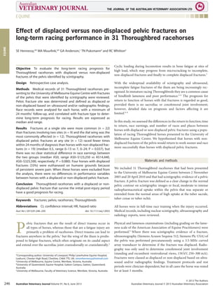
  • 1. Effect of displaced versus non-displaced pelvic fractures on long-term racing performance in 31 Thoroughbred racehorses SE Hennessy,a * MA Muurlink,a,b GA Anderson,c TN Puksmanna and RC Whittona Objective To evaluate the long-term racing prognosis for Thoroughbred racehorses with displaced versus non-displaced fractures of the pelvis identified by scintigraphy. Design Retrospective case analysis. Methods Medical records of 31 Thoroughbred racehorses pre- senting to the University of Melbourne Equine Centre with fractures of the pelvis that were identified by scintigraphy were reviewed. Pelvic fracture site was determined and defined as displaced or non-displaced based on ultrasound and/or radiographic findings. Race records were analysed for each horse, with a minimum of 24 months’ follow-up, and correlated with fracture type to deter- mine long-term prognosis for racing. Results are expressed as median and range. Results Fractures at a single site were more common (n = 22) than fractures involving two sites (n = 9) and the ilial wing was the most commonly affected (n = 12). Thoroughbred racehorses with displaced pelvic fractures at any site (n = 12) raced fewer times within 24 months of diagnosis than horses with non-displaced frac- tures (n = 19) (median 0.5, range 0–13 vs 7, 0–24; P = 0.037), but there was no clear statistical difference in race earnings between the two groups (median A$0, range A$0–$123,250 vs A$14,440, A$0–$325,500, respectively; P = 0.080). Four horses with displaced fractures (33%) were euthanased on humane grounds because of persistent severe pain. When these horses were excluded from the analysis, there were no differences in performance variables between horses with a displaced or non-displaced pelvic fracture. Conclusion Thoroughbred racehorses with a displaced or non- displaced pelvic fracture that survive the initial post-injury period have a good prognosis for racing. Keywords fractures; pelvis; racehorses; Thoroughbreds Abbreviations CI, confidence interval; HR, hazard ratio Aust Vet J 2013;91:246–250 doi: 10.1111/avj.12053 P elvic fractures that are the result of direct trauma occur in all types of horses, whereas those that are a fatigue injury are primarily a problem of racehorses. Direct trauma can lead to fractures anywhere in the pelvis,1 but the wing of the ilium is predis- posed to fatigue fractures, which often originate on its caudal aspect and extend over the sacroiliac joint craniodorsally or craniolaterally.2 Cyclic loading during locomotion results in bone fatigue at sites of high load, which may progress from microcracking to incomplete, non-displaced fractures and finally to complete displaced fractures.3 With the widespread availability of scintigraphy and ultrasound, incomplete fatigue fractures of the ilium are being increasingly rec- ognised.In immature racing Thoroughbreds they are a common cause of hindlimb lameness and poor performance.4–6 The prognosis for return to function of horses with ilial fractures is regarded as good, provided there is no sacroiliac or coxofemoral joint involvement; however, detailed data on prognosis and factors affecting it are limited.6–8 In this study,we assessed the differences in the return to function,time to return, race earnings, and number of races and places between horses with displaced or non-displaced pelvic fractures using a popu- lation of racing Thoroughbred horses presented to the University of Melbourne Equine Centre. We hypothesised that horses with non- displaced fractures of the pelvis would return to work sooner and race more successfully than horses with displaced pelvic fractures. Materials and methods We included 31 Thoroughbred racehorses that had been presented to the University of Melbourne Equine Centre between 2 November 2003 and 20 April 2010 and that had scintigraphic evidence of a pelvic fracture.A pelvic fracture was defined as a clear change in the normal pelvic contour on scintigraphic images or focal, moderate to intense radiopharmaceutical uptake within the pelvis that was separate or extended from the normal areas of high uptake at the tuber sacrale, tuber coxae or tuber ischii. All horses were in full-time race training when the injury occurred. Medical records, including nuclear scintigraphy, ultrasonography and radiology reports, were reviewed. Physical and lameness examinations (including grading on the lame- ness scale of the American Association of Equine Practitioners) were performed.9 Where there was scintigraphic evidence of a fracture, ultrasonography (Siemens Acuson Sequioa 512, Siemens Plc USA) of the pelvis was performed percutaneously using a 3.5-MHz curved array transducer to determine if the fracture was displaced. Radio- graphy was only used to determine coxofemoral joint involvement (standing and recumbent ventrodorsal views, 150 kV, 250–300 mA). Fractures were classed as displaced or non-displaced based on ultra- sound and/or radiographic findings. Treatment protocols and rest periods were clinician-dependent, but in all cases the horse was rested for at least 3 months. *Corresponding author: University of Liverpool, Philip Leverhulme Equine Hospital, Leahurst, Chester High Road, Cheshire, CH64 7TE, UK; simonhennessy@hotmail.com a University of Melbourne, Equine Centre, Werribee, Victoria, Australia b University of Queensland, Equine Hospital, Gatton Campus, Gatton, Queensland, Australia c University of Melbourne, Faculty of Veterinary Science, Werribee, Victoria, Australia bs_bs_banner EQUINE EQUINE © 2013 The Authors Australian Veterinary Journal © 2013 Australian Veterinary AssociationAustralian Veterinary Journal Volume 91, No 6, June 2013246
  • 2. A standard scintigraphic protocol was used for each horse. An 18-gauge catheter was placed in the left jugular vein and 12.7–15 MBq/kg of 99 Tc-HDP administered (standard 7 GBq/14 MBq/kg dose for an average Thoroughbred racehorse). Frusemide (0.7 mg/kg IV) was injected 60 min prior to scanning the pelvis. Dynamic (35 ¥ 2-s frames), delayed-phase standing lateral, dorsal, caudal and oblique pelvis images were acquired 3 h after radiopharmaceutical injection with a large field-of-view gamma camera (Phillips Argus Epic) using a 128 ¥ 128 matrix. Data were transferred to a workstation (Sun Ultra Workstation) and processed with motion correction software (University College London, Institute of Nuclear Medicine). Race and stud book records were accessed in April 2012. Only horses with at least 24 months’ follow-up were included. Data obtained included sex, age, prize money, races and places achieved before and after diagnosis. Statistical analysis Fisher’s exact test was used to compare proportions. Preliminary analysis of the residuals of the post-diagnosis race performance vari- ables showed that almost all were not normally distributed (Shapiro- Wilk’s test). The Mann-Whitney U-test with exact P values was used to compare the mean ranks of post-diagnosis race performance vari- ables between horses with displaced or non-displaced fractures. The log-rank test and the hazard ratio (HR) from a Cox proportional hazard model were used to compare the distribution of time to first race post-diagnosis for displaced and non-displaced fractures, together with the Kaplan-Meier estimate of median time to first race for all horses.10 Two-tailed P < 0.05 was considered to be statistically significant. The statistical software used was Stata 12.1 for Windows (StataCorp, College Station, TX, USA) and IBM SPSS 20 (IBM Corp., New York, NY, USA) was used for Mann-Whitney exact P-values. Results All 31 horses included in the study (mean age 3.3 years, range 1–6 years; 14 females (45%), 13 geldings (42%), 4 entire males (13%)) had been referred for scintigraphic examination. Of them, two horses presented with a history of trauma and both had displaced ischial fractures,six horses presented with a history of poor performance and all had non-displaced fractures of the ilial wing,eight horses presented for lameness immediately following a race or a training gallop, seven horses were referred for mild to moderate hindlimb lameness that could not be localised with distal limb diagnostic analgesia by the attending veterinarian and eight horses presented with severe lame- ness (grade 3/5 or more) without associated trauma,of which four had displaced fractures. All horses had scintigraphic examination of their hindlimbs,pelvis and thoracolumbar spine.Areas of increased radiopharmaceutical uptake, not associated with the pelvis, included the dorsal spinous processes of the thoracic and lumbar vertebrae, the tarsometatarsal joints, the tibial diaphysis, the lateral proximal hind sesamoid bone, the fourth metatarsal bone and the lateral condyle of the third metatarsal bone. There were 22 fractures involving a single site within the pelvis and 9 fractures involving two sites. The most common site of fracture was the ilial wing, followed by the ischium (Table 1); 19 fractures were non-displaced and 12 were displaced (Figures 1, 2). Of those with displaced fractures, four horses (33%) were euthanased at 2, 2, 3 and 18 days after diagnosis on humane grounds because of persistent, severe pain.Of the four horses,two were presented 6–8 weeks after the initial injury, one with fractures of the acetabulum and the ilial wing, and the other with fractures of the acetabulum and ilial shaft. Both of them had severe gluteal muscle atrophy and grade 4/5 lameness that was noted to worsen with box rest. The third horse had bilateral ilial wing fractures with cranial displacement of the pelvis and associated muscle atrophy and severe lameness. The fourth horse presented acutely after fast work with a severe comminuted oblique fracture of the ilial wing and shaft with grade 5/5 lameness. Among the 27 survi- vors, 12 horses were rested for 3 months, 12 for 6 months and 3 were rested for unspecified periods longer than 3 months. With respect to racing, five horses that were in full race training at the time of injury did not race before or after diagnosis, four horses raced Table 1. Pelvic fracture distribution and outcome in 31 Thoroughbred racehorses Fracture site n Fracture type Median no. of races No. euthanased Displaced Non-displaced Pre-diagnosis Post-diagnosis Single site Ilial wing 12 2 10 7.5 13.0 0 Ilial shaft 2 0 2 0.5 5.0 0 Acetabulum 1 0 1 0.0 0.0 0 Ischium 7 5 2 0.0 18.0 0 Multiple sites Bilateral ilial wings 3 1 2 7.0 2.0 1 Ilial wing and shaft 3 1 2 1.0 2.0 1 Acetabulum and ischium 1 1 0 0.0 0.0 0 Acetabulum and ilial wing 1 1 0 0.0 0.0 1 Acetabulum and ilial shaft 1 1 0 8.0 0.0 1 EQUINE EQUINE © 2013 The Authors Australian Veterinary Journal © 2013 Australian Veterinary Association Australian Veterinary Journal Volume 91, No 6, June 2013 247
  • 3. before diagnosis but not after, 9 horses raced after diagnosis but not before, and 13 horses raced before and after diagnosis. The median time to first race based on all horses was 9.9 months (95% confidence interval (CI) 7.7–12.3). The majority of horses with non-displaced pelvic fractures (16/19) raced within 24 months of diagnosis compared with only half of the horses with displaced pelvic fractures (6/12) (Table 2). Horses with a displaced pelvic fracture at any site (n = 12) raced fewer times within 24 months of diagnosis than horses with non-displaced fractures (n = 19) (median 0.5, range 0–13 vs 7, 0–24; P = 0.037), but there was no clear difference in race earnings between the two groups (median A$0, range A$0–$123,250 vs A$14,440, A$0–$325,500, respectively; P = 0.080) (Table 2).When the four horses that were euthanased were excluded from the analysis, there were no differences in the perfor- mance variables between horses with a displaced or non-displaced Figure 1. Left oblique (top) and dorsal scintigraphy images of the pelvis of a horse with an incomplete ilial stress fracture. Note the increased radiopharmaceutical uptake lateral to the left tuber sacrale on the caudal surface of the ilium (black arrows). Ultrasonographic examination revealed no displacement of fracture fragments. Figure 2. Right oblique (top) and dorsal (middle) scintigraphy images of the pelvis of a horse with a complete fracture of the right ilium (black arrows). Ultrasound examination (bottom) revealed marked displace- ment of fracture fragments. EQUINE EQUINE © 2013 The Authors Australian Veterinary Journal © 2013 Australian Veterinary AssociationAustralian Veterinary Journal Volume 91, No 6, June 2013248
  • 4. fracture (Table 3). The median time to first race for horses with displaced pelvic fractures was 10.4 months (95% CI 6.8–16.7) and 9.3 months (95% CI 6.9–11.5) for horses with a non-displaced pelvic fracture (log-rank test P = 0.50).The HR did not differ from 1 for time to first race for horses with a displaced compared with a non-displaced fracture (HR 0.73, 95% CI 0.28–1.86; P = 0.51). Of the 4 horses presenting with fractures involving the acetabulum, none raced post diagnosis (2 were euthanased), whereas 6 of 7 horses with ischial fractures raced post diagnosis and 11 of 12 horses with unilateral ilial wing fractures raced post diagnosis, including both of the horses with displaced fractures. One of two horses that presented with unilateral fractures of the ilial shaft raced post diagnosis. Of the 6 horses with fractures in multiple sites and no acetabular involve- ment, 4 raced post diagnosis (2 were euthanased). Discussion Our study results demonstrated that Thoroughbred racehorses with displaced or non-displaced pelvic fractures that survive the initial post-injury period have a good prognosis for racing. We found no evidence that horses with non-displaced pelvic fractures returned to racing sooner than those with displaced fractures. Pelvic fractures in Thoroughbred racehorses have a variety of presentations and should be considered in the differential diagnosis of poor performance and hindlimb lameness. Our findings are consistent with those of other studies of pelvic fractures, which have demonstrated that 61–75% of horses without acetabular and ilial shaft involvement return to racing after diagno- sis.8,11,12 Less success has been reported for Thoroughbred racehorses with fractures of the ilial wing identified by scintigraphy,with 6 of 10,7 and 1 of 413 returning to racing in two separate studies. Studies that have previously reported a good prognosis for displaced traumatic pelvic fracture relate to successful salvage of animals for potential breeding.11,14 A comparison of displaced and non-displaced pelvic fractures, and the prognosis for return to racing, has not been previ- ously examined. The importance of determining the site of pelvic fracture is highlighted in this study. Similar to other studies, fractures involving the acetabulum had a poor prognosis for return to racing, whereas both ilial and ischial fractures had a good prognosis.8 A number of factors have the potential to affect future race perform- ance in horses with displaced fractures. In the case of ilial fractures, these include changes in the sacroiliac joint and subsequent sacroiliac disease.Many displaced fractures are associated with the development of sacroiliac osteophytes and degenerative changes in the ilial articu- lar surface.2 Following a displaced fracture, the fracture fragments re-attach in a new anatomical arrangement that can influence the time required for return to function as the horse adapts to the new anatomy.15 Also, the greater pain resulting from a displaced fracture will often result in more muscle atrophy,which can delay the return to racing.1,6 The effect of the degree of muscle atrophy on the time to return to racing was not examined in this study. Based on our find- ings, these factors are less important and uncontrollable pain or acetabular involvement are the primary factors affecting long-term outcome. Scintigraphy is commonly used to identify pelvic fractures, but it is rarely possible to determine if a fracture is displaced using this Table 2. Post-diagnosis performance for displaced and non-displaced fractures of 31 Thoroughbred racehorses Outcome Fracture type P value Non-displaced (n = 19) Displaced (n = 12) Raced in 24 months post-diagnosis 16 (84%) 6 (50%) 0.056 Median no. races in 24 months post-diagnosis (mean; range) 7 (7.5; 0–24) 0.5 (3; 0–13) 0.037 Median no. places in 24 months post-diagnosis (mean; range) 2 (2.9; 0–8) 0 (1.6; 0–8) 0.13 Median prize money in 24 months post-diagnosis (A$: mean; range) 14,440 (50,783; 0–325,500) 0 (16,129; 0–123,250) 0.080 Table 3. Post-diagnosis performance for displaced and non-displaced fractures of Thoroughbred racehorses after the exclusion of four horses euthanased on humane grounds Outcome Fracture type P value Non-displaced (n = 19) Displaced (n = 8) Raced in 24 months post-diagnosis 16 (84%) 6 (75%) 0.62 Median no. races in 24 months post-diagnosis (mean; range) 7 (7.5; 0–24) 3.5 (4.5; 0–13) 0.34 Median no. places in 24 months post-diagnosis (mean; range) 2 (2.9; 0–8) 0.5 (2.4; 0–8) 0.61 Median prize money in 24 months post-diagnosis (A$: mean; range) 14,440 (50,783; 0–325,500) 575 (24,194; 0–123,250) 0.41 EQUINE EQUINE © 2013 The Authors Australian Veterinary Journal © 2013 Australian Veterinary Association Australian Veterinary Journal Volume 91, No 6, June 2013 249
  • 5. technique.16 Fracture displacement or callus formation can be observed using ultrasound, in the majority of cases, whereas non- displaced fractures are difficult to identify.17–19 Therefore, the combi- nation of scintigraphy and ultrasound enabled diagnosis in most cases of pelvic fracture in this study. Standing radiography of the pelvis was used in this study to determine coxofemoral joint involve- ment in some cases, because of the difficulty of assessing acetabular involvement using ultrasound alone.17 The overlying sacrum and intestinal contents do not allow a detailed view of the whole ilium on radiographs.14,17,20,21 In the present study, it was often not possible to discriminate between traumatic fractures and displaced fatigue fractures because in only a minority of cases was there a history of trauma or an acute onset of lameness following fast work. However, it is likely that many of the cases were fatigue fractures, as it has been reported that ilial fractures in a British Thoroughbred flat racehorse population accounted for 15.5% of the total fractures seen in the population, with fatigue fractures accounting for 87% of the ilial fractures.22 In the present study, 50% (3/6) of displaced ilial fractures occurred during training of young horses that had never raced. Similarly, it has been reported that in humans the majority of tibial and femoral stress fractures occur early during military training, suggesting that bone adaptation is protective.23 As found in our study, ilial fractures are most commonly observed in young, immature Thoroughbred racehorses aged up to 4 years of age.6,12,15,22 Others have described ilial wing fractures in older horses;1,2,5 however, two of these were post- mortem studies from which it was difficult to determine the time of fracture.2,5 Study limitations We had a low number of cases, resulting in a lack of power, and the study population included four horses that were euthanased after diagnosis, making it impossible to know whether these horses could have returned to racing if the problem had been appropriately managed. However, all four horses were euthanased because of per- sistent, severe pain rather than for economic reasons. Also, not all horses in this study had full-body scintigraphy, so although it is pos- sible that other problems affecting long-term outcome may have been overlooked, the majority were able to perform well. In conclusion, the poorer prognosis for Thoroughbred racehorses with displaced pelvic fractures compared with non-displaced pelvic fractures appears to be because of an inability to manage severe pain in some affected horses and/or involvement of the acetabulum. Accurate localisation of the fracture with a combination of scinti- graphy and ultrasound will allow an appropriate prognosis to be given. References 1. Dyson SJ. Lameness associated with the stifle and pelvic regions. Am Assoc Equine Pract 2002;48:387–411. 2. Haussler KK, Stover SM. Stress fractures of the vertebral lamina and pelvis in Thoroughbred racehorses. Equine Vet J 1998;30:374–381. 3. Riggs CM. Fractures: a preventable hazard of racing Thoroughbreds? Vet J 2002;163:19–29. 4. Bathe AP. 245 fractures in Thoroughbred racehorses: results of a 2-year pro- spective study in Newmarket. Am Assoc Equine Pract 1994;40:175–176. 5. Carrier TK, Estberg L, Stover SM et al. Association between long periods without high–speed workouts and risk of complete humeral or pelvic fracture in Thoroughbred racehorses: 54 cases (1991–1994). J Am Vet Med Assoc 1998;212: 1582–1587. 6. Van Wessum R. The pelvis. In: Baxter GM, editor. Adam’s and Stashak’s lameness in horses. 6th edn. Wiley Blackwell, Oxford, 2011:840–847. 7. Pilsworth RC, Shepherd MC, Herinckx BMB et al. Fractures of the wing of the ilium, adjacent to the sacroiliac joint in Thoroughbred racehorses. Equine Vet J 1994;26:94–99. 8. Fuller A, Beever E, Fraser B et al. A retrospective study of 80 cases of pelvic fracture diagnosed by gamma scintigraphy in Thoroughbred racehorses. Br Equine Vet Assoc 2008;47:142–143. 9. Ross MW. Movement. In: Ross MW, Dyson SJ. Diagnosis and management of lameness in the horse. 1st edn. Saunders, St Louis, 2002:60–73. 10. Hosmer D, Lemeshow S. Applied survival analysis: regression modeling of time to event data. 2nd edn. John Wiley & Sons, New York, 2008. 11. Rutkowski JA, Richardson DW. A retrospective study of 100 pelvic fractures in horses. Equine Vet J 1989;29:256–259. 12. Shepherd MC, Pilsworth RC, Hopes R et al. Clinical signs, diagnosis, manage- ment and outcome of complete and incomplete fracture to the ilium. Am Assoc Equine Pract 1994;40:177–180. 13. Davenport-Goodall CLM, Ross MW. Scintigraphic abnormalities of the pelvic region in horses examined because of lameness or poor performance: 128 cases (1993–2000). J Am Vet Med Assoc 2004;224:88–95. 14. Little C, Hilbert B. Pelvic fractures in horses: 19 cases (1974–1984). J Am Vet Med Assoc 1987;190:1203–1206. 15. Pilsworth RC. Diagnosis and management of pelvic fractures in theThorough- bred racehorse. In: Ross MW, Dyson SJ, editors. Diagnosis and management of lameness in the horse. 2nd edn. Elsevier Saunders, St Louis, 2011:564–571. 16. Hornof WJ, Stover SM, Koblik PD et al. Oblique views of the ilium and the scintigraphic appearance of stress fractures of the ilium. Equine Vet J 1996;28: 355–358. 17. Geburek F, Rotting AK, Stadler PM. Comparison of the diagnostic value of ultrasonography and standing radiography for pelvic-femoral disorders in horses. Vet Surg 2009;38:310–317. 18. Driver AJ, Nagy A. Fracture of the ischium in an eight-year-old Arabian gelding: a diagnostic challenge. Equine Vet Educ 2008;20:127–130. 19. Tomlinson JE, Sage AM, Turner TA et al. Detailed ultrasonographic mapping of the pelvis in clinically normal horses and ponies. Am J Vet Res 2001;62:1768–1775. 20. May SA, Patterson LJ, Peacock PJ et al. Radiographic technique for the pelvis in the standing horse. Equine Vet J 1991;23:312–314. 21. Barrett EL, Talbot AM, Driver AJ et al. A technique for radiography in the standing horse. Equine Vet J 2006;38:266–270. 22. Verheyen KLP, Wood JLN. Descriptive epidemiology of fractures occurring in British Thoroughbred racehorses in training. Equine Vet J 2004;36:167–173. 23. Finestone A, Milgrom C, Wolf O et al. Epidemiology of metatarsal stress frac- tures versus tibial and femoral stress fractures during elite training. Foot Ankle Int 2011;32:16–20. (Accepted for publication 24 September 2012) EQUINE EQUINE © 2013 The Authors Australian Veterinary Journal © 2013 Australian Veterinary AssociationAustralian Veterinary Journal Volume 91, No 6, June 2013250