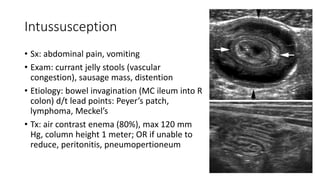
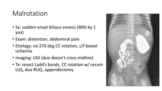
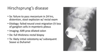
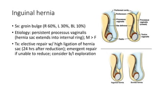
Pediatric Surgery
Fluids & Electrolytes
• Maintenance rate: 4/2/1 cc/kg/hr
• GFR ~21 cc/min/1.73 m2 vs 70 mL/min/1.73 m2 in an adult​
• Urine concentration ~600 mOsm/kg vs ~1200 mOsm/kg​
• Trauma: crystalloid 20 cc/kg bolus x2, if no response pRBC 10 cc/kg bolus
• Adequate UOP: <2 yoa = 2-3 cc/kg/hr, >2 yoa = 1 cc/kg/hr
• Acidosis correction for pH < 7.25:
• Base deficit x weight (kg) x (0.5 in newborns OR 0.4 in small OR 0.3 in older children)
• Dilute to 0.5 mEq/mL d/t hyperosmolarity
• Give ½ as bolus and remaining half slowly
• Adequate UOP: <2 yoa = 2-3 cc/kg/hr, >2 yoa = 1 cc/kg/hr
Hypertrophic Pyloric Stenosis
• Sx: projectile non-bilious emesis after feeds
• Exam: olive like mass in RUQ
• Labs: hypochloremic, hypokalemic, metabolic
alkalosis
• Imaging: US of pylorus >16 mm long, >4 mm
thick
• Tx: fluid resuscitation (NS  D5NS w/ 10 mEq
K), electrolyte optimization, Ramstedt
pyloromyotomy (split layers until submucosa
bulges out)
Intussusception
• Sx: abdominal pain, vomiting
• Exam: currant jelly stools (vascular
congestion), sausage mass, distention
• Etiology: bowel invagination (MC ileum into R
colon) d/t lead points: Peyer’s patch,
lymphoma, Meckel’s
• Tx: air contrast enema (80%), max 120 mm
Hg, column height 1 meter; OR if unable to
reduce, peritonitis, pneumopertioneum
Malrotation
• Sx: sudden onset bilious emesis (90% by 1
yoa)
• Exam: distention, abdominal pain
• Etiology: no 270 deg CC rotation, c/f bowel
ischemia
• Imaging: UGI (duo doesn’t cross midline)
• Tx: resect Ladd’s bands, CC rotation w/ cecum
LLQ, duo RUQ, appendectomy
Gastroschisis
• Sx: intestines through R of umbilicus defect
• Exam: no peritoneal sac, stiff bowel,
associated w/ intestinal atresia
• Tx: saline soaked gauze, resuscitate, TPN,
NPO, repair when stable (silastic mesh silo),
primary closure later if silo (watch ACS)
Omphalocele
• Exam: bowel through midline w/ peritoneal
sac
• Etiology: failure of embryonal development;
associated w/ malro & Down syndrome
• Cantrell Pentalogy: cardiac, pericardium,
sternal, diaphragmatic, omphalocele
• Tx: saline soaked gauze, resuscitate, TPN,
NPO, primary vs mesh repair vs 2-3 month
epithelialization then repair
Hirschsprung’s disease
• Sx: failure to pass meconium in 24 hrs,
distention, stool explosion w/ rectal exam
• Etiology: failed neural crest migration l/t loss
of ganglion cells in myenteric plexus
• Imaging: AXR prox dilated colon
• Dx: full thickness rectal biopsy
• Tx: likely initial colostomy w/ subsequent
Soave vs Duhamel
Umbilical hernia
• Sx: midline bulge
• Etiology: failure of closure of linea alba (most
close by age 3), increased risk in AAs &
prematures
• Tx: surgery at 5 yoa, incarceration, VP shunt
Inguinal hernia
• Sx: groin bulge (R 60%, L 30%, BL 10%)
• Etiology: persistent processus vaginalis
(hernia sac extends into internal ring); M > F
• Tx: elective repair w/ high ligation of hernia
sac (24 hrs after reduction); emergent repair
if unable to reduce; consider b/l exploration
Cryptorchidism
• Sx: groin bulge
• Imaging: MRI if unable to palpate
• Labs: chromosomal studies if bilateral
• Tx: wait 6 months; orchiopexy through
inguinal incision; if unable to get testes down,
wait 6m and try again, if fails, ligate spermatic
vessels (vas deferens blood supply will
collateralize)
• Cancer risk stays same (seminoma)

peds final.pptx

  • 1.
  • 2.
    Fluids & Electrolytes •Maintenance rate: 4/2/1 cc/kg/hr • GFR ~21 cc/min/1.73 m2 vs 70 mL/min/1.73 m2 in an adult​ • Urine concentration ~600 mOsm/kg vs ~1200 mOsm/kg​ • Trauma: crystalloid 20 cc/kg bolus x2, if no response pRBC 10 cc/kg bolus • Adequate UOP: <2 yoa = 2-3 cc/kg/hr, >2 yoa = 1 cc/kg/hr • Acidosis correction for pH < 7.25: • Base deficit x weight (kg) x (0.5 in newborns OR 0.4 in small OR 0.3 in older children) • Dilute to 0.5 mEq/mL d/t hyperosmolarity • Give ½ as bolus and remaining half slowly • Adequate UOP: <2 yoa = 2-3 cc/kg/hr, >2 yoa = 1 cc/kg/hr
  • 3.
    Hypertrophic Pyloric Stenosis •Sx: projectile non-bilious emesis after feeds • Exam: olive like mass in RUQ • Labs: hypochloremic, hypokalemic, metabolic alkalosis • Imaging: US of pylorus >16 mm long, >4 mm thick • Tx: fluid resuscitation (NS  D5NS w/ 10 mEq K), electrolyte optimization, Ramstedt pyloromyotomy (split layers until submucosa bulges out)
  • 4.
    Intussusception • Sx: abdominalpain, vomiting • Exam: currant jelly stools (vascular congestion), sausage mass, distention • Etiology: bowel invagination (MC ileum into R colon) d/t lead points: Peyer’s patch, lymphoma, Meckel’s • Tx: air contrast enema (80%), max 120 mm Hg, column height 1 meter; OR if unable to reduce, peritonitis, pneumopertioneum
  • 5.
    Malrotation • Sx: suddenonset bilious emesis (90% by 1 yoa) • Exam: distention, abdominal pain • Etiology: no 270 deg CC rotation, c/f bowel ischemia • Imaging: UGI (duo doesn’t cross midline) • Tx: resect Ladd’s bands, CC rotation w/ cecum LLQ, duo RUQ, appendectomy
  • 6.
    Gastroschisis • Sx: intestinesthrough R of umbilicus defect • Exam: no peritoneal sac, stiff bowel, associated w/ intestinal atresia • Tx: saline soaked gauze, resuscitate, TPN, NPO, repair when stable (silastic mesh silo), primary closure later if silo (watch ACS)
  • 7.
    Omphalocele • Exam: bowelthrough midline w/ peritoneal sac • Etiology: failure of embryonal development; associated w/ malro & Down syndrome • Cantrell Pentalogy: cardiac, pericardium, sternal, diaphragmatic, omphalocele • Tx: saline soaked gauze, resuscitate, TPN, NPO, primary vs mesh repair vs 2-3 month epithelialization then repair
  • 8.
    Hirschsprung’s disease • Sx:failure to pass meconium in 24 hrs, distention, stool explosion w/ rectal exam • Etiology: failed neural crest migration l/t loss of ganglion cells in myenteric plexus • Imaging: AXR prox dilated colon • Dx: full thickness rectal biopsy • Tx: likely initial colostomy w/ subsequent Soave vs Duhamel
  • 9.
    Umbilical hernia • Sx:midline bulge • Etiology: failure of closure of linea alba (most close by age 3), increased risk in AAs & prematures • Tx: surgery at 5 yoa, incarceration, VP shunt
  • 10.
    Inguinal hernia • Sx:groin bulge (R 60%, L 30%, BL 10%) • Etiology: persistent processus vaginalis (hernia sac extends into internal ring); M > F • Tx: elective repair w/ high ligation of hernia sac (24 hrs after reduction); emergent repair if unable to reduce; consider b/l exploration
  • 11.
    Cryptorchidism • Sx: groinbulge • Imaging: MRI if unable to palpate • Labs: chromosomal studies if bilateral • Tx: wait 6 months; orchiopexy through inguinal incision; if unable to get testes down, wait 6m and try again, if fails, ligate spermatic vessels (vas deferens blood supply will collateralize) • Cancer risk stays same (seminoma)

Editor's Notes

  • #4 1 in 300 live births and commonly in infants between 3 and 6 weeks of age. Male-to-female ratio is nearly 5:1. For most infants, fluid containing 5% dextrose and 0.45% saline with added potassium of 2 to 4 mEq/kg over 24 hours at a rate of approximately 150 to 175 mL/kg per 24 hours will correct the underlying deficit Fredet-Ramstedt pyloromyotom
  • #5 OR – apply pressure to distal limb, usually do not need resection unless associated w/ pathologic lead point