This clinical study evaluated two signs that can indicate a complete release of the transverse carpal ligament (TCL) during carpal tunnel release surgery: the "fat pad" sign and the "little finger pulp" sign. The study reviewed 643 carpal tunnel release procedures performed using a short longitudinal incision. Both signs were observed during surgery to confirm full division of the TCL. Post-operatively, symptoms resolved in nearly all patients, with minimal complications. The signs were useful for ensuring complete TCL release through a small incision.
![PROUBASTA ET AL.
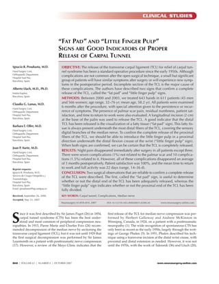
TABLE 1. Results of revision carpal tunnel release after previous open carpal tunnel release surgerya
No. of Intact parts Scar Medical Other No abnor-
Series (ref. no.) Fibrosis
interventions of the TCL tethering injury causes malities
Langloh and Linscheid, 1972 (16) 34 21 (62%) — — — — —
Conolly, 1978 (9) 35 9 (31%) 4 — 7 10 5
Kern et al., 1993 (13) 16 10 (62%) 4 — 1 1 —
Assmus, 1996 (2) 185 91 (49%) 58 — — — 36
Bagatur, 2002 (5) 26 23 (88%) — 1 — 2 —
Unglaub et al., 2005 (37) 38 26 (68%) 8 3 — — 1
Stütz et al., 2006 (35) 200 108 (54%) 46 17 12 4 13
Pülzl et al., 2006 (30) 48 16 (33%) 24 — 8 — —
Assmus et al., 2006 (3) 57 34 (60%) 15 — 3 — 5
Frick and Baumeister, 2006 (11) 63 38 (60%) 21 2 1 1 —
a
TCL, transverse carpal ligament.
Patient satisfaction was 100%, and the mean time period to pad of palmar fat found at the distal end of the TCL and con-
return to work and full activity was 22 days (14–36 d). stitutes a reliable indicator of the complete release of the distal
edge of TCL. This appreciation is the same one that we have
DISCUSSION been using for many years (21). In a comprehensive review of
the anatomy of the carpal tunnel by Skandalakis et al. (34), a
CTS is the best understood and most common of the periph- reference has been made to the specialized palmar fat related to
eral compression neuropathies and, therefore, the most fre- the superficial arterial palmar arch in an area noted as the “dis-
quent hand operation. The common surgical treatment for CTS tal zone,” which was later verified in radiological (25), endo-
in patients who have failed to improve with conservative meas- scopic (12), and surgical (32) studies. For this reason, when the
ures is open carpal tunnel release. Although the longitudinal distal edge of the retinaculum has been completely released,
division of the TCL consistently relieves symptoms in most the fat pad described appears. Our results support the results
patients, some may complain of persistence of symptoms after of most of these studies, as no permanent complications and
surgery due to an incomplete release of the TCL (2, 3, 5, 9, 11, very few transient complications were observed (15).
13, 16, 30, 35, 37) (Table 1). Some authors (8, 23) have stated that
common indications for reoperation include previous incom-
plete surgery and postoperative fibrosis causing recurrence of
CONCLUSION
symptoms. Although the standard longer incision that crosses This study has several limitations because there is no control
the wrist crease allows complete visualization of the TCL from group. However, we have been using this technique without
the forearm fascia proximal to the superficial palmar arch dis- any modifications since 1984, after it was proposed by the sen-
tally, many experienced hand surgeons successfully use shorter ior author (AL), assuring a complete release of the TCL and
longitudinal incisions that do not cross the wrist crease to min- providing immediate and permanent relief of median nerve
imize scar tenderness, scar retraction, and injury to the thenar compression at the carpal tunnel.
sensory branch of the median nerve.
The “fat pad” and “little finger pulp” signs described in this
article have been helpful in confirming complete division of the
REFERENCES
TCL; neither sign is new. In reference to the “little finger pulp” 1. Amadio PC: The first carpal tunnel release? J Hand Surg [Br] 20:40–41, 1995.
sign, Phalen indicates that “after section of the TCL there 2. Assmus H: Correction and reintervention in carpal tunnel syndrome. Report
should be sufficient room in the carpal tunnel to permit a of 185 reoperations [in German]. Nervenarzt 67:998–1002, 1996.
3. Assmus H, Dombert T, Staub F: Reoperations for CTS because of recurrence
curved Kelly hemostat to slide easily into the palm or to allow
or for correction [in German]. Handchir Mikrochir Plast Chir 38:306–311,
the moistened little finger of the surgeon to pass readily along 2006.
the median nerve into the palm” (29, p 223). Although Phalen 4. Avci S, Sayli U: Carpal tunnel release using a short palmar incision and a new
verifies the complete section of the proximal edge of the reti- knife. J Hand Surg [Br] 25:357–360, 2000.
naculum in a proximal-distal direction and we made it in a 5. Bagatur AE: Analysis of the causes of failure in carpal tunnel syndrome sur-
gery and the results of reoperation [in Turkish]. Acta Orthop Traumatol Turc
distal-proximal direction, the principle of the “little pulp sign” 36:346–353, 2002.
is the same. With regard to the “fat pad sign,” in 1997, Ragbir 6. Botte MJ, von Schroeder HP, Abrams RA, Gellman H: Recurrent carpal tun-
et al. (31) described the “yellow fat sign,” which is a constant nel syndrome. Hand Clin 12:731–743, 1996.
812 | VOLUME 61 | NUMBER 4 | OCTOBER 2007 www.neurosurgery-online.com](https://image.slidesharecdn.com/pdf-17-100717120719-phpapp01/85/Fat-pad-and-Little-finger-pulp-3-320.jpg)
![GOOD INDICATORS OF PROPER RELEASE OF THE CARPAL TUNNEL
7. Bromley GS: Minimal-incision open carpal tunnel decompression. J Hand 37. Unglaub F, Goldbach C, Hahn P: Reoperation in carpal tunnel syndrome.
Surg [Am] 19:119–120, 1994. Retrospective analysis [in German]. Nervenarzt 76:1506, 1508–1510,
8. Cobb TK, Amadio PC: Reoperation for carpal tunnel syndrome. Hand Clin 1512–1514, 2005.
12:313–323, 1996. 38. Urbaniak JR, Desai SS: Complications of nonoperative and operative treat-
9. Conolly WB: Pitfalls in carpal tunnel decompression. Aust N Z J Surg ment of carpal tunnel syndrome. Hand Clin 12:325–335, 1996.
48:421–425, 1978.
10. Forman DL, Watson HK, Caulfield KA, Shenko J, Caputo AE, Ashmead D:
Persistent or recurrent carpal tunnel syndrome following prior endoscopic
carpal tunnel release. J Hand Surg [Am] 23:1010–1014, 1998.
COMMENTS
11. Frick A, Baumeister RG: Re-intervention after carpal tunnel release [in
German]. Handchir Mikrochir Plast Chir 38:312–316, 2006.
12. Jimenez DF, Gibbs SR, Clapper AT: Endoscopic treatment of carpal tunnel
syndrome: A critical review. J Neurosurg 88:817–826, 1998.
I n this study, the authors describe two useful observations as part of
their surgical technique for carpal tunnel release surgery. Most
peripheral nerve surgeons are very familiar with the "fat pad" sign and
13. Kern BC, Brock M, Rudolph KH, Logemann H: The recurrent carpal tunnel routinely use it as an indicator for the presence of vascular arcades and,
syndrome. Zentralbl Neurochir 54:80–83, 1993. thus, the proper release of distal transverse carpal ligament. In our
14. Kessler FB: Complications of the management of carpal tunnel syndrome. experience, the more common cause of surgical failure is incomplete
Hand Clin 2:401–406, 1986. division of the proximal aspect of the ligament. This proximal edge of
15. Klein RD, Kotsis SV, Chung KC: Open carpal tunnel release using a the transverse carpal ligament blends imperceptibly into the distal fore-
1-centimeter incision: Technique and outcomes for 104 patients. Plast
arm aponeurosis, and this may be sectioned under direct visualization
Reconstr Surg 111:1616–1622, 2003.
16. Langloh ND, Linscheid RL: Recurrent and unrelieved carpal-tunnel syn-
for up to 1 cm proximal to the skin incision by the assistant elevating
drome. Clin Orthop Relat Res 83:41–47, 1972. the skin edge. We routinely palpate both proximately and distally
17. Learmonth GR: The principle of decompression in the treatment of certain within the incision to make sure that no remaining areas of compres-
diseases of peripheral nerves. Surg Clin N Am 13:905–913, 1933. sion are identified. Proubasta et al. use the term "little finger pulp" to
18. Lee WP, Strickland JW: Safe carpal tunnel release via a limited palmar inci- describe this technique, which is equivalent to our finger palpation
sion. Plast Reconstr Surg 101:418–426, 1998. technique of the proximal aspect of the carpal tunnel. It is commend-
19. Louis DS, Greene TL, Noellert RC: Complications of carpal tunnel surgery. able that they report excellent results in 643 carpal tunnel release pro-
J Neurosurg 62:352–356, 1985. cedures with few complications. This is a reasonably large clinical
20. Lluch A: Palmar approach in carpal tunnel syndrome. Personal revision in
series in the context of this common entrapment neuropathy. It pro-
147 hands [in Spanish]. Rev Esp Cir Mano 12:8–32, 1984.
21. Lluch A: Carpal Tunnel Syndrome [in Spanish]. Barcelona, Editorial Mitre, 1987,
vides important reminders to surgeons who perform carpal tunnel
p 116. release on how to avoid the common mistake of incomplete division of
22. MacDonald RI, Lichtman DM, Hanlon JJ, Wilson JN: Complications of surgi- the transverse carpal ligament.
cal release for carpal tunnel syndrome. J Hand Surg [Am] 3:70–76, 1978.
23. MacKinnon SE, Dellon AL: Painful sequelae of peripheral nerve injury, in Jason H. Huang
Mackinnon SE, Dellon AL (eds): Surgery of the Peripheral Nerve. New York, Eric L. Zager
Meriscola, 1989, pp 455–519. Philadelphia, Pennsylvania
24. Marie F, Foix C: Atrophie isolée de l’eminence thénar d’origin neuritique.
Role du ligament annulaire antérieur du carpe dans la pathogénie de la lesion
[in French]. Rev Neurol 26:647–649, 1913.
25. Mesgarzadeh M, Schneck CD, Bonakdarpour A: Carpal tunnel: MR imaging.
Part I. Normal anatomy. Radiology 171:743–748, 1989.
T his is a brief but worthwhile study describing two useful signs that
demonstrate the complete division of the transverse carpal liga-
ment both distally and proximally. Because incomplete division of the
26. Paget J: Lectures on Surgical Pathology. Philadelphia, Lindsay and Blakiston, ligament is such a common cause of surgical failure, application of
1854, ed 2, p 42. these two intraoperative “tests” may help prevent that outcome. I have
27. Palmer AK, Toivonen DA: Complications of endoscopic and open carpal tun-
been using these two tests for many years since I was taught them by
nel release. J Hand Surg [Am] 24:561–565, 1999.
28. Phalen GS: Spontaneous compression of the median nerve at the wrist. J Am
Dr. Edgar Kahn, one of the early advocates of local anesthesia and the
Med Assoc 145:1128–1133, 1951. longitudinal incision (1). However, I think that these two points have
29. Phalen GS: The carpal-tunnel syndrome. Seventeen years’ experience in diag- been largely handed down as part of our “oral tradition.” It is good and
nosis and treatment of six hundred fifty-four hands. J Bone Joint Surg Am quite useful to see them in print.
48:211–228, 1966. I would add one other helpful technique. When sectioning the liga-
30. Pülzl P, Estermann D, Piza-Katzer H: Surgical treatment of persisting and ment in a proximal direction, the proximal portion of the ligament can
recurrent carpal tunnel syndrome from 1999 to 2003 [in German]. Handchir be seen under direct vision if the wrist is strongly dorsiflexed and the
Mikrochir Plast Chir 38:300–305, 2006. proximal end of the skin incision is retracted and elevated with a vein
31. Ragbir M, Devaraj VS, Evans D: The ‘yellow fat sign’—A reliable indicator of
retractor. A Metzenbaum scissor can then be used to divide the liga-
the completeness of carpal tunnel release. Eur J Plast Surg 20:212–213, 1997.
ment all the way to and through its junction with the forearm fascia.
32. Rodner CM, Katarincic J: Open carpal tunnel release. Tech Orthop 21:3–11,
2006. The little finger pulp can then easily be felt beneath the skin of the fore-
33. Serra JM, Benito JR, Monner J: Carpal release with short incision. Plast arm, proximal to the distal wrist crease.
Reconstr Surg 99:129–135, 1997.
34. Skandalakis JE, Colborn GL, Skandalakis PN, McCollam SM, Skandalakis John E. McGillicuddy
LJ: The carpal tunnel syndrome: Part II. Am Surg 58:77–81, 1992. Ann Arbor, Michigan
35. Stütz N, Gohritz A, van Schoonhoven J, Lanz U: Revision surgery after carpal
tunnel release—Analysis of the pathology in 200 cases during a 2 year period.
J Hand Surg [Br] 31:68–71, 2006.
36. Taleisnik J: The palmar cutaneous branch of the median nerve and the 1. Kahn EA: The surgery of peripheral nerve injuries, in Kahn EA, Crosby EA,
approach to the carpal tunnel. An anatomical study. J Bone Joint Surg Am Schneider RC, Taren JA (eds): Correlative Neurosurgery. Springfield, Charles C.
55:1212–1217, 1973. Thomas, 1969, ed 2, pp 516–518.
NEUROSURGERY VOLUME 61 | NUMBER 4 | OCTOBER 2007 | 813](https://image.slidesharecdn.com/pdf-17-100717120719-phpapp01/85/Fat-pad-and-Little-finger-pulp-4-320.jpg)