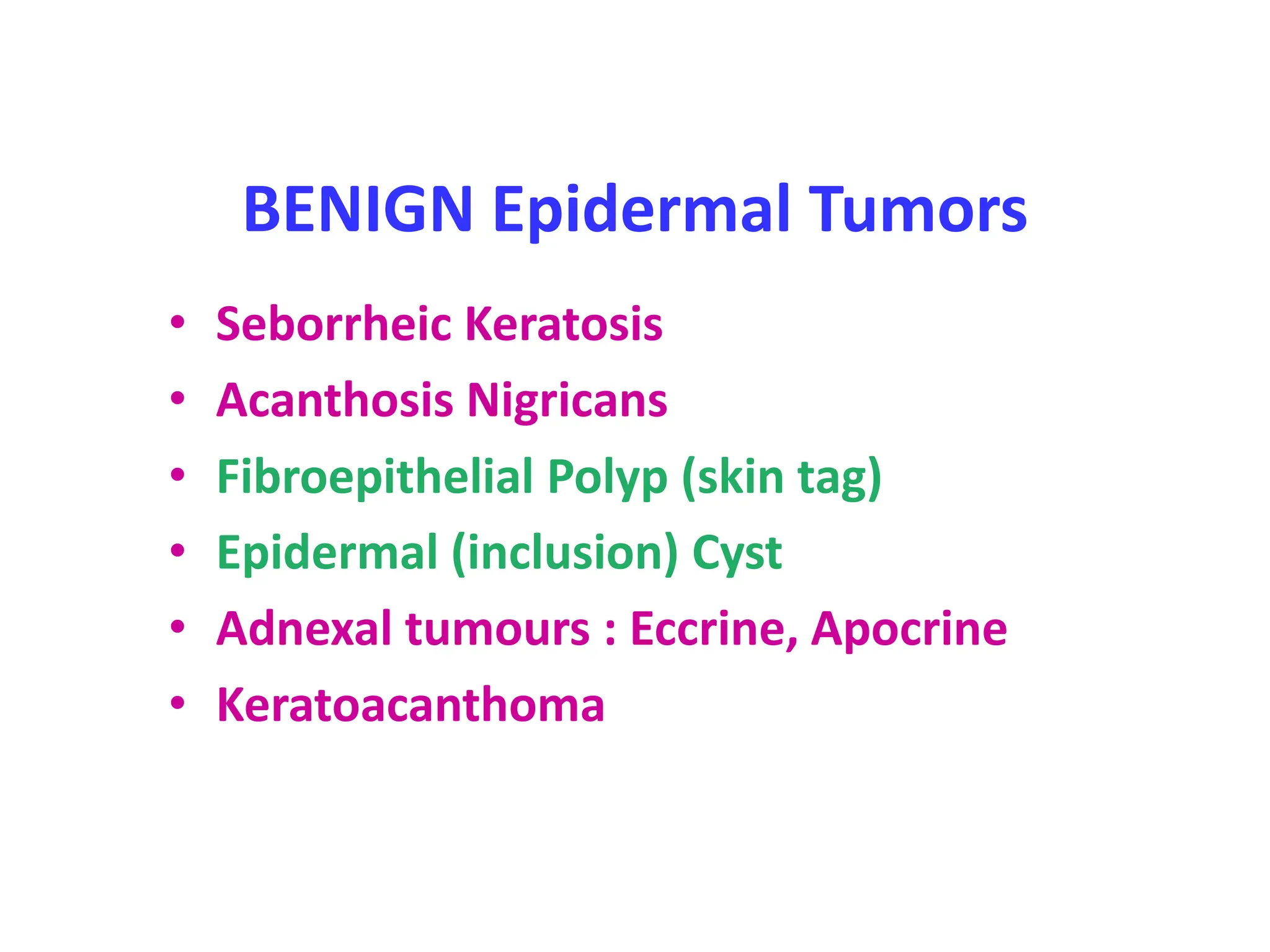
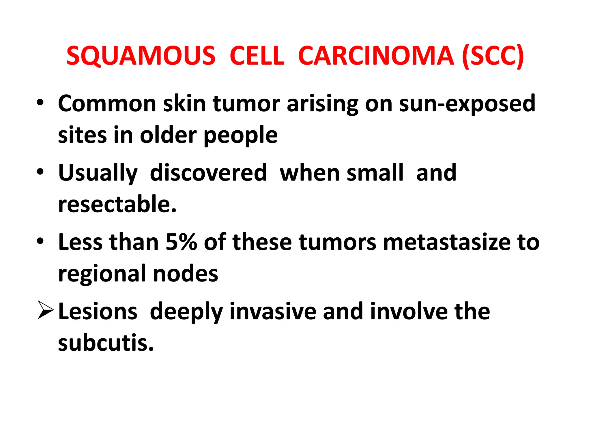
Non- melanocytic tumour
Dr Avinash Thakur
Department of Pathology
SKIN TUMOURS
1. Epidermal tumours
2. Adnexal tumours
3. Melanocytic tumours
4. Soft tissue tumors
5. Lymphomas
BENIGN Epidermal Tumors
• Seborrheic Keratosis
• Acanthosis Nigricans
• Fibroepithelial Polyp (skin tag)
• Epidermal (inclusion) Cyst
• Adnexal tumours : Eccrine, Apocrine
• Keratoacanthoma
SQUAMOUS CELL CARCINOMA (SCC)
• Common skin tumor arising on sun-exposed
sites in older people
• Usually discovered when small and
resectable.
• Less than 5% of these tumors metastasize to
regional nodes
Lesions deeply invasive and involve the
subcutis.
COMMON SITES
• Face
• Pinna of ear
• Back of hands
• Mucocutaneous junction
Lips ,Anal canal , Glans penis
• Preexisting inflammatory and degenerative
lesions .
AETIOLOGY
1. premalignant lesions (actinic keratosis)
2. exposure to ultraviolet (UV) light
3. Xeroderma pigmentosum (inherited defects in
DNA repair)
4. chronic ulcers & draining osteomyelitis
5. old burn scars (Marjolin’s ulcer)
6. ionizing radiation
7. Industrial carcinogens
8. Immunosuppression
GROSS MORPHOLOGY OF
SQUAMOUS CELL CARCINOMA
1. Ulcerated growth
2. Fungated or polypoid
growth
MICROSCOPY OF
SQUAMOUS CELL CARCINOMA
1. Invasion
2. Pleomorphism
3. Keratin pearl
formation
4. Increased mitosis
5. Necrosis
MORPHOLOGY OF SQUAMOUS CELL
CARCINOMA
BASAL CELL CARCINOMA (RODENT ULCER)
• Common skin tumor arising on sun-exposed sites
• Common in middle aged whites
• Are slow growing and locally invasive
• Rarely metastasizes.
• Most common location:-- Face (90%)
AETIOLOGY
1. Exposure to ultraviolet (UV) light
2. Immunosuppression
3. Xeroderma pigmentosum (inherited defects
in DNA repair)
GROSS MORPHOLOGY OF BCC
• BCC may manifest as –Erythematous patch
papule or
nodule with telangiectasis
which is often eroded , or
ulcerated
GROSS MORPHOLOGY OF BASAL CELL CARCINOMA
(CONTD)
• presents as:
1. Telangiectasias
• (pearly papules with
prominent, dilated
subepidermal blood
vessels
GROSS MORPHOLOGY OF
BASAL CELL CARCINOMA
2. Ulcers
• extensive local
invasion of bone or
facial sinuses.
 Rodent ulcers
MICROSCOPY OF
BASAL CELL CARCINOMA
1. Proliferation of
basaloid cells
arising from
epidermis
MICROSCOPY OF
BASAL CELL CARCINOMA
2. Cells arranged in
nests and cords
3. Clefts around the
nests
4. Peripheral nuclear
palisading
THANK YOU

Pathology of skin tumors in detail 12.pptx

  • 1.
    Non- melanocytic tumour DrAvinash Thakur Department of Pathology
  • 2.
    SKIN TUMOURS 1. Epidermaltumours 2. Adnexal tumours 3. Melanocytic tumours 4. Soft tissue tumors 5. Lymphomas
  • 4.
    BENIGN Epidermal Tumors •Seborrheic Keratosis • Acanthosis Nigricans • Fibroepithelial Polyp (skin tag) • Epidermal (inclusion) Cyst • Adnexal tumours : Eccrine, Apocrine • Keratoacanthoma
  • 5.
    SQUAMOUS CELL CARCINOMA(SCC) • Common skin tumor arising on sun-exposed sites in older people • Usually discovered when small and resectable. • Less than 5% of these tumors metastasize to regional nodes Lesions deeply invasive and involve the subcutis.
  • 6.
    COMMON SITES • Face •Pinna of ear • Back of hands • Mucocutaneous junction Lips ,Anal canal , Glans penis • Preexisting inflammatory and degenerative lesions .
  • 7.
    AETIOLOGY 1. premalignant lesions(actinic keratosis) 2. exposure to ultraviolet (UV) light 3. Xeroderma pigmentosum (inherited defects in DNA repair) 4. chronic ulcers & draining osteomyelitis 5. old burn scars (Marjolin’s ulcer) 6. ionizing radiation 7. Industrial carcinogens 8. Immunosuppression
  • 8.
    GROSS MORPHOLOGY OF SQUAMOUSCELL CARCINOMA 1. Ulcerated growth 2. Fungated or polypoid growth
  • 9.
    MICROSCOPY OF SQUAMOUS CELLCARCINOMA 1. Invasion 2. Pleomorphism 3. Keratin pearl formation 4. Increased mitosis 5. Necrosis
  • 11.
    MORPHOLOGY OF SQUAMOUSCELL CARCINOMA
  • 12.
    BASAL CELL CARCINOMA(RODENT ULCER) • Common skin tumor arising on sun-exposed sites • Common in middle aged whites • Are slow growing and locally invasive • Rarely metastasizes. • Most common location:-- Face (90%)
  • 13.
    AETIOLOGY 1. Exposure toultraviolet (UV) light 2. Immunosuppression 3. Xeroderma pigmentosum (inherited defects in DNA repair)
  • 14.
    GROSS MORPHOLOGY OFBCC • BCC may manifest as –Erythematous patch papule or nodule with telangiectasis which is often eroded , or ulcerated
  • 15.
    GROSS MORPHOLOGY OFBASAL CELL CARCINOMA (CONTD) • presents as: 1. Telangiectasias • (pearly papules with prominent, dilated subepidermal blood vessels
  • 16.
    GROSS MORPHOLOGY OF BASALCELL CARCINOMA 2. Ulcers • extensive local invasion of bone or facial sinuses.  Rodent ulcers
  • 17.
    MICROSCOPY OF BASAL CELLCARCINOMA 1. Proliferation of basaloid cells arising from epidermis
  • 18.
    MICROSCOPY OF BASAL CELLCARCINOMA 2. Cells arranged in nests and cords 3. Clefts around the nests 4. Peripheral nuclear palisading
  • 19.

Editor's Notes

  • #12 Ionising radiation: Arsenic Immunosuppression : increases the risk of HPV infections Xeroderma pigmentosa: mutations in genes that are responsible for nucleotide excision repair pathway
  • #13 A firm bump on the skin, called a nodule. The nodule might be the same color as the skin, or it might look different. It can look pink, red, black or brown, depending on skin color. A flat sore with a scaly crust. A new sore or raised area on an old scar or sore. A rough, scaly patch on the lip that may become an open sore. A sore or rough patch inside the mouth. A raised patch or wartlike sore on or in the anus or on the genitals.
  • #19 Waxy papules with central depression. Pearly appearance. Erosion or ulceration, often central. Bleeding, especially when traumatized. Crusting. Rolled (raised) border. Translucency. Telangiectases over the surface.
  • #25 Fair skinned individuals have less amount of melanin production that increases the amount of damage caused by sun exposure
  • #27 ABCDEs of melanoma