ABSTRACT: The parathyroid glands are involved in calcium homeostasis. Primary hyperparathyroidism (PHPT) is the most common cause of elevated parathyroid hormone (PTH) and calcium levels. Parathyroidectomy is excision of one or more parathyroid glands. This is a surgical management for hyperparathyroidism. Objective: Retrospective study of 68 cases over 2 years and literature review to assess if Parathyroidectomy can be safely performed as a daycase surgery. Method: Retrospective study of postoperative outcome over 2 years, of 68 cases who had parathyroidectomy procedure. Postoperative complications, duration of hospital stay and indication for parathyroidectomy was analysed. Effects of change in parathyroid hormone levels in blood postoperatively, importance of imaging to localise the parathyroid lesion in order to achieve minimally invasive surgery and safe postoperative outcome is discussed. Result: 59 patients out of 68 had PHPT. 53 of them were discharged on the same day, that is 89.8%. Remaining 6 patients were admitted overnight to confirm their postoperative normal calcium levels. None of them developed any postoperative complication. 40.7% of these PHPT patients were prescribed calcium supplement prophylactically. 33.3% of secondary and tertiary hyperparathyroidism (HPT) patients were discharged on the same day. All of them were followed up by endocrinologists or renal team postoperatively to regulate plasma calcium levels. Conclusion: Parathyroidectomy for Primary hyperparathyroidism can be performed safely as day case surgery, if the adenoma is accurately localised by dual modality imaging.
2. Parathyroidectomy as a Daycase Surgery: Retrospective Study
*Corresponding Author: Ms.Rupali Sawant1
28 | Page
None of the patients develop hypocalcemia postoperatively. In this study the patients’ age varied from 21years
to 67 years, with average age of 43years. Female patients were more, female: male ratio being 4.66.
Fig.1 Hospital stay of the patients
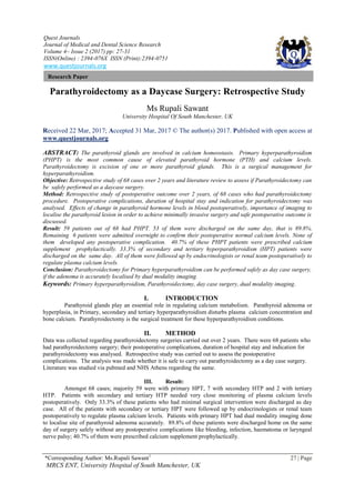
Fig.2 Distribution of patients as per indication of surgery
Number of patients
Primary HPTH
Secondary HPTH
Tertiary HPTH
3. Parathyroidectomy as a Daycase Surgery: Retrospective Study
*Corresponding Author: Ms.Rupali Sawant1
29 | Page
IV. DISCUSSION
The parathyroid glands are involved in calcium homeostasis. They release parathyroid hormone (PTH)
in response to low serum concentrations of ionized calcium, and the release of the hormone is inhibited by an
increase in serum ionized calcium. PTH causes the kidneys to increase the tubular resorption of calcium and
decrease the resorption of phosphorus.[1] PTH also acts on bone and the intestine to increase serum calcium
levels.
Fig.3 Calcium metabolism
IV.I Hyperparathyroidism- Primary hyperparathyroidism (PHPT) is defined as abnormal hypersecretion of
parathyroid hormone (PTH), producing hypercalcemia and hypophosphatemia, even high normal parathyroid
hormone levels are considered pathologic in patients with chronic hypercalcemia.
Primary hyperparathyroidism (PHPT) is the most common cause of elevated parathyroid hormone (PTH) and
alcium levels. Approximately 85% of cases are found to be caused by an isolated parathyroid adenoma, 15%
by diffuse parathyroid hyperplasia, and less than 1% by parathyroid carcinoma. Other causes include neck or
mediastinal parathyroid cysts, which are also uncommon. Rarely, primary hyperparathyroidism (PHPT) may be
related to multiple endocrine neoplasia (MEN); family history or other endocrine tumor warrants screening for
MEN.[2] Localising adenoma: Parathyroid adenomas greater than 3g are called as giant parathyroid adenomas,
preferably they are removed via a large collar incision.[3] If present in the superior mediastinum may require a
sternotomy.[4] 1-3% of parathyroid glands are found in ectopic locations. The most common ectopic site is in
the mediastinum, as many as 25%.[5] The embryological descent of the inferior parathyroid glands accompany
the thymus from the third pharyngeal pouch to the lower neck and superior mediastinum. Therefore, ectopic
parathyroid gland can be seen from the angle of the jaw to the pericardium.[4] One case study has demonstrated
minimally invasive surgery for giant mediastinal parathyroid adenoma, which was performed as a day case
surgery.[6]
A prospective cohort study by Vaidya indicated that a connection exists between low physical activity and
the development of primary hyperparathyroidism. The study, which included 69,621 females, found that the
age-adjusted relative risk of developing primary hyperparathyroidism was reduced by 50% in women in the
highest physical activity quintile, compared with women in the lowest quintile. The investigators also found
that, compared with participants with high physical activity and high calcium intake, the adjusted relative risk of
developing the condition was 2.37-fold greater in women with a combination of low physical activity and low
calcium intake.[7]
Secondary hyperparathyroidism (HPT) is a compensatory hyperfunctioning of the parathyroid glands
caused by hypocalcemia or peripheral resistance to parathyroid hormone. As opposed to primary
hyperparathyroidism (PHPT), treating the underlying cause can reverse secondary hyperparathyroidism (HPT).
The most common setting is in a patient with end-organ failure from chronic renal insufficiency, with
hypocalcemia and hyperphosphatemia. Advanced age and malnutrition are risk factors for developing
secondary hyperparathyroidism in patients with chronic kidney disease. Less commonly, secondary
hyperparathyroidism may be caused by calcium malabsorption, osteomalacia, vitamin D deficiency, or deranged
4. Parathyroidectomy as a Daycase Surgery: Retrospective Study
*Corresponding Author: Ms.Rupali Sawant1
30 | Page
vitamin D metabolism. Tertiary hyperparathyroidism (HPT) occurs in a setting of previous secondary
hyperparathyroidism (HPT) in which the glandular hyperfunction and hypersecretion continue despite correction
of the underlying abnormality, as in renal transplantation.
Indications for parathyroidectomy-
1. Primary Hyperparathyroidism who are symptomatic
2. Refractory secondary hyperparathyroidism
3. Asymptomatic primary hyperparathyrodism with following:
• Serum calcium level more than 1.0 mg/dL above the upper limit of normal
• Marked hypercalciuria (> 400 mg/day) or renal stones
• Creatinine clearance less than 30% of normal
• Marked bone density reduction with a T-score lower than 2.5 at any site
• Age less than 50 years (if the problem is left untreated, many of these younger patients eventually
develop complications of primary hyperparathyroidism)
• A patient who requests surgery or a patient for whom surveillance and follow-up are difficult or
impossible.
Surgical procedure involves smaller incision over neck anteriorly. Dissection continues along the
thyroid capsule, and the thyroid gland is rotated anteriorly and medially. If a preoperative localization study
suggested either a superior or inferior gland, the corresponding area is examined first. The location of the
superior parathyroid gland is sought on the posterior and lateral aspect of the thyroid gland. The middle thyroid
veins are ligated, and the gland is rotated. The superior gland should be located deep to the plane of the
recurrent laryngeal nerve and superior to the intersection of the recurrent laryngeal nerve and the inferior thyroid
artery. The superior gland is often within 1 cm of the cricothyroid cartilage articulation. Care should be taken
to maintain excellent hemostasis. Preoperative imaging plays a major role to increase surgical success via a
minimally invasive approach, a significant reduction in operating time and complications. The imaging used are
ultrasonography and Sestamibi scan as first line imaging; computerised tomography(CT) scans, Four
dimentional computed tomography(4DCT), Magnetic Resonance Imaging(MRI) and Positron emission
tomography and computed tomography(PET-CT) for revision surgeries. USS is a non-invasive, inexpensive
outpatient procedure without radiation to the patient, which helps to localise the adenomatous gland in relation
to adjacent structures with precision. Technetium99m methoxyisobutylisonitrile(Sestamibi)scan picks up the
hyperfunctioning gland, which retains the Technetium longer than the other parathyroids and thyroid gland due
to its increased mitochondrial activity.[8] Sestamibi-single photon emission computed tomography(SPECT-
MIBI) provides a multi-dimentional higher resolution images with a 92-98% sensitivity compared to 71-79% of
using MIBI alone.[9,10]
Complications of Procedure- With parathyroidectomy, as with all surgical procedures, bleeding and
infection are potential complications. Because parathyroidectomy is a clean operation and because meticulous
homeostasis is crucial to its performance, both of these complications should be rare. There is a risk of injury to
the recurrent and superior laryngeal nerves. In difficult cases, where the abnormal gland is not easily found
easily, it is important to identify the recurrent laryngeal nerve, both to protect it from injury and to have it
available as a landmark during the dissection. Failure to cure the hyperparathyroidism, persistent or recurrent
hypercalcemia, and postoperative hypocalcemia are also potential adverse results of parathyroidectomy.
Hungry bone syndrome: Transient hypoparathyroidism after resecting significant amounts of
parathyroid glands, which were secreting excess PTH, due to hyperplasia or an adenoma; resulting in high
alkaline phosphatase and significant bone demineralisation and rapid rebound recalcification of bones after
prolonged hypocalcaemia. Bone hunger is exacerbated by pre-existing renal dysfunction.[11]
V. CONCLUSION
None of the patients had other surgical postoperative complications than hypocalcaemia. Patients with
secondary and tertiary HPT needed monitoring of plasma calcium levels postoperatively. Patients with primary
HPT did not present with any postoperative complication. Parathyroidectomy can be performed as a day-case
surgery, if the adenoma is accurately localised by dual modality imaging. The anatomical findings are reinforced
by performing the Sestamibi (MIBI) scan, which is a functional scan. The location of the adenoma was
determined anatomically via ultrasonography and functionally via sestamibi scan.[12] There was a 96%
concordance between intra-operative location of the adenoma with the pre-operative imaging.
5. Parathyroidectomy as a Daycase Surgery: Retrospective Study
*Corresponding Author: Ms.Rupali Sawant1
31 | Page
REFERENCES
[1]. Blaine J, Chonchol M, Levi M. Renal control of calcium,phosphate and magnesium homeostasis. Clinical Journal of the
American Society of Nephrology 2015; 10(7):1257-72.
[2]. Allerheiligen DA, Schoeber J, Houston RE, et al. Hyperparathyroidism. American family physician 15 April 1998; 57(8):1795-
802,1807-8.
[3]. Christakis I, MichasS, Mouziouras V et al.Minimally invasive techniques for giant parathyroid tumours; an oxymoron? Hell J
Surg 2012;84:220-24.
[4]. Caporale DM, Bobbio A, Accordino R et al. Endoscopic mediastinal parathyroid adenoma. Acta Biomed 2003;74:157-59.
[5]. Cupisti K, Dotzenrath C, Simon D et al. Therapy of suspected intrathoracic parathyroid adenomas.
[6]. Langenbecks Arch Surg 2002; 386: 488- 93.
[7]. A Haldar, A Thapar, S Khan et al. Day case minimally invasive excision of a giant mediastinal parathyroid adenoma. RCS
Annals July 2014; 96(5):21-23.
[8]. A Vaidya,G Curhan, J Paik et al. Physical activity and risk of primary hyperparathyroidism. J Clin Endocrinol Metab
2016;101(4):1590-97.
[9]. Hetrakul N, Civelek AC, Stagg CA et al. In vitro accumulation of technetium-99m-Sestamibi in human parathyroid
mitochondria. Surgery. 2001;130(6):1011-8.
[10]. Civelek AC, Ozalp E, Donovan P et al. Prospective evaluation of delayed technetium-99m Sestamibi SPECT scintigraphy for
preoperative localization of primary hyperparathyroidism. Surgery.2002;131(2):149-57.
[11]. Nichols KJ, Tomas MB, Tronco GG. Preoperative parathyroid scintigraphic lesion localization:accuracy of various types of
readings. Radiology.2008;248(1):221-32.
[12]. Segen’s Medical Dictionary. 2012 Farlex
[13]. Abbas,Yasmin,Quraishi M. Outcomes of minimally invasive parathyroidectomy with dual-modality imaging.Journal of ENT
Masterclass 2014:vol.7:144-48.