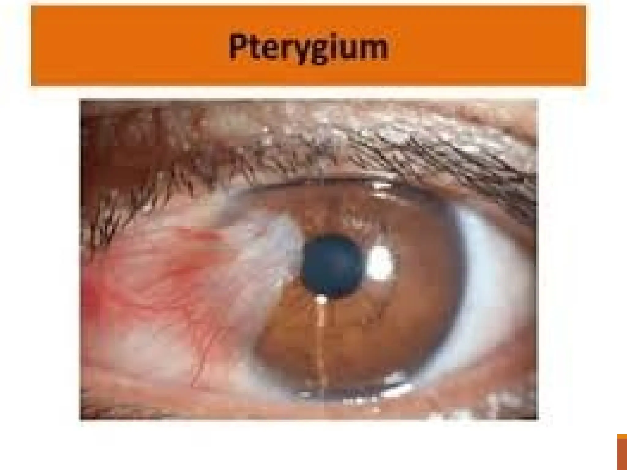
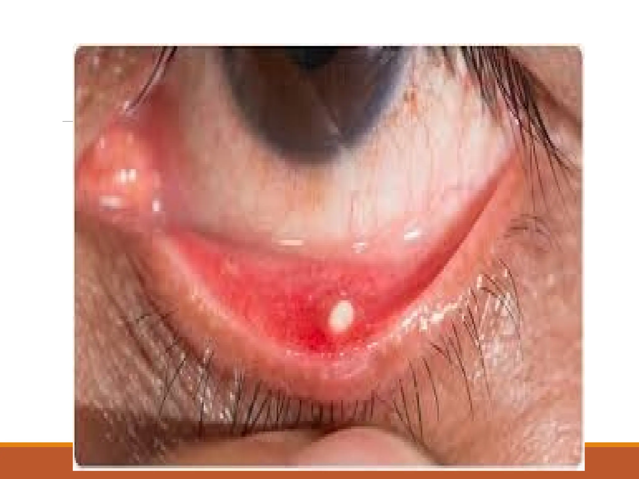
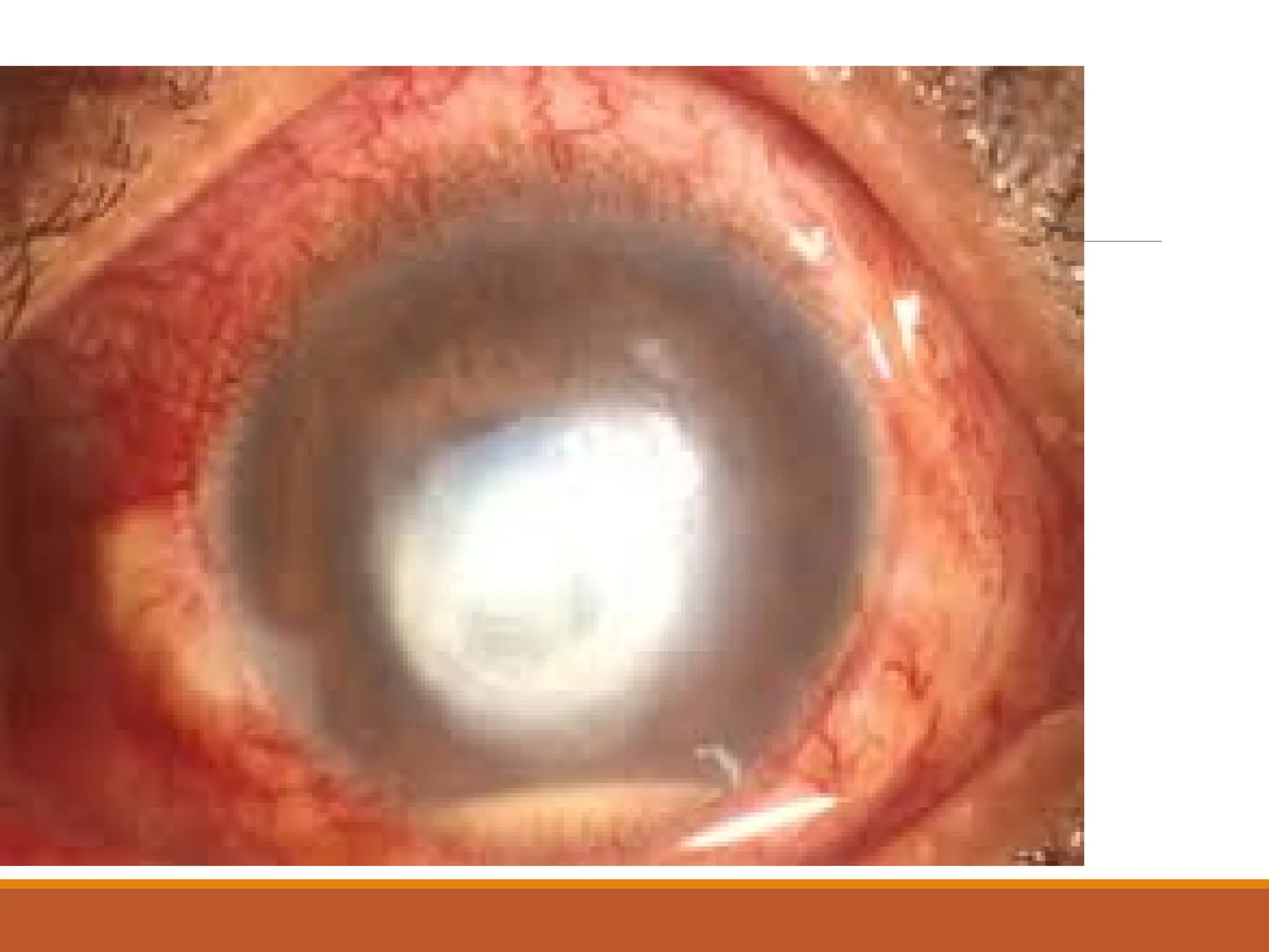
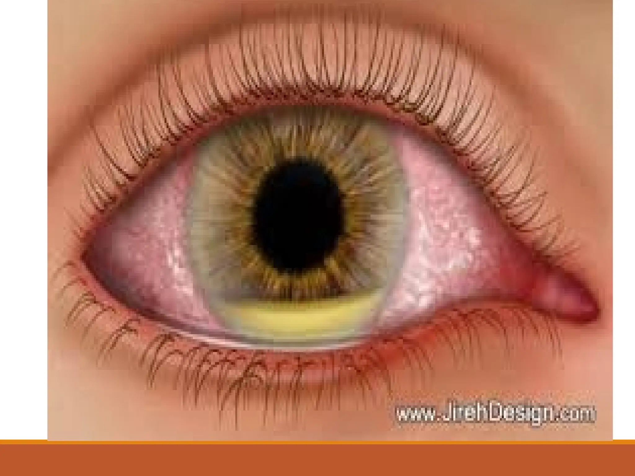
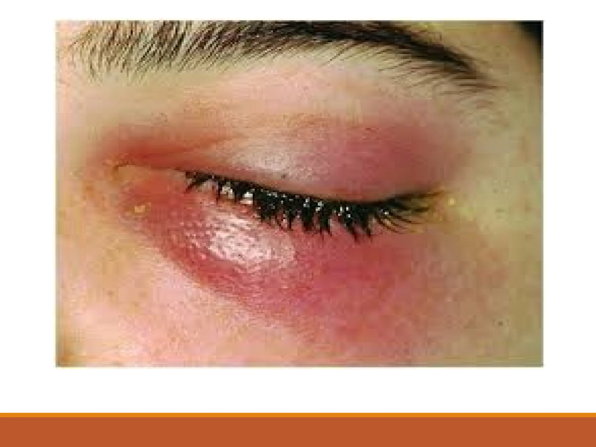
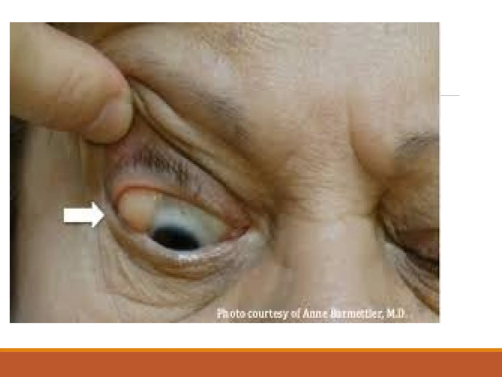
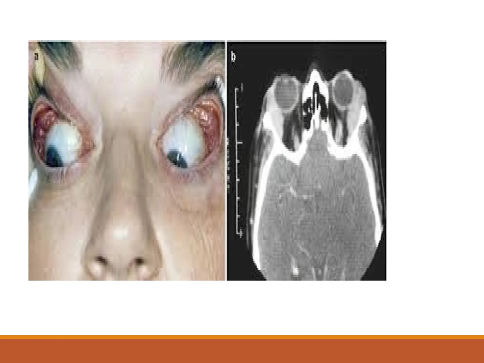
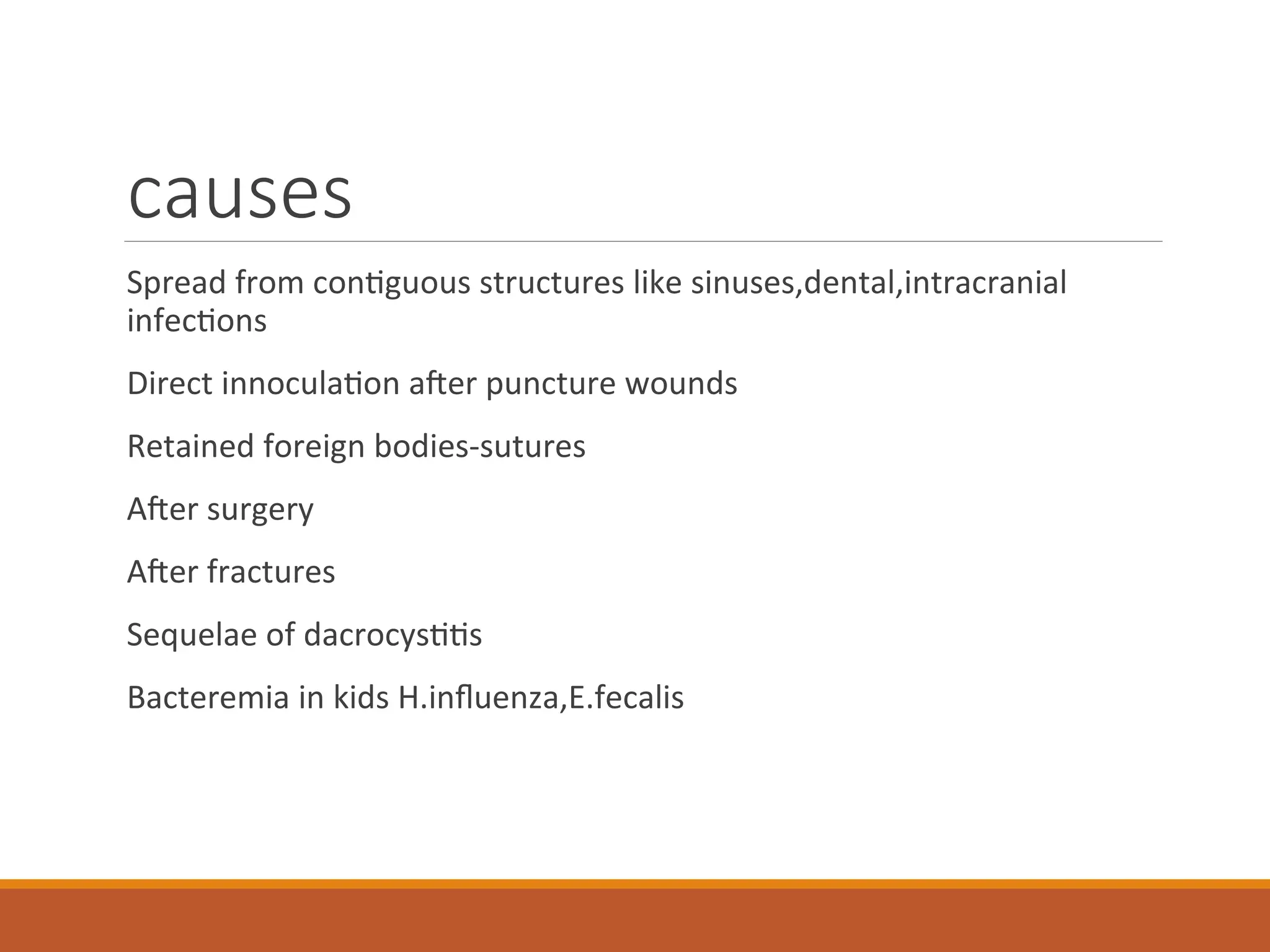
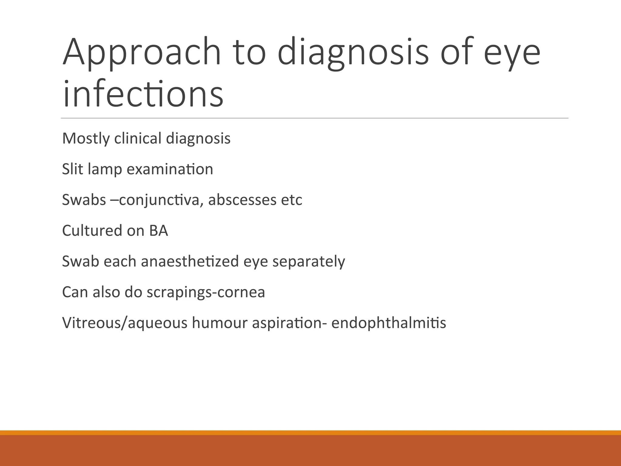
Eye infections
KIBET M.P
MSC MPE
BSC OPHTAL&CATR SURG
DIP CM & SURG
Anatomy
Anatomy
Anatomy
 The anterior chamber is the area bounded in front by the cornea
and in back by the lens, and filled with aqueous.
 The aqueous is a clear, watery solution in the anterior
and posterior chambers.
 The artery is the vessel supplying blood to the eye.
 The canal of Schlemm is the passageway for the aqueous fluid to
leave the eye.
Anatomy
 The choroid , which carries blood vessels, is the inner coat between
the sclera and the retina .
 The ciliary body is an unseen part of the iris , and these together
with the ora serrata form the uveal tract.
 The conjunctiva is a clear membrane covering the white of the eye
(sclera).
 The cornea is a clear, transparent portion of the outer coat of the
eyeball through which light passes to the lens.
Anatomy
 The iris gives our eyes color and it functions like the aperture on a
camera, enlarging in dim light and contracting in bright light. The
aperture itself is known as the pupil
 The lens helps to focus light on the retina.
 The macula is a small area in the retina that provides our most central,
acute vision.
 The optic nerve conducts visual impulses to the brain from the retina.
 The ora serrata and the ciliary body form the uveal tract, an unseen part
of the iris.
Anatomy
 The posterior chamber is the area behind the iris, but in front of the lens, that is
filled with aqueous.
 The pupil is the opening, or aperture, of the iris.
 The rectus medialis is one of the six muscles of the eye.
 The retina is the innermost coat of the back of the eye, formed of light-sensitive
nerve endings that carry the visual impulse to the optic nerve. The retina may be
compared to the film of a camera.
 The sclera is the white of the eye.
 The vein is the vessel that carries blood away from the eye.
 The vitreous is a transparent, colorless mass of soft, gelatinous material filling the
eyeball behind the lens.
anatomy
The eyeball is protected anteriorly by the eyelids
And contained in the orbit
Normal flora of the eye
Predorminant organisms
Diphtheroids
S.epidermidis
Non hemolytic strep
Eye infections
The infections could be:-
Acute
Chronic
Primary
secondary
conjunctiva
Conjunctivitis is the most common ocular inflammation
Clinical manifestations-hyperemia,secretion –due to exudates of
inflammatory cells and fibrin rich edematous fluid-which may be
purulent,mucopurulent,fibrinous or serosanguinous depending on the
cause.
When the exudate dries ,the eyelids stick together
conjunctiva
The normal transparency may be lost
Papillae may form especially in tarsal conjunctiva
Symptoms include gritty eyes,photophobia,diminished vision and pain
organisms
Staph epidermidis
Acinetobacter
Aeromonas hydrophila
Peptostreptococcus
Bartonella
* most common
conjunctivitis
Routes of entry
Routes of entry-hand to eye
-airborne formites
-contact with URTIs
-contact with genital tract
infections
spread from adjacent structures-face
and eyelids,sinuses
-Hematogenous spread -rare
Determinants of infective
agents
Age-
neisseriae /chlamydia-newborns
Children-
influenza,strep pneumo,staph aureus
Young adults-
strep pneumo,staph aureus/epidermidis
Management/control
Mostly self limiting
Px education-hand washing!
Rx-topical gentamicin/tobramycin-gram neg
Neomycin/polymixin-gram pos
Topical quinolones-severe infections
Parenteral ceftriaxone for gonococcal
Erythromycin syrup for chlamydia in neonates/erythromycin ointment.
Cornea
Inflammation of the cornea
Clinically presents as loss of vision,,tearing,photophobia and
blepharospasm,ulceration
Symptoms-foreign body sensation,pain
Organisms implicated
Gram pos cocci- gram neg bacilli
*Staph aureus .*pseudomonas
Staph epidermidis . proteus
Strep viridans .klebsiella
Strep pyogenes .serratia
Strep fecalis .E.coli
Peptostreptococcus * most common
*Strep pneumo
organisms
Gram neg coccobacilli gram-positive bacil
Moraxella corynebacterium
Pasturella c.tetani/c.perfringen
Morganella bacillus cereus
Serratia spirochetes
E.coli treponema
Aeromonas borrelia burgdoferi
mycobateria-tb,mac
Routes of entry/predisposing
factors
Direct penetration-organisms producing toxins/enzymes/virulent
factors-neisseria
Following injury,eyelid abnormalities,tear dysfuntional states,corneal
anesthesia
Immunocompromised states
Use of contact lenses
Treatment
Broad spectrum antibiotics used pending lab results-cephalosporins
+aminoglycosides
Aminoglycosides can be used synergistically with ticarcillin.
Quinolones-pseudomonas and gram negatives
Use topical antibiotics
Parenteral-severe cases
Steroids??
Endophthalmitis
Most cases develop after intraocular surgery-cataract surgery.
Organisms involved-microflora
Clinically-decreased visual acuity,pain,hypopion,hyperemia
organisms
 Staph aureus .E.coli
 Staph epidermidis .H.influenza
 Strep pneumo .klebsiella
 Bacillus cereus .moraxella
 Corynebacteria spp .proteus
 Listeria .pseudomonas
 N.meningitidis .s.typhimurium
 Acinetobacter .serratia
 Enterobacter .clostridium
 Propiono bacterium acnes treponema pallidum
 Actinomyctes israeli .m.tuberculosis/leprae
Treatment
Is according to culture and sensitivity
Iv antibiotics-3G cephalosporins
Intravitreal vancomycin-s.aureus
Sx-vitrectomy
Steroids??
Periocular infections
These involve orbit and cellular adnexa
Principal periocular structure susceptible to infections are eyelids ,the
components of lacrimal apparatus and the orbit.
Eyelids
Inflammation of the lid margins-blepharitis
Often chronic and bilateral
Two types-anterior-staphylococcal
-posterior-meibominitis
Organisms
Staphaureus,epidermidis,pseudomonas,proteus,moraxella
.Mascara used has been implicated
Eyelids
Erysipelas-acute cellulitis –strep pyogenes,staph aureus-invasion of
subcutaneous after trauma
Hordeolum-internal/external depending on glands involved-staph
implicated
Internal-meibomian gland infection
External-stye infection of glands of zeis sebaceous gland of eye lids
Lacrimal apparatus
Produce the aqueous component of tear film
Canaliculitis-chronic inflammation of canaliculi-by
propionibacterium,actinomyces
Dacrocystitis-inflammation of lacrimal sac-
streppneumo,staphaureus,pseudomonas,chlamydia,h.influenza in
children
Clinically-epiphora
Lacrimal app
Dacroadenitis-inflammation of main lacrimal gland-
staph,strep,tuberculosis-chronic
Orbit and carvenous sinus
Cellulitis-pre septal anterior orbit septum and post septal-orbital
contents
Serious-loss of sight and spread to carvenous sinus leading to
thrombosis and death,
causes
Spread from contiguous structures like sinuses,dental,intracranial
infections
Direct innoculation after puncture wounds
Retained foreign bodies-sutures
After surgery
After fractures
Sequelae of dacrocystitis
Bacteremia in kids H.influenza,E.fecalis
organisms
Staph aureus
Strep pyogenes
Strep pneumo
Clostridia
H.influenza-<5s
Tb-hematogenous spread
Clinical
Evidence of trauma-bleedng,fever,lid edema and rhinorrhoea.
Pain,headache,loss of vision
Tenderness,black eye,proptosis
Treatment
Blepharitis-Topical –bacitracin,erthromycin
Steroids-reduce inflammation
Hordeolum-warm compresses and sytsemic antibiotics if multiple or no
response I&D if not responding to rx
Canalliculitis-antibiotic irrigation with penicillin G
Dacrocystitis-oral penicillin+warm compresses
treatment
Dacroadenitis-systemic antibiotics
Cellulitis-cloxacillin,oxacillin,cephalexin
Clindamycin for gram neg
Iv antibiotics orbital cellulitis
Approach to diagnosis of eye
infections
Mostly clinical diagnosis
Slit lamp examination
Swabs –conjunctiva, abscesses etc
Cultured on BA
Swab each anaesthetized eye separately
Can also do scrapings-cornea
Vitreous/aqueous humour aspiration- endophthalmitis
diagnosis
 Gram stain
 ELISA
 Dna/pcr-chlamydia
 Fluorescent microscopy
 u/s,ct,MRI for cellulitis
ANY QUESTION FOR THE
FACULTY?
DANKE

ophthalmic common infections. introduction

  • 1.
    Eye infections KIBET M.P MSCMPE BSC OPHTAL&CATR SURG DIP CM & SURG
  • 2.
  • 3.
  • 4.
    Anatomy  The anteriorchamber is the area bounded in front by the cornea and in back by the lens, and filled with aqueous.  The aqueous is a clear, watery solution in the anterior and posterior chambers.  The artery is the vessel supplying blood to the eye.  The canal of Schlemm is the passageway for the aqueous fluid to leave the eye.
  • 5.
    Anatomy  The choroid, which carries blood vessels, is the inner coat between the sclera and the retina .  The ciliary body is an unseen part of the iris , and these together with the ora serrata form the uveal tract.  The conjunctiva is a clear membrane covering the white of the eye (sclera).  The cornea is a clear, transparent portion of the outer coat of the eyeball through which light passes to the lens.
  • 6.
    Anatomy  The irisgives our eyes color and it functions like the aperture on a camera, enlarging in dim light and contracting in bright light. The aperture itself is known as the pupil  The lens helps to focus light on the retina.  The macula is a small area in the retina that provides our most central, acute vision.  The optic nerve conducts visual impulses to the brain from the retina.  The ora serrata and the ciliary body form the uveal tract, an unseen part of the iris.
  • 7.
    Anatomy  The posteriorchamber is the area behind the iris, but in front of the lens, that is filled with aqueous.  The pupil is the opening, or aperture, of the iris.  The rectus medialis is one of the six muscles of the eye.  The retina is the innermost coat of the back of the eye, formed of light-sensitive nerve endings that carry the visual impulse to the optic nerve. The retina may be compared to the film of a camera.  The sclera is the white of the eye.  The vein is the vessel that carries blood away from the eye.  The vitreous is a transparent, colorless mass of soft, gelatinous material filling the eyeball behind the lens.
  • 8.
    anatomy The eyeball isprotected anteriorly by the eyelids And contained in the orbit
  • 9.
    Normal flora ofthe eye Predorminant organisms Diphtheroids S.epidermidis Non hemolytic strep
  • 10.
    Eye infections The infectionscould be:- Acute Chronic Primary secondary
  • 11.
    conjunctiva Conjunctivitis is themost common ocular inflammation Clinical manifestations-hyperemia,secretion –due to exudates of inflammatory cells and fibrin rich edematous fluid-which may be purulent,mucopurulent,fibrinous or serosanguinous depending on the cause. When the exudate dries ,the eyelids stick together
  • 12.
    conjunctiva The normal transparencymay be lost Papillae may form especially in tarsal conjunctiva Symptoms include gritty eyes,photophobia,diminished vision and pain
  • 13.
  • 18.
  • 19.
    Routes of entry Routesof entry-hand to eye -airborne formites -contact with URTIs -contact with genital tract infections spread from adjacent structures-face and eyelids,sinuses -Hematogenous spread -rare
  • 20.
    Determinants of infective agents Age- neisseriae/chlamydia-newborns Children- influenza,strep pneumo,staph aureus Young adults- strep pneumo,staph aureus/epidermidis
  • 21.
    Management/control Mostly self limiting Pxeducation-hand washing! Rx-topical gentamicin/tobramycin-gram neg Neomycin/polymixin-gram pos Topical quinolones-severe infections Parenteral ceftriaxone for gonococcal Erythromycin syrup for chlamydia in neonates/erythromycin ointment.
  • 22.
    Cornea Inflammation of thecornea Clinically presents as loss of vision,,tearing,photophobia and blepharospasm,ulceration Symptoms-foreign body sensation,pain
  • 26.
    Organisms implicated Gram poscocci- gram neg bacilli *Staph aureus .*pseudomonas Staph epidermidis . proteus Strep viridans .klebsiella Strep pyogenes .serratia Strep fecalis .E.coli Peptostreptococcus * most common *Strep pneumo
  • 27.
    organisms Gram neg coccobacilligram-positive bacil Moraxella corynebacterium Pasturella c.tetani/c.perfringen Morganella bacillus cereus Serratia spirochetes E.coli treponema Aeromonas borrelia burgdoferi mycobateria-tb,mac
  • 28.
    Routes of entry/predisposing factors Directpenetration-organisms producing toxins/enzymes/virulent factors-neisseria Following injury,eyelid abnormalities,tear dysfuntional states,corneal anesthesia Immunocompromised states Use of contact lenses
  • 29.
    Treatment Broad spectrum antibioticsused pending lab results-cephalosporins +aminoglycosides Aminoglycosides can be used synergistically with ticarcillin. Quinolones-pseudomonas and gram negatives Use topical antibiotics Parenteral-severe cases Steroids??
  • 30.
    Endophthalmitis Most cases developafter intraocular surgery-cataract surgery. Organisms involved-microflora Clinically-decreased visual acuity,pain,hypopion,hyperemia
  • 34.
    organisms  Staph aureus.E.coli  Staph epidermidis .H.influenza  Strep pneumo .klebsiella  Bacillus cereus .moraxella  Corynebacteria spp .proteus  Listeria .pseudomonas  N.meningitidis .s.typhimurium  Acinetobacter .serratia  Enterobacter .clostridium  Propiono bacterium acnes treponema pallidum  Actinomyctes israeli .m.tuberculosis/leprae
  • 35.
    Treatment Is according toculture and sensitivity Iv antibiotics-3G cephalosporins Intravitreal vancomycin-s.aureus Sx-vitrectomy Steroids??
  • 36.
    Periocular infections These involveorbit and cellular adnexa Principal periocular structure susceptible to infections are eyelids ,the components of lacrimal apparatus and the orbit.
  • 40.
    Eyelids Inflammation of thelid margins-blepharitis Often chronic and bilateral Two types-anterior-staphylococcal -posterior-meibominitis Organisms Staphaureus,epidermidis,pseudomonas,proteus,moraxella .Mascara used has been implicated
  • 44.
    Eyelids Erysipelas-acute cellulitis –streppyogenes,staph aureus-invasion of subcutaneous after trauma Hordeolum-internal/external depending on glands involved-staph implicated Internal-meibomian gland infection External-stye infection of glands of zeis sebaceous gland of eye lids
  • 45.
    Lacrimal apparatus Produce theaqueous component of tear film Canaliculitis-chronic inflammation of canaliculi-by propionibacterium,actinomyces Dacrocystitis-inflammation of lacrimal sac- streppneumo,staphaureus,pseudomonas,chlamydia,h.influenza in children Clinically-epiphora
  • 46.
    Lacrimal app Dacroadenitis-inflammation ofmain lacrimal gland- staph,strep,tuberculosis-chronic
  • 49.
    Orbit and carvenoussinus Cellulitis-pre septal anterior orbit septum and post septal-orbital contents Serious-loss of sight and spread to carvenous sinus leading to thrombosis and death,
  • 50.
    causes Spread from contiguousstructures like sinuses,dental,intracranial infections Direct innoculation after puncture wounds Retained foreign bodies-sutures After surgery After fractures Sequelae of dacrocystitis Bacteremia in kids H.influenza,E.fecalis
  • 51.
    organisms Staph aureus Strep pyogenes Streppneumo Clostridia H.influenza-<5s Tb-hematogenous spread
  • 52.
    Clinical Evidence of trauma-bleedng,fever,lidedema and rhinorrhoea. Pain,headache,loss of vision Tenderness,black eye,proptosis
  • 53.
    Treatment Blepharitis-Topical –bacitracin,erthromycin Steroids-reduce inflammation Hordeolum-warmcompresses and sytsemic antibiotics if multiple or no response I&D if not responding to rx Canalliculitis-antibiotic irrigation with penicillin G Dacrocystitis-oral penicillin+warm compresses
  • 54.
  • 55.
    Approach to diagnosisof eye infections Mostly clinical diagnosis Slit lamp examination Swabs –conjunctiva, abscesses etc Cultured on BA Swab each anaesthetized eye separately Can also do scrapings-cornea Vitreous/aqueous humour aspiration- endophthalmitis
  • 56.
    diagnosis  Gram stain ELISA  Dna/pcr-chlamydia  Fluorescent microscopy  u/s,ct,MRI for cellulitis
  • 57.
    ANY QUESTION FORTHE FACULTY? DANKE