Download to read offline


![The high prevalence of OME—along with many issues,
including difficulties in diagnosis and assessing its duration,
associated conductive hearing loss, potential impact on
child development, and significant practice variations in
management—makes OME an important condition for up-
to-date clinical practice guidelines.
Purpose
The purpose of this multidisciplinary guideline is to iden-
tify quality improvement opportunities in managing
OME and to create explicit and actionable recommenda-
tions to implement these opportunities in clinical practice.
Specifically, the goals are to improve diagnostic accuracy,
identify children who are most susceptible to developmental
Table 2. Frequently Asked Questions: Understanding Ear Fluid.
Question Answer
What is ear fluid, and
how common is it?
Ear fluid, also called otitis media with effusion (OME), is a buildup of mucus or liquid behind the eardrum,
without symptoms of an ear infection. Nearly all children get ear fluid at least once by school age.
How does ear fluid differ
from an ear infection?
Ear infections (acute otitis media [AOM]) occur when germs (bacteria and/or viruses) enter the middle ear
and cause fever, ear pain, and active (acute) inflammation. Both AOM and OME have fluid in the middle
ear, but with OME the fluid is not actively infected, and pain may be absent or minimal.
If my child gets ear fluid,
how can I tell?
You might not be able to tell. Some children with OME have obvious hearing problems, but other children
may have no symptoms at all or more subtle findings (eg, ear rubbing, clumsiness, selective hearing,
disturbed sleep). Your doctor can detect ear fluid by looking in the ear canal (otoscopy) or by measuring
the movement of the eardrum (tympanometry or pneumatic otoscopy).
What causes ear fluid? OME may be caused by a cold, an ear infection (AOM), or the normal congestion (negative pressure) that
many young children have in their middle ear. Often OME is detected during a routine doctor’s visit, and
the exact cause is unknown.
Should I worry if my child
has ear fluid?
Most fluid goes away on its own in weeks or months, especially if it was caused by a cold or an ear
infection. OME is of more concern if it lasts .3 mo or when your child has other problems that could be
made worse by persistent ear fluid (eg, delays in speech, language, learning, or development). Your doctor
should check the ears periodically until the fluid is gone.
What is the best way to
manage ear fluid?
There are many opinions about managing OME, but the best advice can be found in clinical practice
guidelines, which make recommendations based on best available evidence and by considering the
potential benefits and harms of different strategies.
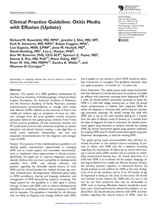
Figure 2. Comparison of otitis media with effusion (top) and acute
otitis media (bottom). The left images show the appearance of the
eardrum on otoscopy, and the right images depict the middle ear
space. For otitis media with effusion, the middle ear space is filled
with mucus or liquid (top right). For acute otitis media, the middle
ear space is filled with pus, and the pressure causes the eardrum to
bulge outward (bottom right). With permission from Rosenfeld 2005.
Figure 3. Position of the eustachian tube (red) as it connects the
middle ear space to the back of the nose, or nasopharynx. The
child’s eustachian tube (right) is shorter, more floppy, and more
horizontal, which makes it less effective in ventilating and protect-
ing the middle ear than the eustachian tube in the adult (left).
S4 Otolaryngology–Head and Neck Surgery 154(1S)
at HINARI - Parent on February 15, 2016oto.sagepub.comDownloaded from](https://image.slidesharecdn.com/omeguidelines-160901151853/85/Ome-guidelines-4-320.jpg)

![Direct and Indirect Costs
Direct costs related to otitis media, which includes OME
and AOM, are $3 billion to $5 billion annually,48-51
and the
true economic impact is likely higher, because indirect costs
are sizable yet difficult to estimate.37,52
Studies of AOM
suggest that the indirect cost of lost caregiver productivity
may far exceed that of the direct cost of medical treat-
ment.52
In addition, the estimated net cost of impaired well-
being from otitis media is $1.1 billion to $2.6 billion.53,54
The direct costs of managing OME include medical ther-
apy, which is largely ineffective. Antibiotics, for example,
have short-term efficacy, but long-term use cannot be justi-
fied because of concerns over adverse events and induced
bacterial resistance.55
Although several studies have shown
an association between gastroesophageal reflux and OME,
the limited evidence regarding antireflux therapy does not
show significant benefits.56
Similarly, despite a high preva-
lence of atopic conditions, such as allergic rhinitis, in chil-
dren with OME,57-59
there are no benefits to routinely
treating with antihistamines, decongestants, or steroids (sys-
temic or topical intranasal).3,60,61
Most studies, however, do
not consider the allergy status of children, and it is unknown
if those with proven allergies might respond differently.
Methods
General Methods and Literature Search
In developing this update of the evidence-based clinical
practice guideline, the methods outlined in the third edition
of the AAO-HNSF’s guideline development manual were
followed explicitly.62
An executive summary of the original OME guideline1
was sent to a panel of expert reviewers from the fields of
general otolaryngology, pediatric otolaryngology, otology,
family practice, pediatrics, nursing, audiology, and speech
language pathology who assessed the key action statements
to decide if they should be kept in their current form, revised,
or removed and to identify new research that might affect the
guideline recommendations. The reviewers concluded that
the original guideline action statements remained valid but
should be updated with major modifications. Suggestions
were also made for new key action statements.
An information specialist conducted 2 systematic litera-
ture searches using a validated filter strategy to identify
clinical practice guidelines, systematic reviews, and RCTs
published since the prior guideline (2004). Search terms
used were ‘‘Otitis Media with Effusion’’[Mesh] OR ‘‘otitis
media with effusion’’[tiab] OR (OME[tiab] AND otitis) OR
‘‘middle ear effusion’’[tiab] OR ‘‘glue ear’’[tiab]; otitis/exp
OR otitis AND media AND (effusion/exp OR effusion);
MH ‘‘Otitis Media with Effusion’’ OR TI (OME and effu-
sion) OR TI ‘‘otitis media with effusion’’; and (DE
‘‘OTITIS MEDIA’’) OR ‘‘otitis media with effusion’’ OR
(OME AND otitis) OR ‘‘middle ear effusion’’ OR ‘‘glue
ear.’’ In certain instances, targeted searches for lower-level
evidence were performed to address gaps from the systema-
tic searches identified in writing the guideline. The original
MEDLINE search was updated from January 2004 to
January 2015 to include Medline, National Guidelines
Clearinghouse, Cochrane Database of Systematic Reviews,
Excerpta Medica database, Cumulative Index to Nursing
and Allied Health, and the Allied and Complimentary
Medicine Database.
1. The initial search for clinical practice guidelines
identified 13 guidelines. Quality criteria for includ-
ing guidelines were (a) an explicit scope and
purpose, (b) multidisciplinary stakeholder involve-
ment, (c) systematic literature review, (d) explicit
system for ranking evidence, and (e) explicit
system for linking evidence to recommendations.
The final data set retained 4 guidelines that met
inclusion criteria.
2. The initial search for systematic reviews identified
138 systematic reviews or meta-analyses that were
distributed to the panel members. Quality criteria
for including reviews were (a) relevance to the
guideline topic, (b) clear objective and methodol-
ogy, (c) explicit search strategy, and (d) valid data
extraction methods. The final data set retained was
20 systematic reviews or meta-analyses that met
inclusion criteria.
3. The initial search for RCTs identified 86 RCTs
that were distributed to panel members for review.
Quality criteria for including RCTs were (a) rele-
vance to the guideline topic, (b) publication in a
peer-reviewed journal, and (c) clear methodology
with randomized allocation to treatment groups.
The total final data set retained 49 RCTs that met
inclusion criteria.
The AAO-HNSF assembled a guideline update group
(GUG) representing the disciplines of otolaryngology–head
and neck surgery, pediatric otolaryngology, otology, pedia-
trics, allergy and immunology, family medicine, audiology,
speech-language pathology, advanced practice nursing, and
consumer advocacy. The GUG had several conference calls
and one in-person meeting during which it defined the
scope and objectives of updating the guideline, reviewed
comments from the expert panel review for each key action
statement, identified other quality improvement opportuni-
ties, and reviewed the literature search results.
The evidence profile for each statement in the earlier
guideline was then converted into an expanded action state-
ment profile for consistency with our current development
standards.62
Information was added to the action statement
profiles regarding the quality improvement opportunity,
level of confidence in the evidence, differences of opinion,
intentional vagueness, and any exclusion to which the
action statement does not apply. New key action statements
were developed with an explicit and transparent a priori pro-
tocol for creating actionable statements based on supporting
evidence and the associated balance of benefit and harm.
Electronic decision support software (BRIDGE-Wiz; Yale
S6 Otolaryngology–Head and Neck Surgery 154(1S)
at HINARI - Parent on February 15, 2016oto.sagepub.comDownloaded from](https://image.slidesharecdn.com/omeguidelines-160901151853/85/Ome-guidelines-6-320.jpg)


This document provides a clinical practice guideline for managing otitis media with effusion (OME), which is fluid in the middle ear without signs of acute ear infection. It is an update of a 2004 guideline. Key changes include adding consumer advocates, incorporating new evidence from clinical practice guidelines and studies, emphasizing patient education and shared decision making, and new/expanded recommendations regarding diagnosis, management of chronic/at-risk cases, and assessing outcomes of OME resolution or improved hearing/quality of life. The guideline provides 11 action statements with strong or recommended approaches to diagnosing and managing OME in children ages 2 months to 12 years.


