Obstetric clinical algorithms2nd Edition
Davidson download
https://ebookgate.com/product/obstetric-clinical-algorithms-2nd-
edition-davidson/
Get Instant Ebook Downloads – Browse at https://ebookgate.com
2.
Get Your DigitalFiles Instantly: PDF, ePub, MOBI and More
Quick Digital Downloads: PDF, ePub, MOBI and Other Formats
Davidson s foundations of clinical practice 1st Edition
Stanley Davidson
https://ebookgate.com/product/davidson-s-foundations-of-clinical-
practice-1st-edition-stanley-davidson/
Obstetric Anesthesia 1st Edition Craig M. Palmer
https://ebookgate.com/product/obstetric-anesthesia-1st-edition-
craig-m-palmer/
Essays on Actions and Events 2nd Edition Donald
Davidson
https://ebookgate.com/product/essays-on-actions-and-events-2nd-
edition-donald-davidson/
Women in Management Worldwide 2nd Edition Marilyn J.
Davidson
https://ebookgate.com/product/women-in-management-worldwide-2nd-
edition-marilyn-j-davidson/
3.
The Obstetric HematologyManual 1st Edition Sue Pavord
https://ebookgate.com/product/the-obstetric-hematology-
manual-1st-edition-sue-pavord/
Graphs algorithms and optimization 2nd ed Edition Kocay
https://ebookgate.com/product/graphs-algorithms-and-
optimization-2nd-ed-edition-kocay/
Analysis And Design Of Algorithms 2nd Edition Amrinder
Arora
https://ebookgate.com/product/analysis-and-design-of-
algorithms-2nd-edition-amrinder-arora/
Ghostlier Demarcations Michael Davidson
https://ebookgate.com/product/ghostlier-demarcations-michael-
davidson/
de Swiet s Medical Disorders in Obstetric Practice
Fifth Edition
https://ebookgate.com/product/de-swiet-s-medical-disorders-in-
obstetric-practice-fifth-edition/
Obstetric Clinical
Algorithms
Second Edition
ErrolR. Norwitz, M.D., Ph.D., M.B.A.
Louis E. Phaneuf Professor of Obstetrics & Gynecology
Tufts University School of Medicine
Chairman., Department of Obstetrics & Gynecology
Tufts Medical Center
Boston, USA
George R. Saade, M.D.
University of Texas Medical Branch
Galveston, TX, USA
Hugh Miller, M.D.
Department of Obstetrics and Gynecology
University of Arizona
Tuscon, AZ, USA
Christina M. Davidson, M.D.
Baylor College of Medicine
Ben Taub Hospital
Houston, TX, USA
vii
Preface
Recent advances inobstetrical practice and
research have resulted in significant improve-
ments in maternal and perinatal outcome. Such
improvements carry with them added responsi-
bility for the obstetric care provider. The deci-
sion to embark on a particular course of
management simply because “that’s the way we
did it when I was in training” or because “it worked
the last time I tried it” is no longer acceptable.
Clinical decisions should, wherever possible, be
evidence‐based. Evidence‐based medicine can
be defined as “the conscientious, explicit, and judi-
cious use of current best evidence in making decisions
about the care of individual patients” [1]. In prac-
tice, evidence‐based medicine requires exper-
tise in retrieving, interpreting, and applying the
results of scientific studies and in effectively
communicating the risks and benefits of differ-
ent courses of action to patients. This daunting
task is compounded by the fact that the volume
of medical literature is doubling every 10–15
years. Even within the relatively narrow field
of Obstetrics & Gynecology, there are more
than five major publications each month con-
taining an excess of 100 original articles and 35
editorials. How then does a busy practitioner
maintain a solid foundation of up‐to‐date
knowledge and synthesize these data into indi-
vidual management plans? New information
can be gleaned from a variety of sources: the
advice of colleagues and consultants, textbooks,
lectures and continuing medical education
courses, original research and review articles,
and from published clinical guidelines and con-
sensus statements. The internet has created an
additional virtual dimension by allowing
instant access to the medical literature to both
providers and patients. It is with this back-
ground in mind that we have written Obstetric
Clinical Algorithms: Management and Evidence, 2nd
edition.
Standardization of management reduces medi-
cal errors and improves patient safety and
obstetrical outcomes [2,3]. In this text, we have
developed a series of obstetric algorithms based
on best practice to mimic the decision‐making
processes that go on in our brains when faced
with a vexing clinical problem. To further facili-
tate decision‐making, we have superimposed
“levels of evidence” as defined by the report of
the US Preventive Services Task Force (USPSTF)
of the Agency for Healthcare Research Quality,
an independent panel of experts appointed and
funded by the US government to systematically
review evidence of effectiveness and develop
recommendations for clinical preventive ser-
vices [4]. The table below summarizes the
‘levels of evidence’ used in this text.
10.
viii Preface
Level I:Evidence obtained from at least one
properly powered and conducted randomized
controlled trial (RCT); also includes well‐
conducted systematic review or meta‐analysis
of homogeneous RCTs.
Level II‐1: Evidence obtained from well‐designed
controlled trials without randomization.
Level II‐2: Evidence obtained from well‐
designed cohort or case‐control analytic studies,
preferably from more than one center or
research group.
Level II‐3: Evidence obtained from multiple
time series with or without the intervention;
dramatic results from uncontrolled trials might
also be regarded as this type of evidence.
Level III: Opinions of respected authorities,
based on clinical experience; descriptive studies
or case reports; or reports of expert committees.
Obstetric care providers can be broadly divided
into two philosophical camps: those who believe
that everything possible should be offered in a
given clinical setting in the hope that something
may help (also called the “we don’t have all the
information we need” or “might as well give it, it
won’t do any harm” group) and those who hold
out until there is consistent and compelling sci-
entific evidence that an individual course of
action is beneficial and has a favorable risk‐to‐
benefit ratio (sometimes referred to as “thera-
peutic nihilists”). As protagonists of the latter
camp, we argue that substantial harm can be
done—both to individual patients and to society
as a whole—by implementing management
plans that have not been the subject of rigorous
scientific investigation followed by thoughtful
introduction into clinical practice. In Obstetric
Clinical Algorithms: Management and Evidence, 2nd
edition, we provide evidence‐based manage-
ment recommendations for common obstetrical
conditions. It is the sincere hope of the authors
that the reader will find this book both practical
and informative. However, individual clinical
decisions should not be based on medical algo-
rithms alone, but should be guided also by pro-
vider experience and judgment.
Errol R. Norwitz
George R. Saade
Hugh Miller
Christina M. Davidson
1. Sackett DL, Rosenberg WM, Gray JA et al.
Evidence based medicine: what it is and
what it isn’t. BMJ 1996;312:71–72.
2. Pettker CM, Thung SF, Norwitz ER et al.
Impact of a comprehensive patient safety
strategy on obstetric adverse events. Am J
Obstet Gynecol 2009;200:492 (e1‐8).
3. Clark SL, Belfort MA, Byrum SL et al.
Improved outcomes, fewer cesarean deliver-
ies, and reduced litigation: results of a new
paradigm in patient safety. Am J Obstet Gynecol
2008;199:105 (e1‐7).
4. Report of the US Preventive Services Task
Force (USPSTF). Available at http://www.
ahrq.gov/clinic/uspstfix.htm (last accessed
on 19 February 2016).
‘Levels of Evidence’ used in Obstetric Clinical Algorithms:
Management and Evidence, 2nd edition:
Color
key
Levels of evidence
available on which
to base
recommendations*
Recommendation/
suggestions for
practice
Red
bold
Level I/II‐1 Definitely offer or
provide this service
Red
regular
Level II‐1/II‐2 Consider offering or
providing this service
Red
italics
Level II‐2/II‐3/III Discuss this service,
but insufficient
evidence to strongly
recommend it
Black
regular
Level II‐3/III Insufficient evidence
to recommend this
service, but may be a
reasonable option
*Levels of evidence are based on the ‘hierarchy of
research design’ used in the report of the 2nd
US
Preventive Services Task Force:
11.
ix
List of Abbreviations
ABGarterial blood gas
AC abdominal circumference
ACA anticardiolipin antibody
ACE angiotensin‐converting enzyme
ACIP Advisory Committee on
Immunization Practices
ACOG American College of Obstetricians
and Gynecologists
AED antiepileptic drug
AED automated external defibrillator
AFE amniotic fluid embolism
AFI Amniotic Fluid Index
AGA appropriate for gestational age
AGC atypical glandular cells
AHA American Heart Association
AIDS acquired immune deficiency
syndrome
AIS adenocarcinoma in situ
AMA advanced maternal age
ANA antinuclear antibodies
APLAS antiphospholipid antibody syndrome
ARB angiotensin receptor blockers
ARDS acute respiratory distress syndrome
ART assisted reproductive technology
ART antiretroviral therapy
ARV antiretroviral
ASCUS atypical squamous cells of
undetermined significance
ATP alloimmune thrombocytopenia
AZT azidothymidine
BCG Bacillus Calmette‐Guérin
BMI body mass index
BP blood pressure
BPD biparietal diameter
BPP biophysical profile
BUN blood urea nitrogen
BV bacterial vaginosis
CAOS chronic abruption‐oligohydramnios
sequence
CBC complete blood count
CDC Centers for Disease Control and
Prevention in the U.S.
CFU colony‐forming units
CI cervical insufficiency
CL cervical length
CMV cytomegalovirus
CO cardiac output
CPD cephalopelvic disproportion
CST contraction stress test
CT computed tomography
CTG cardiotocography
CVS chorionic villous sampling
CXR chest radiograph
DCIS ductal carcinoma in situ
DES diethylstilbestrol
DIC disseminated intravascular
coagulopathy
DKA diabetic ketoacidosis
DVT deep vein thrombosis
ECC endocervical curettage
ECG electrocardiography
ECT electroconvulsant therapy
ECV external cephalic version
EDD estimated date of delivery
EFM electronic fetal monitoring
EFW estimated fetal weight
ELISA enzyme‐linked immunosorbant assay
EMB endometrial biopsy
FEV1 forced expiratory volume in one
second
fFN fetal fibronectin
FFP fresh frozen plasma
12.
x List ofAbbreviations
FL femur length
FSE fetal scalp electrode
FTA‐ABS fluorescent treponemal antibody
absorption
FVC forced vital capacity
GBS Group B β‐hemolytic streptococcus
GCT glucose challenge test
GDM gestational diabetes mellitus
GFR glomerular filtration rate
GLT glucose load test
GTT glucose tolerance test
HBsAb anti‐hepatitis B surface antibodies
HBsAg hepatitis B surface antigen
HBIg hepatitis B immunoglobulin
HBV hepatitis B virus
HC head circumference
hCG human chorionic gonadotropin
HEG hyperemesis gravidarum
HELLP hemolysis, elevated liver enzymes,
low platelets
HGSIL high‐grade squamous
intraepithelial lesions
HIE hypoxic ischemic encephalopathy
HIV human immunodeficiency virus
HPV human papilloma virus
HSV herpes simplex virus
IAI intraamniotic infection
ICP intrahepatic cholestasis of
pregnancy
ICU intensive care unit
IgA immunoglobulin A
IgG immunoglobulin G
IGRA interferon gamma release assay
INH isoniazid
IOL induction of labor
IOM Institute of Medicine
ITP immune thrombocytopenic
purpura
IUFD intrauterine fetal demise
IUGR intrauterine growth restriction
IUPC intrauterine pressure catheter
IV intravenous
IVIG intravenous immune globulin
LAC lupus anticoagulant
LEEP loop electrosurgical excision
procedure
LFT liver function test
LGA large‐for‐gestational age
LGSIL low‐grade squamous
intraepithelial lesions
LMP last menstrual period
LMWH low molecular weight heparin
LTL laparoscopic tubal ligation
MCA middle cerebral artery
MDI metered dose inhaler
MFM maternal‐fetal medicine
MFPR multifetal pregnancy reduction
MHA‐TP microhemagglutination assay for
antibodies to T. pallidum
MoM multiples of the median
MRCP MR cholangiopancreatography
MRI magnetic resonance imaging
MS‐AFP maternal serum α‐fetoprotein
MTX methotrexate
NIDDM non‐insulin‐dependent diabetes
mellitus
NIPT noninvasive prenatal testing
NR‐NST non‐reactive NST
NSAIDs non‐steroidal anti‐inflammatory
drugs
NST non‐stress testing
NT nuchal translucency
NTD neural tube defect
NVP nausea and vomiting in
pregnancy
OCT oxytocin challenge test
OST oxytocin stimulation test
PCOS polycystic ovarian syndrome
PCP pneumocystis carinii pneumonia
PCR polymerase chain reaction
PE pulmonary embolism
PEFR peak expiratory flow rate
PKU phenylketonuria
po per os (orally)
POC products of conception
PPD purified protein derivative
PPH postpartum hemorrhage
pPROM preterm PROM
PRBC packed red blood cell
PROM premature rupture of membranes
PTT partial thromboplastin time
PTU propylthiouracil
13.
List of Abbreviationsxi
PUBS percutaneous umbilical blood
sampling
q every
QFT‐GIT QuantiFERON®
‐TB Gold In‐Tube
test
RhoGAM anti‐Rh[D]‐immunoglobulin
R‐NST reactive NST
RPL recurrent pregnancy loss
RPR rapid plasma reagin
SC subcuticular
SGA small for gestational age
SIADH syndrome of inappropriate ADH
secretion
SLE systemic lupus erythematosus
SMA spinal muscular atrophy
SSI surgical site infection
STI sexually transmitted infection
TB tuberculosis
TBG thyroxine‐binding globulin
TFT thyroid function test
TORCH toxoplasmosis, rubella,
cytomegalovirus, herpes
TPPA T. pallidum particle agglutination
assay
TRAP twin reverse arterial perfusion
TST tuberculin skin testing
TTP/HUS thrombotic thrombocytopenic
purpura/hemolytic uremic
syndrome
TTTS twin‐to‐twin transfusion
syndrome
UA C&S urine culture and sensitivity
UDCA ursodeoxycholic acid
UFH unfractionated heparin
UTI urinary tract infection
VAS vibroacoustic stimulation
VBAC vaginal birth after cesarean
VDRL Venereal Disease Research
Laboratory
VL viral load
V/Q ventilation‐perfusion
VTE venous thromboembolism
ZDV zidovudine
14.
Levels of evidence
Thelevels of evidence used in this book are those recommended by the U.S. Preventive
Services Task Force, an independent panel of experts responsible for developing
evidence-based recommendations for primary care and prevention, in 2007
(http://www.ahrq.gov/clinic/uspstmeth.htm):
Level I: Evidence obtained from at least one properly designed randomized controlled trial.
Level II: Evidence obtained from controlled trials without randomization or cohort / case-
controlled studies that include a comparison group.
Level III: Evidence from uncontrolled descriptive studies (including case series) or opinions of
respected authorities or expert committees.
Level IV: Evidence from uncontrolled descriptive studies (including case series) or opinions of
respected authorities or expert committees.
Preventative Health
Section 1
1
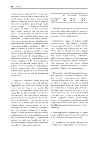
Abnormal Pap Smearin Pregnancy 3
is very low among adolescents, but they should
be encouraged to receive human papilloma virus
(HPV) vaccination and counseled about safe sex
practices to limit exposure to sexually transmitted
infections. Women between the age of 21–29
years should be screened with cervical cytology
alone. Women >30years of age should be screened
with cytology and HPV testing every 5 years (or
with cytology alone every 3 years). Women with
a history of cervical cancer, HIV or other risk
factors (such as immunocompromise) should
continue annual screening. These guidelines and
the associated algorithm are based on a large
database of patients including adolescents who
were managed using former criteria in the Kaiser
Healthcare system. The American Society of
Colposcopy and Cervical Pathology (ASCCP) has
developed an updated free App that can assist
with the current recommendations.
2. Women who have risk factors for cervical/
vaginal cancer (such as a history of in utero
diethylstilbestrol (DES) exposure, HIV, women
who are immunocompromised, or those on
chronic steroids) should be screened annually.
3. Women aged 21–29 with normal cytology
but absent or insufficient endocervical–transfor
mation zone elements can continue regular
screening, which should not include HPV test
ing. In women >30years with a similar cytology
result, HPV testing is recommended. Positive
HPV results should prompt repeat co‐testing in
one year, unless the HPV genotype is known to
be 16 or 18, in which case, immediate colpos
copy is recommended. A negative HPV result in
a woman >30years means that she can go back
to routine screening.
4. Unsatisfactory cytology is less common in cur
rent practice with the use of liquid‐based media
for cervical screening. Insufficient squamous
cells to detect epithelial abnormalities generally
arise from blood or inflammation that obscures
the result. Repeat cytology is recommended in
2–4 months. Colposcopy can be considered in
women >30years with positive HPV, and is rec
ommended in those women who have had two
consecutive unsatisfactory cytology test results.
5. Women should always be informed of an
abnormal Pap result by her physician or another
healthcare professional who can answer basic
questions and allay anxiety. Verbal notification
should be followed with written information
and clear recommendations for follow‐up.
Additionally, if there is evidence of infection
along with cellular abnormalities, the infection
should be treated.
6. The 2012 criteria substantially clarify the
management of ASCUS, which is guided by HPV
test results whether obtained reflexively or as a
co‐test. The management in pregnancy differs
only in that colposcopy and endocervical curet
tage (ECC) should be deferred until 6 weeks
postpartum unless a CIN 2+ lesion is suspected.
Women >25years old with a negative HPV test
should be returned to a regular three‐year
follow‐
up cycle. Following pregnancy colposcopy is
recommended in women who are HPV+ with
annual co‐test
follow‐up. Similarly, an endocer
vical curettage (ECC) should be obtained
whenever possible and excisional
procedures
should be avoided to prevent over‐treatment.
In women 21–24 years old, cytology should be
repeated in one year. A positive HPV result does
not change the recommended
follow‐up, but
a negative result should return the woman to a
three‐year follow‐up cycle.
7. Atypical squamous cells cannot exclude
high‐grade squamous intraepithelial lesions
(HSIL) (ASC‐H), which is associated with a
higher risk of CIN 3+ regardless of patient age
and a five‐year invasive cancer risk of 2%
regardless of HPV status. That said, HPV is highly
correlated with ASC‐H, but the cancer risk
demands that all women receive immediate col
poscopy, including those 21–24 years of age.
Colposcopy with directed biopsies of any area
that might be concerning for micro invasion
17.
4 Abnormal PapSmear in Pregnancy
should be done by a highly trained clinician.
Treatment should be dictated by histologic eval
uation of the biopsied lesions.
8. Atypical glandular cells (AGC) or adenocar
cinoma in situ (AIS) warrant aggressive investi
gation and close follow‐up. Although the risk of
cancer is lower in younger age groups, women
>30years have a 9% risk of CIN3+ and 2% risk
of invasive cancer. All such women of all ages
should have antenatal colposcopy with 6‐weeks
postpartum follow‐up to include colposcopy,
ECC and endometrial biopsy (EMB). Subsequent
treatment and follow‐up are dictated by the
biopsy results, maternal age, and the histologic
evaluation of the glandular elements.
9. Approximately 60% of low‐grade squamous
intraepithelial lesions (LGSIL) will regress spon
taneously without treatment depending on the
age of the patient, HPV status, and HPV geno
type. For women >25years old in whom HPV
testing is negative, repeat co‐testing in ome year
is preferred but colposcopy is acceptable.
However, if the HPV is positive, then colposcopy
is preferred. If colposcopy is not part of the ini
tial evaluation, subsequent co‐testing needs to
be entirely normal to allow patients to return to
three‐year follow‐up. Any abnormality at the
one‐year follow‐up visit should result in colpos
copy. In women 21–24 years old, annual repeat
cytology without HPV testing is preferred and
colposcopy should be avoided unless the results
recur for two consecutive years or if one of the
following lesions is detected: ASC‐H, AGC, or
HSIL. Pregnant women >25years old with low‐
grade squamous intraepithelial lesions should
undergo immediate colposcopy without ECC,
while those 21–24 years old should be evalu
ated postpartum.
10. High‐grade squamous intraepithelial
lesions (HGSIL) are associated with a 60% risk
of CIN2+ and a 2% risk of invasive cervical
cancer. Immediate colposcopy with directed
biopsies of any area that might be concerning
for micro invasion is recommended, regardless
of maternal age. The antepartum diagnosed of
HGSIL should prompt a 6‐weeks postpartum
follow‐up colposcopy with ECC and treatment
as dictated by the biopsy results. If diagnosed
early in pregnancy, colposcopy can be repeated
every 12 weeks. Treatment during pregnancy
should be reserved for invasive carcinoma and
should be managed in concert with a gyneco
logic oncologist.
6 Immunization
therefore notprogrammed, and subsequent
exposure to vaccine‐preventable infections can
lead to active infection.
2. Vaccination works by inducing antibodies in
recipients that protects them against infection
after future exposure to specific disease‐causing
microbes. The level of protection varies accord-
ing to the strength and durability of the immune
response induced by the vaccine as well as the
virulence, prevalence, and ease of transmission
of the infection itself. Vaccination programs
may have different goals: (i) to protect at‐risk
individuals (e.g., meningococcal disease); (ii) to
establish control by minimizing the overall
prevalence of the infection (e.g., measles, vari-
cella); or (iii) to attain global elimination of an
infection (e.g., neonatal tetanus, polio).
3. Vaccination in pregnancy is of benefit and at
times poses concern relative to the increased
vulnerability of the mother and fetus.
Inactivated vaccines are approved for use in
pregnancy. The inactivated influenza vaccine
should be given to all pregnant women dur-
ing the influenza season (October through May
in the northern hemisphere), regardless of ges-
tational age. It is clear that there are significant
maternal benefits including fewer cases of fever
and respiratory illness and substantial neonatal
protection through the transplacental passage
of antibodies that provide months of protection
at a time when the infant is vulnerable and
could not be directly vaccinated. However, live‐
attenuated vaccines (including rubella, MMR,
varicella) are not recommended for pregnant
women despite the fact that no cases of con-
genital anomalies have been documented.
Exceptions include yellow fever and polio,
which can be given to pregnant women when
traveling to high prevalence areas. In addition,
women should be advised not to get pregnant
within 1 month of receiving a live‐attenuated
vaccine. The live‐attenuated influenza vaccine
is available as an intranasal spray, which is con-
sidered safe in the postpartum period. Vaccines
considered safe in pregnancy include tetanus,
diphtheria, hepatitis B, and influenza. Tetanus
immunization during pregnancy is a common
strategy used in the developing world to com-
bat neonatal tetanus
4. Risk factors for specific vaccine‐preventable
illnesses include:
• illicit drug users (hepatitis A and B, tetanus)
• men who have sex with men (hepatitis A) or
>1 sexual partner in the past 6 months (hep-
atitis A, human papilloma virus)
• travel to or immigration from areas where
infection is endemic (hepatitis A and B, measles,
meningococcus, rubella, tetanus, varicella)
• healthcare workers (hepatitis B, influenza,
varicella)
• nursing home residents (meningococcus,
pneumococcus, varicella) or ≥50years of age
(influenza)
• chronic medical diseases: diabetes, asthma,
HIV, liver disease and/or renal disease (hepa-
titis A, influenza, pneumococcus)
• adults who have had their spleens removed
(meningococcus, pneumococcus)
• accidental or intentional puncture wounds
(tetanus)
5. One of the ongoing controversies about
vaccination in pregnancy is whether vaccines
containing thimerosal pose a risk to the fetus.
Thimerosal is a mercury‐containing preserva-
tive that has been used in multidose vaccines
since the 1930s. Although there has been con-
cern about the cumulative levels of mercury,
the current scientific evidence does not consider
thimerosal to be associated with adverse out-
comes in children exposed in utero. The Centers
for Disease Control and Prevention’s Advisory
Committee on Immunization Practices (ACIP)
does not recommend avoiding thimerosal con-
taining vaccines. Although the ACIP does not
recommend any specific formulation, there are
newer trivalent and quadrivalent influenza vac-
cines (containing two A and two B influenza
strains) that are available for use. The following
20.
Immunization 7
adult vaccinesare thimerosal‐free: Tdap (but
not Td), Recombivax hepatitis B vaccine (but
not Engerix‐B), and some influenza vaccines
(Fluzone with no thimerosal).
6. Tetanus toxoid, reduced diphtheria toxoid
and acellular pertussis vaccine (Tdap) may be
given at any time of pregnancy or the postpar-
tum period but ideally is administered between
27–36 weeks to confer the best passive immu-
nity through the transfer of antibodies to the
fetus. This recommendation has developed to
address the significant impact of pertussis dis-
ease in the newborn.
Preconception Care 9
2.Discuss social, financial, and psychological
issues in preparation for pregnancy.
3. Maternal alcohol use is the leading known
cause of congenital mental retardation and is
the leading preventable cause of birth defects in
the Western world. An accurate drinking his-
tory is best elicited using a tool that employs
standardized screening questions (such as the
CAGE questionnaire). The adverse effects of
alcohol may be compounded with abuse of
other drugs. Cigarette smoking, cocaine, and
other drug use should be included in the his-
tory. Patients at risk should be provided educa-
tion, contraceptive counselling, and referral for
treatment as necessary.
4. Screen for domestic violence. Be aware of
available state and local resources and state
laws regarding mandatory reporting. Risk
increases with pregnancy. Domestic violence is
not isolated to any particular risk group in
pregnancy; it cuts across socio‐economic and
ethnic lines.
5. Take an occupational history that will allow
assessment of workplace risks to the pregnancy.
Elicit information about any exposures to haz-
ardous materials or biologic hazards (HIV, cyto-
megalovirus (CMV), toxoplasmosis) and review
the use of safety equipment. Talk to patients
about the appropriate and correct use of seat
belts while in a moving vehicle.
6. Counsel patients with a history of preec-
lampsia, placental abruption, unexplained
fetal death, or severe intrauterine growth
restriction (IUGR) about the risks of recur-
rence. Low‐dose aspirin starting at the end of
the first trimester is recommended to prevent
recurrent preeclampsia. The use of low‐dose
aspirin, calcium supplementation, and/or anti-
coagulation for women with documented
inherited thrombophilias to prevent adverse
pregnancy outcome is controversial, and cannot
be routinely recommended.
7. Personal and family histories should be exam-
ined for evidence of genetic diseases. Genetic
testing is available to determine a patient’s carrier
status for some autosomal recessive conditions
such as Tay–Sachs, Canavan disease, sickle cell
disease, and the thalassemias. Consider referral
for further genetic counselling if patients are at
high risk. ACOG currently recommends that all
couples be offered prenatal testing for cystic
fibrosis. ACMG (but not ACOG) recommends
that all couples also be offered genetic testing for
spinal muscular atrophy (SMA).
8. Emphasize the importance of nutrition.
Assess appropriateness of patient’s weight for
height, special diets and nutrition patterns such
as vegetarianism, fasting, pica, bulimia, and
vitamin supplementation. Recommend folic
acid supplementation as necessary: 0.4 mg per
day for all pregnant women or women consid-
ering pregnancy, 4.0 mg per day if the woman
has a personal/family history of a child with a
neural tube defect or is on anticonvulsant medi-
cations (especially valproic acid). Counsel to
avoid oversupplementation (such as vitamin A).
Review the recommendations on dietary fish
ingestion (<12 ounces per week of cooked fish)
to minimize mercury intake, and steps for pre-
vention of listeriosis (avoiding raw or under-
cooked meat/fish, unpasteurized milk and soft
cheeses, unwashed fruit and vegetables) and
toxoplasmosis (exposure to cat feces).
9. A thorough immunization history should be
obtained that addresses vaccination. Women
should be tested for immunity to rubella and vac-
cinated prior to pregnancy if not immune.
Women without a history of chickenpox (vari-
cella) should be tested and offered vaccination
prior to pregnancy. Hepatitis B vaccination should
be offered to all women at high risk, and screen-
ing for other sexually transmitted infections
should be offered as needed. The U.S. Centers for
Disease Control and Prevention (CDC) recom-
mends that pregnancy be delayed for at least 1
month after receiving a live‐attenuated vaccine
23.
10 Preconception Care
(suchas MMR, varicella, live‐attenuated influ-
enza, BCG).
10. Discuss birth spacing and the options avail-
able for postpartum contraception.
11. Effects of the pregnancy on any medical
conditions for both mother and fetus should be
discussed. Pregnancy outcomes can be improved
by optimizing control of chronic medical con-
ditions prior to pregnancy (such as glycemic
control in patients with diabetes and blood
pressure control in patients with hypertension).
Medications should be reviewed, and patients
counselled regarding alternatives that may be
safer in pregnancy. Close communication with
the patient’s primary care and subspecialty physi-
cians should always be maintained.
12 Prenatal Care
1.The goal of prenatal care is to promote the
health and well‐being of the pregnant woman,
fetus, infant, and family up to 1 year after birth.
To achieve these aims, prenatal care must be
available and accessible. The three major com-
ponents are: (i) early and continuing risk assess-
ment, including preconception assessment (see
Chapter 3, Preconception Care); (ii) continued
health promotion; and (iii) both medical and
psychosocial assessment and intervention.
2. Routine prenatal tests that should be com-
pleted for all pregnant women include complete
blood count (CBC), blood group type and screen
(Rh status), rubella serology, HIV, hepatitis B,
syphilis serology (VDRL/RPR), Pap smear, cystic
fibrosis (CF) carrier status, chlamydia/gonor-
rhea cultures, and urine culture and sensitivity
(UA C&S).
3. Approximately 20% (1 in 5) of pregnancies
are considered high risk. Risk factors for adverse
pregnancy outcome may exist prior to preg-
nancy or develop during pregnancy or even
during labor (examples are listed below,
although this list should not be regarded as
comprehensive).
4. The frequency and timing of prenatal visits
will vary depending on the risk status of the
pregnant woman and her fetus. In low‐risk
women, prenatal visits are typically recom-
mended q 4 weeks to 28 weeks, q 2 weeks to 36
weeks, and then weekly until delivery.
5. See Chapter 12 (Preeclampsia).
6. See Chapter 53 (Prenatal diagnosis).
7. See Chapter 10 (Gestational diabetes mellitus)
8. See Chapter 24 (GBS)
9. See Chapter 55 (Screening for preterm birth)
High‐Risk Pregnancies
Maternal factors
• Pre‐existing medical conditions
(diabetes, chronic hypertension,
cardiac disease, renal disease,
pulmonary disease)
• Preeclampsia
• Gestational diabetes
• Morbid obesity
• Extremes of maternal age
• Active venous
thromboembolic disease
• Poor obstetric history (prior
preterm birth, preterm PROM,
stillbirth, IUGR, placental
abruption, preeclampsia,
recurrent miscarriage)
Fetal factors
• Fetal structural or chromosomal
anomaly
• History of a prior baby with a
structural or chromosomal
anomaly
• Family or personal history of a
genetic syndrome
• Toxic exposure (to
environmental toxins,
medications, illicit drugs)
• IUGR
• Fetal macrosomia
• Multiple pregnancy
(esp. if monochorionic)
• Isoimmunization
• Intra‐amniotic infection
(chorioamnionitis)
• Nonreassuring fetal testing
Uteroplacental factors
• Preterm premature rupture of
membranes
• Unexplained oligohydramnios
• Large uterine fibroids (esp. if
submucosal)
• Prior cervical insufficiency
• Prior uterine surgery
(especially prior “classic”
hysterotomy)
• Placental abruption
• Placenta previa
• Uterine anomaly (didelphys,
septate)
• Abnormal placentation
(placenta accreta, increta or
percreta)
• Vasa previa
26.
Levels of evidence
Thelevels of evidence used in this book are those recommended by the U.S. Preventive
Services Task Force, an independent panel of experts responsible for developing
evidence-based recommendations for primary care and prevention, in 2007
(http://www.ahrq.gov/clinic/uspstmeth.htm):
Level I: Evidence obtained from at least one properly designed randomized controlled trial.
Level II: Evidence obtained from controlled trials without randomization or cohort / case-
controlled studies that include a comparison group.
Level III: Evidence from uncontrolled descriptive studies (including case series) or opinions of
respected authorities or expert committees.
Level IV: Evidence from uncontrolled descriptive studies (including case series) or opinions of
respected authorities or expert committees.
Maternal Disorders
Section 2
13
Antiphospholipid Antibody Syndrome15
3. Clinical manifestations of APLAS include:
(i) recurrent pregnancy loss (defined as
≥ 3
unexplained first‐trimester pregnancy losses or ≥1
unexplained second‐trimester pregnancy loss); (ii)
unexplained thrombosis (venous, arterial, cerebro-
vascular accident or myocardial infarction); and/or
(iii) autoimmune thrombocytopenia (platelets
<100,000/mm3
). Recent consensus opinions sug-
gest that such clinical conditions as unexplained
intrauterine growth restriction (IUGR), massive
placental abruption, and
recurrent early‐onset
severe pre‐eclampsia be included.
4. At least one of three serologic tests confirm-
ing the presence of circulating antiphospholipid
antibodies (below) is required to make the
diagnosis of APLAS. Moreover, the diagnosis
requires the persistence of such antibodies
as confirmed by two or more positive tests at
least 12 weeks apart.
• Lupus anticoagulant (LAC) is an unidentified
antiphospholipid antibody (or antibodies)
that causes prolongation of phospholipid‐
dependent coagulation tests in vitro by bind-
ing to the prothrombin–activator complex.
Examples of tests that can confirm the pres-
ence of LAC include the activated PTT test,
dilute Russel viper venom test, kaolin clot-
ting time, and recalcification time. In vivo,
however, LAC causes thrombosis. LAC results
are reported as present or absent (no titers
are given). The term LAC is a misnomer: it is
not specific to lupus (SLE) and it acts in vivo
as a procoagulant and not an anticoagulant.
• Antibodies against specific phospholipids as
measured by enzyme‐linked immunosorbant
assay (ELISA). These high‐avidity IgG anti-
bodies have anticoagulant activity in vitro, but
procoagulant activity in vivo. The most com-
monly used ELISA test is the anticardiolipin
antibody (ACA). Cardiolipin is a negatively
charged phospholipid isolated from ox heart.
ACA ELISA is at best semi‐quantitative.
Results have traditionally been reported as
low, medium or high titers. More recently,
standardization of the phospholipid extract
has allowed for standard units to be developed
(GPL units for IgG, MPL units for IgM). ACA
IgM alone, IgA alone, and/or
low‐
positive IgG
may be a nonspecific (incidental) finding
since they are present in 2–4% of asympto-
matic pregnant women. As such, moder-
ate‐to‐high levels of ACA IgG (>40 GPL
units) are required to make the diagnosis of
APLAS.
• The presence of anti‐β2‐glycoprotein I
antibodies.
5. A number of additional antiphospholipid
antibodies are described, including antiphos-
phatidylserine, antiphosphatidylethanolamine,
antiphosphatidylcholine, anti‐Ro, and anti‐La,
but these are not sufficient to make the diagno-
sis. A false‐positive test for syphilis (defined as a
positive rapid plasma reagin (RPR) or Venereal
Disease Research Laboratory (VDRL) test, but
negative definitive test for syphilis) is another
common finding in women with APLAS, but is
nonspecific and is not sufficient to confirm the
diagnosis. Antinuclear antibodies (ANA) are not
antiphospholipid antibodies, and may suggest
the diagnosis of SLE but not APLAS.
6. Treatment for APLAS depends on the clinical
features:
• For women with thrombosis (such as stroke
or pulmonary embolism), therapeutic antico-
agulation is indicated with either unfraction-
ated heparin (UFH) or low molecular weight
heparin (LMWH) during pregnancy followed
by oral anticoagulation (coumadin) postpar-
tum because of a 5–15% risk of recurrence.
In pregnancy, regular blood tests are required
4 hours after administration of the drug to
ensure that anticoagulation is therapeutic:
the PTT should be 1.5‐ to 2.5‐fold normal and
anti‐Xa activity levels should be 0.6–1.0 U/
mL. Side‐effects include hemorrhage, throm-
bocytopenia, and osteopenia and fractures.
Such women may need lifelong treatment.
For women with recurrent pregnancy loss,
treatment should include prophylactic UFH
29.
16 Antiphospholipid AntibodySyndrome
(5000–10,000 units sc bid) or LMWH (enoxa-
parin (Lovenox) 30–40 mg sc daily or daltepa-
rin (Fragmin) 2500–5000 U sc daily) starting in
the first trimester of pregnancy. Although pro-
phylactic dosing does not change PTT, it will
increase anti‐Xa activity to 0.1–0.2 U/mL.
However, it is not necessary to follow serial
anti‐Xa activity in such patients. The goal of this
treatment is to prevent pregnancy loss and to
prevent VTE, which is possible in women with
APLAS in pregnancy even if they have not had
a VTE in the past. Therefore, anticoagulation
should be administered throughout pregnancy
and typically for 6–12 weeks after delivery.
• For women with autoimmune thrombocyto-
penia or a history of severe pre‐eclampsia,
IUGR or placental abruption, the optimal
treatment is unknown. Consider treating as
for recurrent pregnancy loss. Postpartum
anticoagulation is probably not necessary.
18 Asthma
which correlateswell with FEV1, can be meas-
ured using a hand‐held peak flow meter and is a
useful measure in the clinic or home setting.
PEFR is determined for each patient (personal
best) or by using charts adjusted for age, height,
gender, and race. PEFR results are categorized
into green (80–100% of normal or personal
best), yellow (50–80%), and red (less than
50%). Usually, the green zone means that the
asthma is well controlled, yellow means that
adjustments to medications and/or environ-
ment are needed, and red is a medical alert that
needs immediate attention. Findings consistent
with asthma include a variability of >20% in
PEFR, a reduction in FEV1 and FEV1/FVC ratio
on spirometry, an increase in FEV1 of more
than 15% from the baseline following adminis-
tration of 2–4 puffs of a bronchodilator, and
heightened sensitivity to bronchoprovocation.
Asthma complicates 1–4% of all pregnancies.
Pregnancy has a variable effect on asthma (25%
improve, 25% worsen, 50% are unchanged). In
general, women with mild, well‐controlled
asthma tolerate pregnancy well. Women with
severe asthma are at risk of symptomatic
deterioration.
2. Respiratory adaptations during pregnancy
are designed to optimize maternal and fetal
oxygenation, and to facilitate transfer of CO2
waste from the fetus to the mother. The
mechanics of respiration change with preg-
nancy. The ribs flare outward and the level of
the diaphragm rises 4cm. During pregnancy,
tidal volume increases by 200mL (40%) result-
ing in a 100–200mL (5%) increase in vital
capacity and a 200mL (20%) decrease in the
residual volume, thereby leaving less air in
the lungs at the end of expiration. The respira-
tory rate remains unchanged or increases
slightly. The end result is an increase in minute
ventilation and a drop in arterial PCO2
. Arterial
PO2
is essentially unchanged. A compensatory
decrease in bicarbonate enables the pH to
remain unchanged. Pregnancy thus represents a
state of compensated respiratory alkalosis.
pH Po2
(mmHg) Pco2
(mmHg)
Non‐pregnant 7.40 93–100 35–40
Pregnant 7.40 100–105 28–30
3. The differential diagnosis of asthma includes
pneumonia, pulmonary embolism, pneumo-
thorax, congestive cardiac failure, pericarditis,
pulmonary edema, and rib fracture.
4. Characteristic triggers for asthma include
exercise, cold air, and exposure to allergens.
Exercise‐triggered symptoms typically develop
10–15 minutes after exertion and are more
intense when the inhaled air is cold. Allergens
that typically trigger asthma symptoms include
dust, molds, furred animals, cockroaches,
pollens, and other irritant‐type exposures (ciga-
rette smoke, strong fumes, airborne chemicals).
Viral infections can also trigger asthma
symptoms. Influenza vaccination is recom-
mended (see Chapter 2 on Immunization).
5. The principal goals of treatment are to mini-
mize symptoms, normalize pulmonary func-
tion, prevent exacerbations, and improve
health‐related quality of life. Initial treatment
for relief of symptoms should be an inhaled
short‐acting beta‐agonist used on an as‐needed
basis rather than at regularly scheduled inter-
vals. The most commonly used agent is an
albuterol inhaler at a dose of 2–4 puffs as needed
every 4–6 hours. If this is not adequate to
control symptoms, inhaled glucocorticoids
(such as beclomethasone dipropionate) should
be given by metered dose inhaler (MDI) and
should be taken at regular intervals two to three
times daily.
6. Pregnancy‐related complications of severe
asthma include intrauterine growth restriction
(IUGR), stillbirth, and maternal mortality.
7. A number of alternative therapies are
available on an outpatient basis. These include a
32.
Asthma 19
short courseof oral glucocorticoids. A typical
regimen is prednisone 0.5mg per kg body
weight given orally each day and tapered over a
period of one to two weeks. This can be given
alone or in combination with a leukotriene
modifying agent––such as the leukotriene D4
receptor antagonists zafirlukast (Accolate) and
montelukast (Singulair) or the 5‐lipoxygenase
inhibitor zileuton (Zyflo)––or a slow‐release
theophylline.
8. SeeChapter75(AcuteAsthmaExacerbation).
Cholestasis of Pregnancy21
include cholestasis in a prior pregnancy (recur-
rence rate is >90%), multiple gestation, preg-
nancies conceived through IVF, and underlying
liver, renal, and/or bowel disease.
2. Cholestasis of pregnancy is a clinical/
biochemical diagnosis. Patients typically present
with complaints of acute onset of severe pruri-
tus in the latter half of pregnancy, usually
>30weeks’ gestation. Physical examination may
reveal jaundice and/or skin excoriations, but is
often unremarkable. Bile acids will usually be
elevated at values >10–14 micromoles/L (6–10
micromoles/L in the fasting state). Bile acids are
derived from hepatic cholesterol metabolism.
Cholic and chenodeoxycholic acid are the domi-
nant fractionated constituents. Liver function
tests, specifically serum transaminases, will
frequently be elevated.
3. The differential diagnosis of cholestasis
includes skin allergy, parasitic infections, sys-
temic lupus erythematosis (SLE), syphilis, viral/
drug‐induced hepatitis, preeclampsia, metabolic
disorders, and gall bladder diseases.
4. Cholestasis of pregnancy is associated with
adverse perinatal outcome, including increase
perinatal mortality (unexplained stillbirth), pre-
mature birth, and meconium passage and
aspiration. Many of these adverse outcomes are
directly associated with elevated bile acids,
particularly with values ≥40 micromol/L. The
association with IUGR is less clear. For these
reasons, regular (weekly or twice weekly) fetal
surveillance is recommended after 32 weeks of
gestation. However, it is not clear whether fetal
testing is associated with an improvement in
perinatal outcome.
5. Ursodeoxycholic acid (ursodiol (UDCA)) has
become the preferred therapeutic intervention
for ICP. Although treatment with UDCA has not
been conclusively shown to improve perinatal
outcome, it does significantly reduce pruritis and
normalize LFTs. The recommended initial dose
of 300mg TID can be upward adjusted as needed
to a maximum of 2 gms/day in divided doses.
Other agents that have been used for sympto-
matic relief include hydroxyzine (25–50mg/day,
which may have significant somnolent side
effects) and cholestyramine (a foul‐tasting resin
that binds bile acids in the
gastrointestinal sys-
tem). Response to such medications may take
several weeks and is highly variable. Alternative
treatment options that are less well established
include ultraviolet light, rifampicin, phenobarbi-
tone, epomediol, or S‐adenosyl‐L‐methionine.
6. In contrast to the effects on the fetus, choles-
tasis of pregnancy is not associated with adverse
maternal outcome. Many patients remain symp-
tomatic despite treatment and live with anxiety
relative to the small risk of fetal death, which is
seen most commonly >38weeks. As a conse-
quence, some consensus bodies recommend
delivery at 37–38 weeks even with reassuring
fetal testing. Earlier delivery is reserved for fetal
indications or maternal jaundice despite treat-
ment. Mode of delivery should be governed by
routine obstetric indications. Patients should be
aware that symptoms generally resolve in the
immediate postpartum period, but recurrence in
future pregnancies can be as high as 90%.
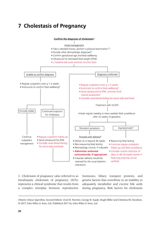
Chronic Hypertension 23
thediagnosis should also be suspected in women
with a sustained elevation in BP prior to 20
weeks of gestation. However, if BP was normal in
the first trimester and then increases before 20
weeks of gestation, early preeclampsia should
also be considered.
2. All women with pre‐existing hypertension
should be assessed either before pregnancy or
early in pregnancy to rule out secondary (and
potentially curable) hypertension, and to evaluate
for evidence of target organ damage. Most women
with chronic hypertension have essential (pri-
mary) hypertension. Up to 10% of women have
secondary hypertension, due most commonly to
chronic kidney disease. Other causes may include
renal artery stenosis and an underlying endo-
crinopathy (such as primary hyperaldosteronism,
pheochromocytoma, and Cushing syndrome).
3. Baseline evaluation should include serum
analysis for creatinine, electrolytes, uric acid,
liver enzymes, and platelet count as well as uri-
nary protein estimation. These values can be
used for comparison if superimposed preec-
lampsia is suspected later in pregnancy. Left
ventricular function should be assessed in
women with severe hypertension of more than
4 years duration either by electrocardiography
(ECG) or echocardiography.
4. Chronic hypertension is associated with an
increased risk of superimposed preeclampsia
and higher rates of adverse maternal‐fetal
outcome, such as severe hypertension, cerebro-
vascular accident (stroke), uteroplacental insuf-
ficiency leading to fetal growth restriction,
placental abruption, and stillbirth.
5. Pharmacologic treatment of mild hyperten-
sion has not been shown to improve pregnancy
outcome. The goals of treatment during preg-
nancy are to minimize acute maternal and fetal
risks of severe hypertension. As such, it is rarely
necessary to initiate antihypertensive therapy in
early pregnancy. If a patient is well controlled
on medications prior to pregnancy, it is usual to
leave her medications unchanged. The excep-
tions are the angiotensin‐converting enzyme
(ACE) inhibitors and angiotensin receptor
blockers (ARB), which should be discontinued
as soon as a positive pregnancy test is attained.
First trimester exposure has been associated
with cardiac and central nervous system anom-
alies, and use in the second and third trimester
can result in progressive and irreversible renal
injury as well as oligohydramnios and fetal
growth restriction. Drugs of choice include
α‐methyldopa, β‐blockers (labetalol) or calcium
channel blockers (nifedipine). Diuretic therapy
is generally discouraged in pregnancy.
6. In the absence of maternal end organ dam-
age, treatment is not recommended if the sys-
tolic BP remains <160mmHg and the diastolic
BP <105mmHg. If end organ damage is present,
BP goals are more strict (systolic BP <140mmHg
and diastolic BP <90mmHg) to avoid progres-
sion of disease and its associated complications
during pregnancy.
7. When antihypertensive therapy is initiated
during pregnancy, it is suggested that BP be
maintained between 120–160mmHg systolic
and 80–105mmHg diastolic. Acute‐onset,
severe systolic hypertension (>160mmHg) and/
or severe diastolic hypertension (>110mmHg)
should be treated with antihypertensive ther-
apy with the aim to achieve BP of 140–150/
90–100mmHg. First‐line treatment for the
management of acute‐onset, severe hyperten-
sion includes intravenous labetalol, intravenous
hydralazine, or oral nifedipine.
8. Preeclampsia is superimposed on chronic
hypertension when there is a sudden increase
in BP that was previously well controlled or
an escalation of antihypertensive medications
needed to control BP; or new onset of proteinu-
ria or a sudden increase in proteinuria in
pregnancy. Superimposed preeclampsia can be
further classified into with or without severe
37.
24 Chronic Hypertension
features.The diagnosis of superimposed preec-
lampsia with severe features is established when
any of the following are present: (i) severe‐
range BP despite escalation of antihyperten-
sive therapy; (ii) thrombocytopenia (platelet
count <100,000/microliter); (iii) elevated liver
transaminases to twice normal concentrations;
(iv) new‐onset/worsening renal insufficiency
(serum creatinine >1.1mg/dL or a doubling of
the creatinine in the absence of other renal dis-
ease); (v) pulmonary edema; or (vi) persistent
cerebral or visual disturbances.
9. Intravenous magnesium sulfate is the drug of
choice for seizure prophylaxis and should be
given intrapartum and for at least 24 hours post-
partum to prevent eclampsia. An IV loading dose
of 4–6g should be followed by a maintenance
dose of 1–2g/h. The use of magnesium sulfate
therapy in superimposed preeclampsia without
severe features is not recommended, but should
be initiated if there is progression to severe dis-
ease either intrapartum or postpartum.
10. Delivery is the only effective treatment for
superimposed preeclampsia. It is recommended
in women with superimposed preeclampsia
without severe features at or beyond
37–0/7weeks. It is recommended in women
with superimposed preeclampsia with severe
features if the gestational age is at least
34–0/7weeks. If the gestational age is <34–0/7
weeks and the diagnosis is superimposed preec-
lampsia by BP criteria alone, expectant man-
agement can be considered (see Chapter 12).
There is no proven benefit to routine delivery
by cesarean; however, the probability of vaginal
delivery decreases with decreasing gestational
age. With labor induction, the likelihood of
cesarean delivery is 93–97% at <28 weeks,
53–65% at 28–32 weeks, and 31–38% at 32–34
weeks. Preeclampsia and its complications
always resolve following delivery (with the
exception of stroke). Diuresis (>4L/day) is the
most accurate clinical indicator of resolution.
Fetal prognosis is dependent largely on gesta-
tional age at delivery.
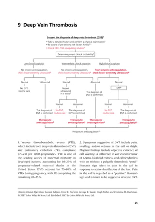
26 Deep VeinThrombosis
However, a “positive” Homan’s sign is only
around 30–40% sensitive, and a “negative”
Homan’s sign does not exclude the diagnosis.
Isolated iliac‐vein thrombosis may present with
abdominal pain, back pain, and swelling of the
entire leg.
3. A personal history of VTE is the single most
important risk factor for VTE during pregnancy.
The risk of recurrent VTE during pregnancy
is increased three–fourfold, and 15–25% of all
cases of VTE in pregnancy are recurrent events.
The second most important individual risk
factor for VTE in pregnancy is the presence of
an inherited thrombophilia (such as factor V
Leiden mutation, prothrombin gene mutation,
protein S/protein C/antithrombin deficiency) or
acquired thrombophilia (antiphospholipid anti-
body syndrome, see Chapter 5), which is pre-
sent in 20–50% of women who experience VTE
during pregnancy and the puerperium. Other
risk factors for VTE include advanced maternal
age, black race, heart disease, sickle cell disease,
diabetes, lupus, hypertension, hemoglobinopa-
thies, smoking, multiple pregnancy, obesity,
prolonged immobility (bedrest), trauma, preg-
nancy (due to its hypercoagulable state), and
cesarean delivery (especially an intrapartum
emergency cesarean). VTE is fourfold more
common in pregnancy than in nonpregnant
women. Two‐thirds of DVTs occur antepartum,
with these events distributed throughout all
three trimesters.
4. Laboratory tests (including circulating
D‐dimer levels) are generally unhelpful in con-
firming the diagnosis of DVT, but baseline
coagulation studies should be sent if the patient
requires anticoagulation.
5. If the clinical suspicion of DVT is high, con-
sider starting anticoagulation immediately to
avoid DVT propagation and possible PE.
6. The diagnosis of DVT is usually confirmed
noninvasively by compression ultrasonography
of the proximal veins, including Doppler duplex.
Compression ultrasonography is reliable at
detecting proximal lower extremity DVT with a
sensitivity of 97%, specificity of 94%, using
contrast venography as the gold standard, but is
less effective at diagnosing isolated calf and iliac
vein thrombosis.
7. If compression ultrasonography is negative,
future management depends in large part on
the clinical suspicion for DVT. The minimal
requirement in a symptomatic patient with a
negative lower extremity compression ultra-
sound examination is to repeat the test in
1 week.
8. If the clinical setting is highly suspicious but
compression ultrasound is negative or equivo-
cal, an MRI should be performed. Contrast
venography, once the standard technique for
DVT diagnosis, has now been replaced by com-
pression ultrasound and an MRI due to the less
invasive nature and lack of radiation exposure.
9. Deep vein thrombosis in pregnancy should
be treated to prevent propagation of the throm-
bus and PE. If untreated, 25% of patients with
DVT will have a PE as compared with 5% of
treated patients. Admission is generally war-
ranted for initiation of treatment in pregnancy.
Therapeutic subcutaneous low molecular
weight-heparin (LMWH) is now the treatment
of choice for DVT in pregnancy. The advantages
of LMWH over unfractionated heparin (UFH)
include a reduced risk of bleeding, predictable
pharmacokinetics allowing weight‐based dos-
ing without the need for monitoring, and a
reduced risk of heparin‐induced thrombocyto-
penia and heparin‐induced osteoporotic frac-
tures. A twice‐daily weight‐adjusted dosing
regimen should be used, such as enoxaparin
(Lovenox) 1mg/kg sc q12h. LMWH does not
significantly alter PTT, but serum anti‐factor Xa
activity can be measured. Therapeutic antico-
agulation is achieved with a circulating anti‐Xa
activity of 0.6–1.0 U/mL, however, routine
40.
Deep Vein Thrombosis27
monitoring of anti‐Xa activity may not be justi-
fied due to the absence of large studies using
clinical end points that demonstrate an optimal
therapeutic anti‐Xa LMWH range or that dose
adjustments increase the safety or efficacy of
therapy, the lack of accuracy and reliability of
the measurement, the lack of correlation with
risk of bleeding and recurrence, and the cost of
the assay. The management of isolated calf vein
thrombosis is controversial, with no established
guidelines. Since most iliofemoral thromboses
originate from calf vein thromboses, full antico-
agulation with LMWH is suggested for sympto-
matic patients.
• When LMWH cannot be used or when UFH
is preferred (e.g., in patients with renal dys-
function and when delivery or surgery may
be necessary), UFH can be administered as an
initial IV therapy followed by subcutaneous
UFH. IV treatment should be initiated with a
loading dose of 80 U/kg followed by an initial
infusion of 18 U/kg/h. Serum PTT should be
checked every 4–6 hours, and the infusion
adjusted to maintain PTT at 1.5–2.5 times
control. Once a steady state has been
achieved, PTT levels should be measured
daily. After 5–10 days, IV heparin can be
changed to SC injection (not IM injection
because of the risk of hematoma) as follows:
begin with 10,000 U SC three times daily and
titrate dosage upward depending on the results
of the mid‐interval PTT; aim for PTT 1.5–2.5
times control. When subcutaneous UFH is
used during pregnancy, higher doses and three
times daily dosing are usually required to
maintain adequate anticoagulation.
• Alternative therapies (fibrinolytic agents,
surgical intervention) are associated with a
high incidence of complications in pregnancy
and, as such, are best avoided. The use of
vena caval filters should be considered only
for patients in whom anticoagulation is con-
traindicated or in whom extensive venous
thromboembolism develops within 2 weeks
before delivery.
• Treatment for acute DVT should be continued
throughout pregnancy and for at least 6
weeks postpartum (for a minimum total
duration of therapy of 3 months).
10. Women receiving therapeutic LMWH may
be switched to therapeutic UFH in the last
month of pregnancy or if delivery appears
imminent due to the shorter half‐life of UFH,
although the benefit of this approach has not
been validated by clinical studies. Alternately,
therapeutic LMWH can be discontinued 24
hours prior to induction of labor. The purpose of
conversion to UFH is primarily to reduce the
risk of an epidural or spinal hematoma with
regional anesthesia. The pharmacokinetics of
subcutaneous UFH and LMWH are quite simi-
lar, though, which may limit the benefit of this
approach. The American Society of Regional
Anesthesia and Pain Medicine guidelines
recommend withholding neuraxial blockade for
24 hours after the last therapeutic dose of
LMWH. These guidelines support the use of
neuraxial anesthesia in patients receiving dos-
ages of 5,000 units of UFH twice daily, but the
safety in patients receiving 10,000 units twice
daily or more is unknown. Pregnant women at
the highest risk of recurrence (e.g., proximal
DVT or PE within 2 weeks) can be switched to
therapeutic IV UFH, which is then discontinued
4–6h prior to the expected time of delivery or
epidural insertion.
According to the American Society of
Regional Anesthesia and Pain Medicine,
resumption of therapeutic UFH or LMWH
should be delayed for 24 hours after delivery,
vaginal or cesarean, in women who have
received neuraxial anesthesia. Otherwise, it
can be restarted no sooner than 4–6 hours after
a vaginal delivery or 6–12 hours after cesarean
delivery. Women who require more than 6
weeks of postpartum therapeutic anticoagula-
tion may be bridged to warfarin. Warfarin,
UFH and LMWH are all compatible with
breastfeeding.
Gestational Diabetes Mellitus29
and human chorionic somatomammotropins
(previously known as human placental lacto-
gens). These mechanisms ensure a continuous
supply of glucose for the fetus. In some women,
these changes unmask an underlying predispo-
sition to insulin resistance leading to GDM.
Depending on the population and the screening
method used, up to 18% of pregnancies may be
complicated by GDM. Up to 7% of pregnant
women will be diagnosed with GDM using the
screening approach recommended above.
3. Risk factors for GDM include: a prior history
of GDM, a family history (first degree relative)
of diabetes, a prior macrosomic or large‐for‐ges-
tational age (LGA) infant, sustained glycosuria,
a prior unexplained late intrauterine fetal
demise (IUFD), hypertension, or obesity. Such
patients should have early testing for GDM at
16–20 weeks. If the early testing is negative, this
should be repeated at 24–28 weeks.
4. Glucose load test (GLT) – also known as a
glucose challenge test (GCT) – is a non‐fasting
test, but the woman should not eat after her
50‐g glucose load until a venous blood sample is
drawn one hour later. A plasma value of
≥140mg/dL is considered positive and should be
followed with a 3‐hour glucose tolerance test
(GTT). <2% of women with a GLT <140mg/dL
will have a positive GTT.
5. A definitive diagnosis of GDM requires a
3‐hour GTT. There is no GLT cut‐off that is diag-
nostic of GDM. However, almost all women
with a GLT value ≥240mg/dL will have an
abnormal GTT and it is acceptable to manage
them as GDM without the GTT. To perform a
GTT, a 100‐g glucose load is administered after
an overnight fast. Venous plasma glucose is
measured fasting at 1 hour, 2 hours, and 3
hours. GDM requires two or more abnormal
values defined as either ≥95, ≥180, ≥155, and
≥140mg/dL, respectively (Carpenter & Coustan
criteria) or ≥105, ≥190, ≥165, and ≥145mg/dL,
respectively (NDDG criteria). There is no place
for HbA1c to diagnose GDM.
6. The goal of antepartum management is to
maintain strict glycemic control throughout ges-
tation, defined as fasting blood glucose <95mg/dL
and 1‐hour post‐prandial <140mg/dL (or a 2‐
hour post‐prandial <120mg/dL). A diabetic diet is
recommended (defined as 36kcal/kg or 15kcal/lb
of ideal body weight+100kcal per
trimester given
as 40–50% carbohydrate, 20% protein, 30–40%
fat) but, if diet alone does not maintain blood glu-
cose at the desirable levels, additional treatment
may be needed. Insulin remains the gold stand-
ard, although oral hypoglycemic agents (glybur-
ide, glipizide) appear to be safe and effective and
are being used more commonly as first line
agents. If fasting glucose levels are >95mg/dL,
treatment can be started right away because you
“can’t diet more than fasting.”
7. Fetal testing is recommended for insulin‐
requiring GDM (class A2‐GDM) after 32 weeks’
gestation because of the risks of abnormal fetal
growth (IUGR or macrosomia) and fetal demise.
Testing should include daily fetal kickcharts,
weekly non‐stress testing, and serial ultrasound
q 3–4 weeks for fetal growth.
8. If an elective delivery is planned prior to
39‐0/7weeks’ gestation, ACOG recommends
that fetal lung maturity is documented by amni-
ocentesis prior to delivery using diabetes‐
specific cut‐offs.
9. During labor, patients are typically starved.
Glucose should therefore be administered (5%
dextrose IV at 75–100mL/h) and blood glucose
checked every 1–2 hours. Regular insulin is
given as needed (either by subcutaneous
injection or IV infusion) to maintain glucose at
100–120mg/dL.
10. Delivery of the fetus and placenta removes
the source of the anti‐insulin hormones that
43.
30 Gestational DiabetesMellitus
causes GDM. As such, no further management
is required in the immediate postpartum period.
GDM likely unmasks an underlying predisposi-
tion for insulin resistance, and 40–60% of
women with GDM will develop type II diabetes
later in life. All women with GDM should
therefore have a standard, non‐pregnant 75‐g,
2‐hour oral GTT 6–8 weeks after delivery to
exclude diabetes. Since the risk of developing
type II diabetes remains elevated even if the
6 weeks GTT is negative, it should be repeated at
least every 3 years.
32 Gestational Hypertension
20weeks of gestation, and likely represents an
exaggerated physiologic response of the mater-
nal cardiovascular system to pregnancy.
4. Gestational hypertension may progress to
preeclampsia, but preeclampsia can be excluded
initially by the absence of maternal symptoms
of preeclampsia and laboratory abnormalities.
The following tests should be sent: urinalysis,
24‐hour urine collection for protein quantita-
tion and creatinine clearance, CBC with plate-
lets, liver and renal function tests, and uric acid.
It may be necessary to consider hospitalization
for approximately 24 hours to exclude preec-
lampsia. If the systolic BP is ≥160mmHg and/or
diastolic BP is ≥110mmHg on two occasions at
least 4 hours apart, the diagnosis is severe gesta-
tional hypertension. Mild gestational hyperten-
sion is rarely associated with adverse maternal
or fetal outcome. However, severe gestational
hypertension has been associated with out-
comes similar to women with preeclampsia. Of
note, 14–20% of women with eclampsia (and
hence severe preeclampsia) do not have pro-
teinuria prior to their seizure.
5. Such testing should include a nonstress test
(looking for evidence of uteroplacental insuffi-
ciency), a biophysical profile (BPP) or amniotic
fluid estimation, ultrasound for estimated fetal
weight (EFW) or a combination of these modal-
ities. Umbilical artery Doppler velocimetry is
only useful in the setting of fetal growth
restriction.
6. Treatment of mild hypertension has not
been shown to improve pregnancy outcome.
Antihypertensive therapy is used to prevent
severe gestational hypertension and maternal
hemorrhagic stroke. There are only three
indications for antihypertensive therapy in the
setting of preeclampsia: (i) underlying chronic
hypertension; (ii) to achieve BP control to pre-
vent cerebrovascular accident while effecting
delivery; and/or (iii) expectant management of
severe preeclampsia by BP criteria alone (Sibai
protocol).
7. In women with mild gestational hyperten-
sion, the progression to severe gestational
hypertension or preeclampsia often develops
within 1–3 weeks after diagnosis. Elevated con-
centrations of uric acid (>5.2mg/dL) might be
predictive of progression to preeclampsia and a
risk factor for adverse maternal‐fetal outcome.
8. Delivery is the only effective treatment for
gestational hypertension and preeclampsia. It is
recommended in women with mild gestational
hypertension and preeclampsia without severe
features at or beyond 37‐0/7weeks or at or
beyond 34‐0/7weeks of gestation if there is evi-
dence of fetal growth restriction <5th percen-
tile. If severe gestational hypertension develops,
management is similar to women with preec-
lampsia with severe features and delivery is
recommended at 34‐0/7weeks of gestation.
There is no proven benefit to routine delivery
by cesarean; however, the probability of vaginal
delivery decreases with decreasing gestational
age. With labor induction, the likelihood of
cesarean delivery is 93–97% at <28weeks,
53–65% at 28–32 weeks, and 31–38% at 32–34
weeks.
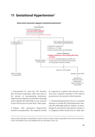
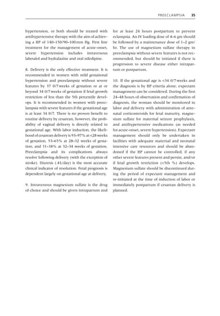
34 Preeclampsia
should onlybe made after 20 weeks of gesta-
tion, and likely represents an exaggerated phys-
iologic response of the maternal cardiovascular
system to pregnancy.
3. Preeclampsia is a multisystem disorder spe-
cific to pregnancy and the puerperium. More
precisely, it is a disease of the placenta since it
occurs in pregnancies where there is trophoblast
but no fetal tissue (complete molar pregnancies).
Preeclampsia usually occurs after 20 weeks of
gestation, most often near term. Evidence of
gestational proteinuric hypertension prior to
20 weeks should raise the possibility of an
underlying molar pregnancy, drug withdrawal
or (rarely) chromosomal abnormality in the
fetus. Diagnostic criteria for preeclampsia include
the following: hypertension after 20 weeks of
gestation in a woman with a previously normal
BP and proteinuria (>300mg of protein in a
24‐hour urine collection or this amount extrap-
olated from a timed collection; or protein/creati-
nine ratio >0.3; or urine dipstick test of 1+). In
the absence of proteinuria, preeclampsia is diag-
nosed in the setting of new‐onset hypertension
and any of the following: thrombocytopenia
(platelet count <100,000/microliter); renal
insufficiency (serum creatinine >1.1mg/dL or a
doubling of the creatinine in the absence of
other renal disease); impaired liver function
(elevated serum transaminases to twice normal
concentration and/or severe, persistent right
upper quadrant or epigastric pain); pulmonary
edema; cerebral or visual disturbances.
Preeclampsia is classified as “mild” or “severe.”
The terminology has recently undergone revi-
sion and “mild” preeclampsia is now referred to
as “preeclampsia without severe features,” while
“severe” preeclampsia is preeclampsia with
severe features. Proteinuria is no longer required
for the diagnosis of preeclampsia and massive
proteinuria (>5 gm) has been eliminated from
the consideration of preeclampsia as severe.
4. Refers to all women with only mild hyper-
tension (systolic BP of>140mm Hg but <160mm
Hg and/or diastolic BP of >90mmHg but
<110mm Hg) and proteinuria.
5. Refers to all women with severe hyperten-
sion (systolic BP of >160mm Hg and/or diastolic
BP of >110mmHg on two occasions at least 4
hours apart, or sooner if antihypertensive medi-
cation is administered) and proteinuria or
women with new‐onset hypertension (mild or
severe) and any of the features outlined above.
6. Outpatient management of preeclampsia
without severe features is an option for women
as long as both patient and physician are com-
fortable with the plan and understand the risks.
The patient will report symptoms of severe
preeclampsia immediately, and the patient can
comply with regular visits that include all of the
following:
• Maternal evaluation: once‐weekly clinic visits
with assessment for symptoms of preeclamp-
sia and lab evaluation (CBC with platelets,
AST, ALT, serum creatinine).
• Fetal evaluation: daily kick counts, once‐
weekly antenatal testing (i.e., BPP), ultrasound
assessment of fetal growth every 3 weeks.
7. Antihypertensive therapy is used to prevent
severe gestational hypertension and maternal
hemorrhagic strokes. There are only three
indications for antihypertensive therapy in the
setting of preeclampsia: (i) underlying chronic
hypertension; (ii) to achieve BP control to pre-
vent cerebrovascular accident while effecting
delivery; and/or (iii) expectant management of
severe preeclampsia by BP criteria alone (Sibai
protocol). BP control is important to prevent
cerebrovascular accident, but does not affect the
natural course of preeclampsia. The degree of
systolic hypertension (as opposed to the level of
diastolic hypertension or relative increase or
rate of increase of mean arterial pressure from
baseline levels) may be the most important
predictor of cerebral injury and infarction.
Acute‐onset, severe systolic (>160mm Hg)
hypertension; severe diastolic (>110mm Hg)
48.
Preeclampsia 35
hypertension, orboth should be treated with
antihypertensive therapy with the aim of achiev-
ing a BP of 140–150/90–100mm Hg. First line
treatment for the management of acute‐onset,
severe hypertension includes intravenous
labetalol and hydralazine and oral nifedipine.
8. Delivery is the only effective treatment. It is
recommended in women with mild gestational
hypertension and preeclampsia without severe
features by 37 0/7weeks of gestation or at or
beyond 34 0/7weeks of gestation if fetal growth
restriction of less than the 5th percentile devel-
ops. It is recommended in women with preec-
lampsia with severe features if the gestational age
is at least 34 0/7. There is no proven benefit to
routine delivery by cesarean, however, the prob-
ability of vaginal delivery is directly related to
gestational age. With labor induction, the likeli-
hood of cesarean delivery is 93–97% at <28weeks
of gestation, 53–65% at 28–32 weeks of gesta-
tion, and 31–38% at 32–34 weeks of gestation.
Preeclampsia and its complications always
resolve following delivery (with the exception of
stroke). Diuresis (.4L/day) is the most accurate
clinical indicator of resolution. Fetal prognosis is
dependent largely on gestational age at delivery.
9. Intravenous magnesium sulfate is the drug
of choice and should be given intrapartum and
for at least 24 hours postpartum to prevent
eclampsia. An IV loading dose of 4–6 gm should
be followed by a maintenance dose of 1–2 gm/
hr. The use of magnesium sulfate therapy in
preeclampsia without severe features is not rec-
ommended, but should be initiated if there is
progression to severe disease either intrapar-
tum or postpartum.
10. If the gestational age is <34 0/7weeks and
the diagnosis is by BP criteria alone, expectant
management can be considered. During the first
24–48 hours of observation and confirmation of
diagnosis, the woman should be monitored in
labor and delivery with administration of ante-
natal corticosteroids for fetal maturity, magne-
sium sulfate for maternal seizure prophylaxis,
and antihypertensive medications (as needed
for acute‐onset, severe hypertension). Expectant
management should only be undertaken in
facilities with adequate maternal and neonatal
intensive care resources and should be aban-
doned if the BP cannot be controlled, if any
other severe features present and persist, and/or
if fetal growth restriction (<5th %) develops.
Magnesium sulfate should be discontinued dur-
ing the period of expectant management and
re‐initiated at the time of induction of labor or
immediately postpartum if cesarean delivery is
planned.
executed ‘Abraham andthe Angels’; and Ribera’s ‘Birth of Christ’ and
‘Æsop’ are here. But more important than these is the work of Velazquez,
the ‘Sons of Jacob,’ the only picture of the great master among the Escorial
collection. This was one of three pictures which Velazquez painted at Rome
and sent to his father-in-law, the versatile Pacheco, artist, canon, and
historian.
The pictures by Bassano were probably among those brought from Italy
by Velazquez when he went on a mission for Philip.
In the Prior’s Hall there are several examples of the work of the Italian
illuminators, some of which came from the collection of Charles I. of
England. There are an ‘Entombment’ and ‘Christ in the House of the
Pharisee’ by Tintoretto. Three of Titian’s works adorn the hall: ‘Our Lady of
Grief,’ the ‘Last Supper,’ and the ‘Prayer in the Garden.’
The original church has an altar of marble, with paintings by Titian of
the ‘Adoration of the Magi’ and ‘Ecce Homo.’ The ‘Entombment’ is a copy
of Titian’s picture in the Prado Gallery at Madrid. Many of the paintings
have been removed from the old church to the Prado, but among the notable
canvases remaining are the ‘Martyrdom of St. James,’ an ‘Annunciation,’
by Paul Veronese, and Zuccaro’s ‘Birth of Christ.’ The most important is
perhaps the picture by El Greco of ‘St. Maurice.’
On the handsome staircase there are more pictures from the brush of the
facile Giordano, one of the most rapid of painters. One of these frescoes
represents the founding of the Escorial, and it has a figure of Philip II.
inspecting the designs of the architects, Juan Bautista de Toledo, Herrera,
and Antonio Villacastin. There is also a portrait of the king’s jester,
D’Antona. San Lorenzo and the Virgin are depicted in the centre of the
dome, and there are portraits of Charles V., San Fernando, and San
Geronimo in the group. The portraits of the Emperor Charles, Philip II.,
Charles II., and other royal personages are said to be excellent likenesses.
The Upper Gallery of the Cloisters is decorated with several paintings by
Barrocci, Giordano, Carducci, Juan de Gomez, Navarrete, and Sebastian
Herrera. Navarrete’s works are of principal interest; they are ‘San
Geronimo,’ the ‘Birth of Christ,’ and the ‘Appearance of Christ to His
Mother.’
The Lecture Hall contains a ‘Resurrection,’ painted by Paul Veronese,
and some copies of Titian and Rubens. Navarrete’s ‘Burial of San Lorenzo’
51.
is a fairinstance of this artist’s manner, and may be considered the most
interesting picture in the room.
In an adjoining small apartment there are many treasures, curiosities, and
objects of art. This collection was despoiled by the French soldiery, but a
considerable number of relics have been preserved. Among them is a
manuscript by St. Augustine concerning the christening of children, dating
from early in the seventh century. St. Teresa of Avila, who was a guest at
the Escorial, wrote the four works which are kept here. One of the volumes
is an Autobiography of the saint, and another is a work upon The Road to
Perfection. The two other books are codes and regulations concerning the
religious houses which Santa Teresa founded.
The full title of the celebrated Spanish saint is Maestra Serafica Madre
Santa Teresa de Jesus. She was born in 1515, and took the veil in her youth.
Philip II. collected and preserved her writings at the Escorial, and Philip III.
urged the canonisation of the saint in 1622. At the death of the patroness of
Spain, the souls of thousands of martyrs appeared at her bedside. Santa
Teresa’s writing-desk and ink-bottle, and an autograph of the Saint are
among the most treasured relics preserved in the Escorial.
Among the sacred relics are a portion of the gridiron of San Lorenzo, a
piece of his clothing, and the veil of St. Agata.
The statue of San Juan is by Nicolas. A jar which was placed here in the
time of Philip II. is reputed to be one of the original vessels which held the
wine at the marriage feast of Cana.
There are several pictures of the French and Flemish schools in this
room, including ‘San Geronimo,’ by Holbein. Bassano and Zuccaro are
represented, and there are a few paintings in the Florentine style, but none
of especial merit. Maëlla, a Spanish painter, who worked between 1739 and
1819, had some reputation as a fresco designer, and became a Court artist
under Charles IV. at the time when Goya was producing some of his finest
pictures. Goya subsequently took the place of Mariano Maëlla as the king’s
painter. In this room there is an ‘Immaculate Conception’ by Maëlla.
A brother of the Order of San Geronimo painted the ceiling, and another
monk carved the figure of their patron saint upon the altar screen.
Portraits of the fathers of the Monastery are hung in the Upper Prior’s
Chamber. The paintings are mostly by minor Spanish artists and Italians
who worked at the Escorial. Sanchez Coello’s portrait of Padre Sigüenza,
52.
the earliest chroniclerof the Monastery, has been copied and hangs here. In
an ante-chamber are oil-paintings by Giordano and copies of pictures by
Reni, Raphael, and Rubens. The ‘Conception’ is by Francisco Bayeu, a
contemporary of Maëlla, who painted frescoes in many of the Castilian
churches. Bayeu’s daughter, Josefa, was the wife of the brilliant Goya.
The Prior’s Oratory contains portraits of Charles III. and Maria of Saxony
and an old German picture of saints. There is also a sculpture of the
‘Conception.’ The Lower Prior’s Chamber or cell has the one work of
Francisco Urbino to be seen in the Escorial, a fresco of some interest. There
is a picture by Pantoja, a portrait of the Emperor Charles in his youth. The
portraits of Maria Luisa and of Charles IV. are copies from Goya’s works.
Vicente López, who painted the portrait of Maria, wife of Ferdinand VII.,
was a painter of some eminence and a contemporary of Goya. López
executed one of the best portraits of Goya.
The Carreño portraits in this apartment are good examples of the work of
this artist, who belonged to the school of Madrid. Carreño de Miranda was a
pupil of Pedro de los Cuevas, and a follower of Velazquez, who obtained
for him a commission to paint pictures of the Royal Palace of Madrid.
Carreño was made a Court painter after the death of the great Velazquez,
and many of his pictures are to be seen in the churches of Spain and at the
Escorial.
Doña Luisa Roldan’s figure of San Miguel is in the vestry. This is a
specimen of the wood carving for which the sculptors of Spain were
famous. The art of carving effigies in wood was revived successfully in
Andalusia by Martinez Montañéz, who died in 1649; and among his
followers were Pedro Roldan and Hernandez, who produced a large number
of carved images for churches and for exhibition in religious processions.
Doña Luisa Roldan, a daughter of Pedro, owed her inspiration to the master
of this art, but her achievements fall short of the perfection which he
attained.
There is a story concerning this señora’s work in the Escorial. It appears
that after a tiff with her husband, Luisa Roldan carved the figure of San
Miguel, and represented her own seraphic countenance in that of the saint,
while the scowling demon at his feet exhibits the features of the irascible
husband. The pictures in the vestry are mostly copies of Titian and
53.
Tintoretto, but the‘Martyrdom of San Pedro’ is said to be an original work
of Caravaggio.
In the four Minor Cloisters are several pictures. One is an anonymous
work, a landscape, and the others, of no great merit, are portraits of saints.
The crucifix to be seen here was the work of an Indian convert to
Christianity.
The spacious Refectory has dining tables of wood on stone pillars. Over
the foundation stone is the Prior’s seat, and there are two pulpits in the hall.
The kitchens, wine cellars, and other domestic offices are in this part of the
monastery.
The Real Monasterio is the most characteristic portion of the huge pile
of the Escorial, and its austerity and atmosphere of contemplation and piety
testify to the religious and ascetic spirit of the royal recluse who founded it.
This is no palace of mere delight and of luxury. It is a temple and a retreat, a
sanctuary from the world of strife and unrest, and an asylum for the penitent
and the devout. It is a monument of the Catholic faith, built with the
devotion of artists and labourers inspired by a deep zeal for religion, erected
without regard to the immense cost of its construction and furnishing, and
dedicated in the name of the pious San Lorenzo to the worship of God and
the Holy Virgin. Truly a strangely interesting memorial.
Here, in the odour of piety, surrounded by friars and monks, Philip II.
lived the life of renunciation and of calm meditation. This tabernacle in the
wilderness is a symbol of the mind of Spain in the days of her power, the
manifestation of her profound faith, and a tribute to the seriousness and
quietism which were the ideals of one of the most remarkable and complex
of her rulers. We feel that the very stones of the building reveal the nature
of Philip, the king who would be saint, the ambitious patriot who longed for
power and vivid life, and yet realised that the placid existence in the cloister
is more excellent than the fevered life of courts.
54.
V
THE LIBRARY
The impressof the intellect of Philip II. is no less manifest in the great
storehouse of books in the Escorial than in the Church and in the
Monastery. It accords with the character of the founder that he should desire
to possess a vast collection of the world’s choicest volumes of theology and
philosophy, for, like Solomon, Philip esteemed wisdom as highly as the
supremacy of rulership and the possession of great riches. His ambition as a
student and an art collector was as keen as his craving for sovereignty and
might. We have seen how he had applied himself in his youth to the study
of literature and of languages, and the respect which he yielded to the
sciences, arts, and letters.
The civilised world was searched; the libraries of all nations were
overhauled to stock the Library of the Escorial with great books and
precious manuscripts. Greece, Arabia, and Palestine contributed to the
Bibliotheca of the kingly scholar, and the collection of volumes was at one
time the finest in Europe. Philip himself took the greatest interest in the
Library. He worked at the catalogue and annotated the list of volumes. The
original collection was greatly augmented from time to time by the
purchases made on the king’s behalf by agents, who travelled in various
countries, by the seizure of volumes belonging to heretics, and by works
presented by loyal and wealthy subjects. Castillo was one of Philip’s book-
hunters; the catalogue of Arabic books and documents was prepared by the
learned Father Sigüenza.
A suitable repository for the books was erected at the Escorial under the
supervision of Philip. The vaulted ceiling was painted by Vicente Carducci,
an Italian artist of great distinction, who may be ranked as the chief of the
Italian designers employed to embellish the Escorial. Carducci’s mural
paintings in the Library are perhaps the finest of all the fresco works in the
building. He was assisted by Tibaldi of Bologna, an artist of meagre talent,
who represented Philosophy, Grammar, Arithmetic, Astronomy, and other
sciences and certain of the arts, upon his portion of the ceiling.
55.
The Library iswell lighted by windows. Even the shelves display the
taste of the founder, for they are beautifully carved by Flecha. The tables
are of marble and jasper, and the floor is paved with marble. Between the
rows of shelves are some portraits of Spanish sovereigns, and among them
is Carreño’s picture of Charles II. at the age of fourteen. Pantoja painted the
Emperor Charles V. and the portraits of Philip II. and Philip III. The pictures
represent the subjects in life-size.
Juan de Herrera, the famous architect of the Escorial, is presented on one
of the canvases by an anonymous artist. Isabella of Portugal, wife of
Charles V., is portrayed here, together with another portrait of Charles V. in
boyhood, which hangs in the same room. The bust of Cicero in the Library
was said to have been unearthed at Herculaneum.
Upon the entrance to the Library are the words of anathema uttered by
the Pope upon any one who should dare to purloin books from the
collection. There have been, however, many losses. The French plundered
the Library, which, long before the invasion, was devastated by the fire of
1671, and from time to time the hand of the pilferer has been laid upon
many of the volumes.
From a nucleus of four thousand volumes, collected by Philip II. and
placed in the Escorial, the Library grew in the number of its books, and in
the costly manuscripts in Arabic and other Eastern tongues. At one period
this was the most notable and valuable collection of Arab works in the
whole of Europe. The gift of Don Diego de Mendoza’s private library
further enriched the collection. Mendoza was Philip’s ambassador to Italy,
and a man of wide culture and with a love of books. When the king
inherited this splendid library, he satisfied all his ambassador’s creditors.
Mendoza’s volumes were presented in 1576. They were bound in an
unusual manner, one cover being red and the other black, and sometimes
the leaf edges are decorated in two colours. Among other donors to the
Library were Augustin, Archbishop of Tarragona, an eminent writer, Ponce
de Léon, and Geronimo de Zurita, a historian. The Balearic Islands
contributed about three hundred volumes, some of them being the writings
of the remarkable Raymond Lully. From the Inquisition the Library
received about one hundred and forty books. Authors of repute frequently
presented their manuscripts to the great collection at the Escorial.
56.
In 1583 aMoorish interpreter, in the employment of Philip, was
commissioned to buy all the Arab books that he could discover in Granada
and Cordova. This bibliographer, Alonso de Castillo, appears to have
devoted great labour to the extension of the Library and catalogue-making.
About seventy manuscripts in Greek, Hebrew, and Arabic, were presented
by Father Montano, who seems to have acted as librarian, though the first to
hold that office was Father Juan de San Geronimo.
Philip III. increased the Library by the addition of confiscated books
seized from Don Ramuz del Prado; and in the reign of the same monarch,
the collection of three thousand volumes belonging to the Emperor of
Morocco was acquired.
Anticipating the rule of our biggest public library, the British Museum, it
was decreed that a copy of every book issued in Spain should be presented
to the Escorial. Besides volumes of philosophy, history, theology, science,
and poetry, there was a collection of church music in the Library, some of
which was composed by the monks, del Valle, Torrijos, and Cordova. Many
of the compositions of the renowned choir-master, Antonio Soler, are in the
Library.
Among the many valuable manuscripts in the Escorial collection are the
Gospels, illuminated in gold on vellum. The Missal is bound in red leather
and wood, with silver clasps. Bound up with these manuscripts are the
‘Epistles of San Geronimo,’ which are still in splendid preservation. Philip
II. and many other collectors presented several breviaries and illuminated
manuscripts to the Library. The relics are mostly well preserved, and in
some cases the bindings are remarkably new in appearance. Some of the
manuscripts are in Persian, others are in the Chinese and Arabic languages.
Several of the Latin manuscripts are exceedingly interesting, and date
from A.D. 976 and the eleventh century. The Bibles, which number nineteen,
are of the fourteenth century, and beautifully bound in parchment. One of
the choicest treasures of the Library is the Apocalypse of St. John, richly
illustrated. The Greek manuscripts contain several works of the early
Fathers, which have never been printed. Another valuable object is the
ancient copy of the Koran, most exquisitely written and ornamented.
The great ‘Chronicle’ of Alfonso is to be seen here, as well as the king’s
treatises on ‘Hunting,’ ‘Chess,’ and ‘Draughts,’ which contain illustrations
and diagrams. A number of old Castilian Bibles, dating from the early part
57.
of the fifteenthcentury, are of especial interest. ‘The Census of Spain,’
contributed by Philip II., is also preserved in sundry volumes.
Among the sketches are works by El Mudo, Tibaldi, and Urbini. A large
number of fine engravings and drawings by Michael Angelo, Titian,
Raphael, and Albert Dürer form part of the collection of prints in the
Library.
The Reading-Room contains over fifty pictures, mostly portraits of little
value in the artistic sense. An inkstand which belonged to Father Sigüenza
is shown here. The most notable treasure is a portrait by the vigorous
Zurbarán, perhaps the most distinctively Spanish painter of the realistic
school. According to Lord Leighton, Francisco de Zurbarán represented ‘all
Spain’ in his art.
Among the portraits of illustrious persons are those of Quevedo, Father
Sigüenza, Torquemada, Francisco Ximenes, Luis de Gongora, and Cardinal
Mendoza.
58.
VI
THE UNIVERSITY
An importantpart of the great building of the Escorial was the Colegio,
designed by Philip as an institution for the instruction of monks and
preachers. We have seen how from his earliest manhood the king valued
culture and esteemed the arts, and the University of the Escorial was his
tribute to learning, just as the Monastery and the Church bore testimony of
his zeal for the Faith of Rome.
The Colegio is approached from the King’s Court by steps through a
vestibule. Beneath the tower is an apartment known as the Hall of Secrets,
on account of its construction permitting whispers to be heard from one side
of the chamber to the other. There is a spacious Refectory to the College,
containing a pulpit. The arched cloister or promenade was used for dramatic
displays, and it has an upper gallery with balconies and railings. Upon the
ceiling are paintings by Francisco Llamas, a minor artist.
In the Chapel of the College, a building over 60 feet in length and about
30 in width, there is a handsome altar by Lorenzo Berni, carved and gilded
with considerable taste. In front of the altar is a carved seat of pine and
walnut, and there are seats for the students along the walls.
The Seminary is attached to the University, and resembles the Colegio in
its structure. It was used by an Order of the devout who practised severe
restraints, and applied themselves to diligent study. Evidence of their
austerity is shown in the absence of windows. This part of the Escorial
contains an infirmary, an oratory, a refectory, and cells.
Passing along the Invalids’ Gallery we reach the Compana, in which
there are various offices and a marble bath. The Dispensary was formerly in
this part of the Escorial. There is a viaduct from the court of the Compana,
beneath which runs another road. Around the smaller patios in this portion
of the pile are the bakeries, storehouses, stables, and various workshops.
To supply the Escorial with water for its seventy-six fountains, its baths,
and taps, several ingenious conduits were constructed before the first stone
59.
of the edificewas laid. This water supply is excellent, and there is scarcely
an apartment in the Escorial which is without pure water from the mountain
springs.
60.
VII
THE PALACE
The apartmentsoccupied by Philip II. were in the north-east corner of the
edifice, and they are entered by the north façade. This was the retreat of the
moody monarch, and it accords with his love of seclusion and the simplicity
of his desires, though the additions of succeeding sovereigns have rendered
the Palace more luxurious and richer in decoration and art treasures.
Philip’s own apartment, where he died, is more like the cell of a hermit than
the chamber of a king. There is a tiled floor to the room, an absence of
ornamentation, and an atmosphere of severe gloom.
Here are some relics of the king—his bookcase, writing-table, a few
chairs, and the stools upon which he laid his foot when attacked by the
gout. From this chamber Philip could peep into the Church during the
celebration of services and hear the voices of the friars at their devotions.
By opening a panel, the king was able to look upon the High Altar and to
catch the odour of the incense.
The fine staircase of the Palace was the work of Ventura Rodriguez, and
it was built in the reign of Carlos IV. In the Lower Gallery there are pictures
of historical interest, some of them being the original works painted for the
founder of the Escorial. They are chiefly battlepieces; and one is a copy of
the fresco in the Hall of Battles. Among the pictures are the ‘Siege of
Grave,’ the ‘Siege of Calais,’ the ‘Christian Fleet attacking the Turks,’
‘Battle at Amiens,’ and the ‘Fleet entering the Port of Messina.’
The most important tapestries of the Escorial are in the Palace. They
were designed by the gifted Goya and by Maëlla, one of the lesser painters
of the eighteenth century. Goya’s designs are typically Spanish, and deserve
the closest attention. The tapestries were mostly made in Madrid, though
those from designs by Teniers were woven in Holland, and a few are of
French and Italian manufacture. The tapestry works are of exceptional
design and are scarcely excelled by any in Europe. Goya’s success as a
designer of tapestry cartoons gained him election to the Académia de San
61.
Fernando, and CharlesIV. appointed him pintor de cámara del Rey, or Court
artist.
In the Apartments of the Prince there are some noteworthy pictures of
the Italian School, and an old Dutch tapestry adorns the wall of one room.
But finer paintings are in the Apartments of the Princess, where Alonso
Cano’s genius is shown in the Virgin and the Infant Christ. Pantoja’s
portrait of Philip II., painted on wood, is here. The Head of Christ by Guido
Reni is in the bedroom; and another sleeping apartment is decorated with a
tapestry from the designs of a Flemish artist. The drawing-room contains a
tapestry of Spanish design, and there are tapestries in the nurse-room and
play-room.
The chief picture in the workroom is one of San Antonio by the powerful
Ribera. Three portraits by Mengs hang here: one of Charles III. in mail, and
the others of Maria Josephine, daughter of Charles, and Maria Luisa, wife
of Leopold of Austria, the Archduke.
In the Reception Hall there is a portrait of the Duque de Olivares, said to
be by Velazquez, but the authenticity is dubious.
The picture of Pope Innocent X. is a copy of Velazquez’s portrait. Among
the other portraits in this hall are Isabel, wife of Philip II. There are a few
landscape paintings, and one or two works of minor painters of the Italian
school.
The corridor of the Hall of the Caryatides is supposed to represent the
handle of the gridiron upon which the patron saint of the Escorial was
martyred for his faith. This is an almost square apartment, designed in the
Doric style. There are two fountains in marble, with figures of Caryatides
pouring water into the basins.
The Hall of Battles is adorned with a number of fresco-paintings
representing various combats. The battle of Higueruela, which was fought
against the Moors by Don John in 1431, was painted by two Italians,
Granelo and Fabricio, from an old battlepiece which was found at Segovia.
Philip II. was much interested in this picture, and he ordered the two artists
to reproduce it in a huge mural painting. The work was divided into eight
sections, depicting various scenes in the great encounter with the infidels,
and the king was well satisfied with the fresco. There are two pictures of
Philip’s battleships in action at either end of the ‘Battle of Higueruela.’
Other martial subjects are treated. They are: ‘A Review by Philip II.,’ the
62.
‘Battle of Lisbonunder the Duke of Alba,’ the ‘Capture of Noyon,’ the
‘Firing of a City,’ the ‘Departure from St. Quintin,’ the ‘Capture of a Fort,’
the ‘Attack on St. Quintin,’ the ‘Spanish Troops under Philibert,’ ‘A Battle,’
and ‘Before the Siege of St. Quintin.’
The ceiling of the Hall of Battles is adorned with quaint pictures.
The Apartments of the Queen are daintily decorated with tapestries,
some by Spanish and others by Dutch designers, while the hangings are of
amber silk. In the Oratory of the Queen there is an altar with a painting of
the ‘Virgin and the Infant Jesus.’ The painter was Juan de Juanes,
sometimes called Vicente Joanes, or Juan Macip. According to C.
Gasquoine Hartley, in A Record of Spanish Painting, this artist was ‘the first
great exponent’ of the art of the Valencian School. ‘He is one of the national
painters of Spain. To some extent his painting was imbued with the Italian
ideal, and it is possible that he received his artistic training in Italy; but the
Spanish personality of his work is rarely obscured. The intense religious
solemnity, the decorous purity, the vigorous handling, the careful painting
of details, the luminous warmth of colour, the lack of creative imagination,
the disregard of beauty, the tendency to exaggeration, all the virtue and all
the limitation of the painters of Spain are outlined in his work.’
Little is known of the life of Juanes. He was a Valencian by birth, and he
painted several pictures in the churches of his Province. Juanes was a
devoutly religious man, and his work shows a strong bias for mysticism and
warm piety. He was an industrious painter; his work ranges from portraits to
large subject pictures, and there are many of his paintings in Valencia.
Juanes died in 1579, and his style is seen in the pictures produced by his
pupils, who are, however, unimbued with his genius. ‘With Juanes,’ writes
C. Gasquoine Hartley, ‘we close the record of the early Hispano-Italian
painters.’
In the Bedroom of the Queen there is a tapestry and a ceiling by López,
who also designed the ceiling of the dressing-room.
We have already noted the tapestries in the Apartments of the Princess.
The Carved Rooms demand inspection, for they contain examples of rare
decoration in woods. The carvings and the inlaid work of these rooms cost a
very large sum. Woods of the rarest and finest were employed for the
adornment of the apartments, and gilt and gold were lavished upon the
walls. The pictures painted on copper are by Montalbo, and the ceiling
63.
painting was intrustedto Maëlla. Blue upholstery decks the seats. There are
four of these sumptuously decorated chambers. The third of them has a
ceiling-painting by Galvez. In the fourth room, Maëlla painted the ceiling,
and the walls are adorned in green and gold. There are a few pictures here,
but none of striking worth. The Oratory contains a tapestry of Spanish
design. On the altar there is a picture by Giordano representing Santa Anna,
St. Joachim, and the Holy Mother.
The Reception Room of this portion of the Palace is decorated with
tapestry, and the ceiling is painted by López. In the Antechamber, Billiard
Room, and the offices adjacent, there are more tapestries.
We have made our survey of this remarkable pile, which contains a
treasure house of works of art, an institution of learning, a convent, a
palace, and a church. Surely such a composite building is not to be seen in
any other quarter of the world. Nor is there any other edifice of such
importance and grandeur amid surroundings so savage and primitive. The
Royal Monastery of the Escorial is indeed an unique structure, historically
instructive, and of deep interest for the architect, the art student, and the
philosopher. It is a reflection and a symbol of Spain and of Philip II.; the
building, in its sombre setting of grey crags and shadowy woods, has an
indefinable atmosphere, a potent fascination.
64.
VIII
APPENDIX I
THE FIRSTCHRONICLER OF THE ESCORIAL
We have it on the authority of Padre Fray José de Sigüenza, the librarian of
the Monasterio, and the first historian of Philip II., that the king desired the
building at the Escorial to excel the majesty of Solomon’s Temple, and to
astonish the whole world. When we consider that the edifice was erected
and adorned with very little aid from machinery, we cannot but marvel at
the expedition in carrying out the architects’ and designers’ plans. A great
army of sawyers, carpenters, quarrymen, stonemasons, and craft-workers
found employment upon this great pile. It is recorded that the labourers
complained of the cold, searching winds in winter, and that they suffered
from the scorching heat in summer.
The rules applying to the workmen were stringent. The labourers appear
to have suffered from a scarcity of wine, which they desired in the hot
weather. No doubt the rigour of the climate in winter increased the
difficulties attending upon the undertaking.
Cranes and levers were employed for lifting the huge blocks of stone
quarried from the surrounding mountains. Even the rough work was allotted
only to workmen of approved ability and reputed industry, while the task of
decoration was given to masters of the arts of painting, illuminating, and
carving in wood, metal, and ivory. Upon this bleak highland a busy colony
of toilers from many parts of Europe sprang up in a few months. Forges,
workshops, and saw-pits were set up or constructed amongst the scoriæ or
shale of the mountains, and the desert rang with the beating of hammers on
metal, the clicking of the mason’s chisel, and the grating of the saws.
An immense number of carts and horses were employed, besides
pannier-mules and asses, to bring the stone from the quarries and the metal
from distant furnaces.
65.
Only a partof the work was performed at the Escorial. The bronze was
compounded in Zaragoza; the white marble came from the Sierras de
Filabres, and the green, black, and variegated marbles were brought from
the mountains of Andalusia, while Cuenca and Segovia furnished part of the
pine wood. Many of the figures of bronze for the altars were designed and
made in Florence and Milan. Toledo supplied lamps, candlesticks, crosses,
and incense-burners. Holland provided heavy bronze candle-holders. Spain,
Italy, and Flanders were searched for precious stones and metals to beautify
the building, and artists and craftsmen were imported from many countries.
Juan Bautista de Toledo, and his pupil Juan de Herrera, expended great
care and pains in the designs. Models of the various parts of the Escorial
were made in wood, and the designs were altered and improved by both
architects from time to time. Under the direction of Juan de Herrera every
detail of the building was submitted to him for his approval before it was
finished. Even the designs of Peregrino and Flecha, the Italian masters,
were submitted to the inspection of the master-architect. Juan de Herrera
was succeeded by Francisco de Mora as chief architect.
The construction of the Principal Choir was a part of the work upon
which much forethought was expended. Father Sigüenza describes the
Corinthian style of the choir as the most beautiful feature of the Church. He
mentions one hundred and twenty-eight as the number of the seats.
The Retablo was a costly piece of work, as a large quantity of jasper and
precious metal was used in its embellishment, and much gilt employed. The
columns are of the Doric order, and the bases are formed of gilded bronze.
Father Sigüenza does not stint his praise for the ‘judicious architects and
singular painters’ employed by Philip II. at the Escorial, and his book is
characterised by a high enthusiasm for the king, and admiration for the
great work, ‘this sumptuous fabric,’ in which the worthy padre spent so
many years of his life as Director of the Royal Library.
Coello painted the strong features of Father Sigüenza upon a canvas that
may be seen in the Prior’s Chamber of the Monastery.
66.
APPENDIX II
LIST OFTHE PRINCIPAL PAINTERS WHOSE
WORKS ARE IN THE ESCORIAL
ARTIST. SUBJECT. SITUATION.
Cano. Virgin and Infant Christ. Apartments of Princess.
Carducci. Portraits of Saints. Relicario of Church.
Arts and Sciences (fresco). Library.
Coello. Altarpieces. In Chapels.
Father Sigüenza. Upper Prior’s Chamber.
El Greco. St. Maurice. The Old Church.
Giordano. Life of David. Ceiling of Choir of Church.
Eight Frescoes. Dome of Church.
San Juan. Ante-Sacristy.
Frescoes. Staircase of Monastery.
Goya. Frescoes of Spanish Customs.Palace.
Holbein. San Geronimo. Lecture Hall.
Juanes. Virgin and Infant. Apartments of the Queen.
Navarrete Our Lady and San Juan. In Church near Prior’s Seat.
(El Mudo).Crucifixion. Library of Church.
Abraham and Angels. Salas de los Capitulos.
San Geronimo. ”
Birth of Christ. Upper Gallery of Cloisters.
Appearance of Jesus. ”
Burial of San Lorenzo. Lecture Hall.
67.
Pantoja. Various. Oratoriesof Church.
Carlos V. Lower Prior’s Chamber.
Philip II. Library.
Philip III. Apartments of Princess.
Reni. Scriptural. Sacristy of Church.
Ribera. San Geronimo. Ante-Sacristy of Church.
Birth of Christ. Salas de los Capitulos.
Philip II. Apartments of the Prince.
San Antonio. Workroom of Palace.
Teniers. Various Frescoes. Palace.
Tibaldi. Birth of Christ.
Adoration of the Magi. Altar Screen of Church.
Elias.
Old Testament Scenes. In Chapels.
Altarpieces. ”
Frescoes of Sciences. Library.
Tintoretto. Entombment. Prior’s Hall.
Washing the Apostles’ Feet. Sacristy of Church.
Titian. San Geronimo.
Agony in the Garden. Salas de los Capitulos.
Last Supper. ”
Prayer in the Garden. ”
Our Lady of Sorrows. ”
Adoration of the Magi. ”
Ecce Homo. ”
Velazquez. Sons of Jacob. Sacristy of Church.
Zuccaro. The Flagellation. Altar Screen of Church.
Birth of Christ. Old Church.
Zurbarán. Sons of Jacob. Sacristy of Church.
A Portrait. Reading-Room.
Welcome to OurBookstore - The Ultimate Destination for Book Lovers
Are you passionate about books and eager to explore new worlds of
knowledge? At our website, we offer a vast collection of books that
cater to every interest and age group. From classic literature to
specialized publications, self-help books, and children’s stories, we
have it all! Each book is a gateway to new adventures, helping you
expand your knowledge and nourish your soul
Experience Convenient and Enjoyable Book Shopping Our website is more
than just an online bookstore—it’s a bridge connecting readers to the
timeless values of culture and wisdom. With a sleek and user-friendly
interface and a smart search system, you can find your favorite books
quickly and easily. Enjoy special promotions, fast home delivery, and
a seamless shopping experience that saves you time and enhances your
love for reading.
Let us accompany you on the journey of exploring knowledge and
personal growth!
ebookgate.com


![This edition first published 2017 © 2010, 2017 by John Wiley & Sons, Ltd.
Registered Office
John Wiley & Sons, Ltd, The Atrium, Southern Gate, Chichester, West Sussex, PO19 8SQ, UK
Editorial Offices
9600 Garsington Road, Oxford, OX4 2DQ, UK
The Atrium, Southern Gate, Chichester, West Sussex, PO19 8SQ, UK
111 River Street, Hoboken, NJ 07030‐5774, USA
For details of our global editorial offices, for customer services and for information about how to apply for permission
to reuse the copyright material in this book please see our website at www.wiley.com/wiley‐blackwell.
The right of the author to be identified as the author of this work has been asserted in accordance with the UK
Copyright, Designs and Patents Act 1988.
All rights reserved. No part of this publication may be reproduced, stored in a retrieval system, or transmitted, in any
form or by any means, electronic, mechanical, photocopying, recording or otherwise, except as permitted by the UK
Copyright, Designs and Patents Act 1988, without the prior permission of the publisher.
Designations used by companies to distinguish their products are often claimed as trademarks. All brand names
and product names used in this book are trade names, service marks, trademarks or registered trademarks of their
respective owners. The publisher is not associated with any product or vendor mentioned in this book. It is sold on
the understanding that the publisher is not engaged in rendering professional services. If professional advice or other
expert assistance is required, the services of a competent professional should be sought.
The contents of this work are intended to further general scientific research, understanding, and discussion only
and are not intended and should not be relied upon as recommending or promoting a specific method, diagnosis,
or treatment by health science practitioners for any particular patient. The publisher and the author make no
representations or warranties with respect to the accuracy or completeness of the contents of this work and specifically
disclaim all warranties, including without limitation any implied warranties of fitness for a particular purpose. In
view of ongoing research, equipment modifications, changes in governmental regulations, and the constant flow of
information relating to the use of medicines, equipment, and devices, the reader is urged to review and evaluate the
information provided in the package insert or instructions for each medicine, equipment, or device for, among other
things, any changes in the instructions or indication of usage and for added warnings and precautions. Readers should
consult with a specialist where appropriate. The fact that an organization or Website is referred to in this work as a
citation and/or a potential source of further information does not mean that the author or the publisher endorses
the information the organization or Website may provide or recommendations it may make. Further, readers should
be aware that Internet Websites listed in this work may have changed or disappeared between when this work was
written and when it is read. No warranty may be created or extended by any promotional statements for this work.
Neither the publisher nor the author shall be liable for any damages arising herefrom.
Library of Congress Cataloging‐in‐Publication Data
Names: Norwitz, Errol R., author. | Saade, George R., 1960– author. |
Miller, Hugh S. (Hugh Stephen), author. | Davidson, Christina (Christina Marie) author.
Title: Obstetric clinical algorithms / Errol R. Norwitz, George R. Saade, Hugh Miller, Christina M. Davidson.
Description: 2nd edition. | Chichester, West Sussex, UK ; Hoboken, NJ : John Wiley & Sons, Inc., 2017. |
Preceded by: Obstetric clinical algorithms : management and evidence / Errol R. Norwitz ... [et al.]. 2010. |
Includes bibliographical references and index.
Identifiers: LCCN 2016024815 (print) | LCCN 2016026085 (ebook) | ISBN 9781118849903 (pbk.) | ISBN
9781118849873 (pdf) | ISBN 9781118849880 (epub)
Subjects: | MESH: Pregnancy Complications–diagnosis | Pregnancy Complications–therapy | Algorithms |
Decision Support Techniques
Classification: LCC RG571 (print) | LCC RG571 (ebook) | NLM WQ 240 | DDC 618.3–dc23
LC record available at https://lccn.loc.gov/2016024815
A catalogue record for this book is available from the British Library.
Wiley also publishes its books in a variety of electronic formats. Some content that appears in print may not be
available in electronic books.
Cover image: Jasmina/Gettyimages
Set in 8.5/12pt Meridien by SPi Global, Pondicherry, India
1 2017](https://image.slidesharecdn.com/10312-250506024242-87a5880c/85/Obstetric-clinical-algorithms-2nd-Edition-Davidson-6-320.jpg)

![vii
Preface
Recent advances in obstetrical practice and
research have resulted in significant improve-
ments in maternal and perinatal outcome. Such
improvements carry with them added responsi-
bility for the obstetric care provider. The deci-
sion to embark on a particular course of
management simply because “that’s the way we
did it when I was in training” or because “it worked
the last time I tried it” is no longer acceptable.
Clinical decisions should, wherever possible, be
evidence‐based. Evidence‐based medicine can
be defined as “the conscientious, explicit, and judi-
cious use of current best evidence in making decisions
about the care of individual patients” [1]. In prac-
tice, evidence‐based medicine requires exper-
tise in retrieving, interpreting, and applying the
results of scientific studies and in effectively
communicating the risks and benefits of differ-
ent courses of action to patients. This daunting
task is compounded by the fact that the volume
of medical literature is doubling every 10–15
years. Even within the relatively narrow field
of Obstetrics & Gynecology, there are more
than five major publications each month con-
taining an excess of 100 original articles and 35
editorials. How then does a busy practitioner
maintain a solid foundation of up‐to‐date
knowledge and synthesize these data into indi-
vidual management plans? New information
can be gleaned from a variety of sources: the
advice of colleagues and consultants, textbooks,
lectures and continuing medical education
courses, original research and review articles,
and from published clinical guidelines and con-
sensus statements. The internet has created an
additional virtual dimension by allowing
instant access to the medical literature to both
providers and patients. It is with this back-
ground in mind that we have written Obstetric
Clinical Algorithms: Management and Evidence, 2nd
edition.
Standardization of management reduces medi-
cal errors and improves patient safety and
obstetrical outcomes [2,3]. In this text, we have
developed a series of obstetric algorithms based
on best practice to mimic the decision‐making
processes that go on in our brains when faced
with a vexing clinical problem. To further facili-
tate decision‐making, we have superimposed
“levels of evidence” as defined by the report of
the US Preventive Services Task Force (USPSTF)
of the Agency for Healthcare Research Quality,
an independent panel of experts appointed and
funded by the US government to systematically
review evidence of effectiveness and develop
recommendations for clinical preventive ser-
vices [4]. The table below summarizes the
‘levels of evidence’ used in this text.](https://image.slidesharecdn.com/10312-250506024242-87a5880c/85/Obstetric-clinical-algorithms-2nd-Edition-Davidson-9-320.jpg)
![List of Abbreviations xi
PUBS percutaneous umbilical blood
sampling
q every
QFT‐GIT QuantiFERON®
‐TB Gold In‐Tube
test
RhoGAM anti‐Rh[D]‐immunoglobulin
R‐NST reactive NST
RPL recurrent pregnancy loss
RPR rapid plasma reagin
SC subcuticular
SGA small for gestational age
SIADH syndrome of inappropriate ADH
secretion
SLE systemic lupus erythematosus
SMA spinal muscular atrophy
SSI surgical site infection
STI sexually transmitted infection
TB tuberculosis
TBG thyroxine‐binding globulin
TFT thyroid function test
TORCH toxoplasmosis, rubella,
cytomegalovirus, herpes
TPPA T. pallidum particle agglutination
assay
TRAP twin reverse arterial perfusion
TST tuberculin skin testing
TTP/HUS thrombotic thrombocytopenic
purpura/hemolytic uremic
syndrome
TTTS twin‐to‐twin transfusion
syndrome
UA C&S urine culture and sensitivity
UDCA ursodeoxycholic acid
UFH unfractionated heparin
UTI urinary tract infection
VAS vibroacoustic stimulation
VBAC vaginal birth after cesarean
VDRL Venereal Disease Research
Laboratory
VL viral load
V/Q ventilation‐perfusion
VTE venous thromboembolism
ZDV zidovudine](https://image.slidesharecdn.com/10312-250506024242-87a5880c/85/Obstetric-clinical-algorithms-2nd-Edition-Davidson-13-320.jpg)

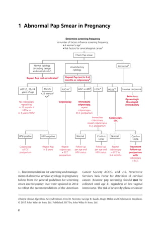
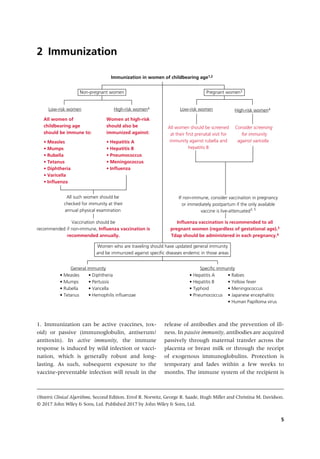


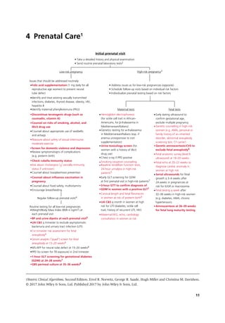
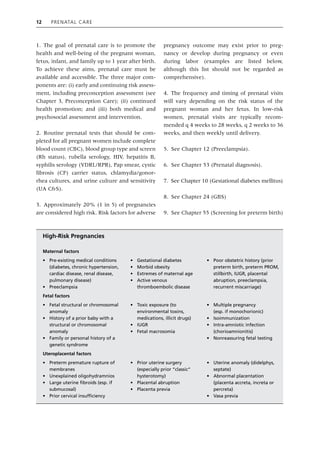

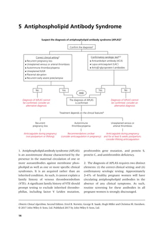

![Obstetric Clinical Algorithms, Second Edition. Errol R. Norwitz, George R. Saade, Hugh Miller and Christina M. Davidson.
© 2017 John Wiley & Sons, Ltd. Published 2017 by John Wiley & Sons, Ltd.
17
Asthma
6
1. Asthma is a chronic inflammatory disorder
of the airways characterized by intermittent
episodes of reversible bronchospasm. The
“
classic” signs and symptoms of asthma are
intermittent dyspnea, cough, and wheezing.
Pulmonary function tests that are most helpful
in diagnosing asthma are peak expiratory flow
rate (PEFR), spirometry (which includes meas-
urement of forced expiratory volume in one
second [FEV1] and forced vital capacity [FVC]),
and bronchoprovocation testing (such as with
a metacholine or exercise challenge). PEFR,
Confirm the diagnosis of asthma
• Take a detailed history, perform a physical examination, and
perform relevant pulmonary tests1
• Be aware of the normal physiologic changes in the respiratory
system during pregnancy2
• Consider differential diagnosis3
What is the patient’s baseline asthma status?
Mild asthma Moderate asthma Severe asthma
Initial prenatal visit Initial prenatal visit
• Identify and eliminate triggers for bronchospasm4
• Check baseline peak flow rate
• Continue baseline therapy5
• Consider pulmonary consultation
• Consider baseline pulmonary function tests
• Identify and eliminate triggers for bronchospasm4
• Check baseline peak flow rate
• Continue baseline therapy5
• Insist on pulmonary consultation
• Check baseline pulmonary function tests
• Consider baseline chest X-ray, EKG, and ABG
(arterial blood gas)2
Subsequent prenatal visits
Subsequent prenatal visits
• Ask about symptoms
• Check peak flow rate
• Clinical assessment of fetal growth
• Routine obstetric care
Consider hospitalization,
intubation, and delivery,
if necessary8
• Ask about symptoms
• Check peak flow rate
• Evaluate for pregnancy complications6
• Consider serial ultrasound growth scans
• Consider adjusting medications, as needed7
• Routine obstetric care
Allow spontaneous labor
and normal vaginal
delivery at term
No symptomatic
deterioration
Symptomatic
deterioration](https://image.slidesharecdn.com/10312-250506024242-87a5880c/85/Obstetric-clinical-algorithms-2nd-Edition-Davidson-30-320.jpg)