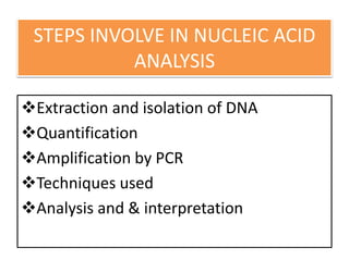
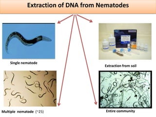
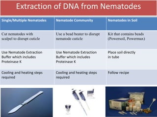
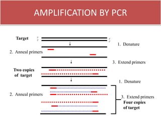
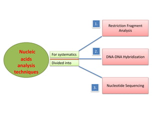
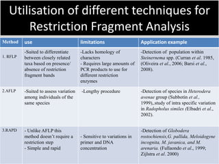
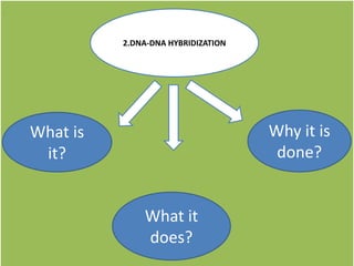
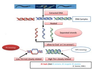
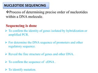
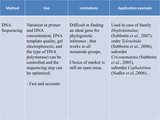
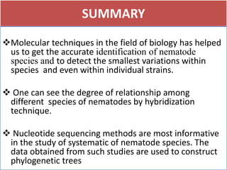
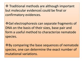
.
CREDIT SEMINAR
NUCLEIC ACID ANALYSIS IN NEMATODE
SYSTEMATICS
Presented By:
Mahnur Ali
A.A.U.
Deptt. of Nematology.
Introduction
Pathway to Molecular Diagnostics
Molecular identification & Morphological
identification
Steps involve in nucleic acid analysis
Extraction, Quantification, Amplification,
Separation, Analysis and interpretation
Utilization of different molecular
techniques for nematode systematic
Summary
.
INTRODUCTION
 Due to release of various metabolites by different (host &
non-host) crops and environmental effects there may be
change in the morphological characteristic of nematodes.
To reveal the actual variation and true identification, nucleic
acid analysis is important.
Variation present in nucleotide sequence cannot be observe
under microscope.
.
NUCLEIC ACIDS
DNA
RNA
Genome sizes
Caenorhabditis elegens = 100 MB
Meloidogyne incognita = 82 MB
Meloidogyne hapla = 54 MB
•Nucleic acids are polymers
•Nucleotides are monomers
Nitrogenous bases
-Purines
-Pyrimidine
Sugar
-Ribose
-Deoxyribose
Phosphates +
Nucleoside=Nucleotides
Nucleosides
.
.
Pathway to Molecular Diagnostics
1865 – G. Mendel, Law of Heredity
1866 –Johann Miescher, , Purification of DNA
1953 – Watson and Crick, Structure of DNA
1977 – DNA sequencing
1985 – Kary Mullis, in vitro Amplification of
DNA (PCR)
1998 – C. elegans the first multi-
cellular organism sequenced
Why Molecular Identification ?
. Morphological Identification Molecular Identification
Set-up cost Low High
Long-term cost High Low
Employee requirement Highly trained, experienced Minimal training
Length of process Slower More rapid
Morphological characters Variable NA
Females required Yes No
Mixed species
populations
Difficult to distinguish No
STEPS INVOLVE IN NUCLEIC ACID
ANALYSIS
Extraction and isolation of DNA
Quantification
Amplification by PCR
Techniques used
Analysis and & interpretation
Extraction of DNA from Nematodes
.
Single nematode
Entire communityMultiple nematode (~25)
Extraction from soil
Extraction of DNA from Nematodes
Single/Multiple Nematodes Nematode Community Nematodes in Soil
Cut nematodes with
scalpel to disrupt cuticle
Use a bead beater to disrupt
nematode cuticle
Kit that contains beads
(Powersoil, Powermax)
Use Nematode Extraction
Buffer which includes
Proteinase K
Use Nematode Extraction
Buffer which includes
Proteinase K
Place soil directly
in tube
Cooling and heating steps
required
Cooling and heating steps
required
Follow recipe
AMPLIFICATION BY PCR
5’
3’
3’
5’
Target
1. Denature
2. Anneal primers
3. Extend primers
Two copies
of target
1. Denature
2. Anneal primers
3. Extend primers
Four copies
of target
Restriction Fragment
Analysis
Nucleic
acids
analysis
techniques
DNA-DNA Hybridization
Nucleotide Sequencing
For systematics
Divided into
1.
3.
2.
1.Restriction Fragment Analysis
• This technique uses restriction enzymes to cleave DNA at
specific sites along the DNA molecule.
• Enzyme will digest DNA at specific sequence to generate
fragments of DNA
• Fragments are separated through gel electrophoresis.
• The banding patterns are visualized under florescent stain
(ethidium bromid) or by autoradiography.
Techniques involved in Restriction
Fragment Analysis
1. RFLP ( Restriction Fragment Length Polymorphism )
2. RAPD ( Random Amplified Polymorphic DNA )
3. AFLP ( Amplified Fragment Length Polymorphism )
RFLPs involves fragmenting
a sample of DNA by a
restriction enzyme, which can
recognize and cut DNA
wherever a specific short
sequence occurs.
A RFLP occurs when the
length of a detected fragment
varies between individuals.
 PCR based product but
the segments of DNA that
are amplified are random
we can amplify restricted
fragments and reduces the
complexity of material to be
analyzed (approx 1000
folds).
it can be used for
comparison between closely
related species only.
1 32
Enzyme Site Recognition in
RFLP
.
Restriction site Palindrome
Fragment 1 Fragment 2
• enzyme will digests (cuts) DNA at a
specific sequence = restriction site
• Enzymes recognize 4- or 6- base
pair, palindromic sequences
(e.g. GAATTC)
.
Power Supply
Agarose gel
Electrophoresis
 Electrical current
carries negatively-
charged DNA
through gel
towards positive
(red) electrode.
 Small fragments
move faster than
large fragments
.
Gel running
.
.
Utilisation of different techniques for
Restriction Fragment Analysis
Method use limitations Application example
1. RFLP
-Suited to differentiate
between closely related
taxa based on presence/
absence of restriction
fragment bands
-Lacks homology of
characters
- Requires large amounts of
PCR products to use for
different restriction
enzymes
-Detection of population within
Steinernema spp. (Curran et al. 1985,
(Oliveira et al., 2006; Barsi et al.,
2008).
2.AFLP -Suited to assess variation
among individuals of the
same species
-Lengthy procedure -Detection of species in Heterodera
avenae group (Subbotin et al.,
1999),.study of intra specific variation
in Radopholus similes (Elbadri et al.,
2002).
3.RAPD - Unlike AFLP this
method doesn’t require a
restriction step
- Simple and rapid
- Sensitive to variations in
primer and DNA
concentration
-Detection of Globodera
rostochiensis,G. pallida, Meloidogyne
incognita, M. javanica, and M.
arenaria. (Fullaondo et al., 1999;
Zijlstra et al. 2000)
.
.
2.DNA-DNA HYBRIDIZATION
What is
it?
What it
does?
Why it is
done?
Extracted DNA
..
DNA Samples
Separated strands
allow to Cool or ( re-anneal )
Heated
Low Tm=not closely related High Tm= closely related
0% homology
100% homology
M. hapla from M. arenaria , M. incognita M. Javanica
(C. Sereno, 2006 )
Process of determining precise order of nucleotides
within a DNA molecule.
Sequencing is done
NUCLEOTIDE SEQUENCING
 To confirm the identity of genes isolated by hybridization or
amplified PCR.
 For determine the DNA sequence of promoters and other
regulatory sequence.
 Reveal the fine structure of genes and other DNA.
 To confirm the sequence of cDNA .
 To identify mutation.
METHOD
 Principle- Use of dideoxynucleotide triphosphate
(ddNTP’s) as DNA chain terminator.
Method requires :-
1. Single stranded DNA template.
2. DNA primer.
3. DNA polymerase.
4. Normal dideoxynucleotide phosphates (dNTP’s ).
5. Modified nucleotides (ddNTP’s) that terminate DNA
strand elongation.
Chain-termination
method(Sanger method). F. Sanger in 1977By
.
Complementary strand
Template strand3’
3’5’
5’
primer
5’3’
1. DNA
polymerase
2. dNTP’s
3. ddNTP’s
+ Dideoxy ATP + Dideoxy TTP + Dideoxy CTP + Dideoxy GTP
DNA strand
.
1 2 3 4
3’
5’
T
G
G
T
A
C
G
Position of nucleotides from 5’ to 3’
.
Method Use Limitations Application example
DNA
Sequencing
Variation in primer
and DNA
concentration, DNA
template quality, gel
electrophoresis, and
the type of DNA
polymerase) can be
controlled and the
sequencing step can
be optimized.
- Fast and accurate
Difficult in finding
an ideal gene for
phylogenetic
inference , that
works in all
nematode groups.
Choice of marker is
still an open issue.
Used in case of family
Hoplolaimidae,
(Subbotin et al., 2007),
order Tylenchida
(Subbotin et al., 2006),
suborder
Criconematina (Subbotin
et al., 2005),
suborder Cephalobina
(Nadler et al.,2006) , .
.
,
M. arenaria TCGGCGATAGAGGTAAATGAC
TCGAGGGCATCTAATAAAGG
420 bp
950bp
Zijlstra et al., 2000
Dong et al., 2001
M. chitwoodi CCAATGATAGAGATAGGAAC
GATCTATGGCAGATGGTATGGA
TGGAGAGCAGCAGGAGAAAGA
400 bp
900 bp
800 bp
Williamson et al., 1997
Petersen et al., 1997
Zijlstra, 2000
M. exigua CATCCGTGCTGTAGCTGCGAG
CTCCGTGGGAAGAAAGACTG
562 bp Randig et al., 2002
M. hapla CAGGCCCTTCCAGCTAAAGA
TGACGGCGGTGAGTGCGA
GGCTGAGCATAGTAGATGATGTT
GGATGGCGTGCTTTCAAC
960 bp
610 bp
1500 bp
440 bp
Williamson et al., 1997
Zijlstra, 2000
Dong et al., 2001bp
Wishart et al., 2002
M. incognita CTCTGCCCAATGAGCTGTCC
TAGGCAGTAGGTTGTCGGG
GGGATGTGTAAATGCTCCTG
GTGAGGATTCAGCTCCCCAG
1200 bp
1350 bp
399 bp
955 bp
Zijlstra et al., 2000
Dong et al., 2001
Randig et al., 2002
Meng et al., 2004
M. javanica CCTTAATGTCAACACTAGAGCC
GGTGCGCGATTGAACTGAGC
ACGCTAGAATTCGACCCTGG
1650 bp
670 bp
519 bp
Dong et al., 2001b
Zijlstra et al., 2000
Meng et al., 2004
M. mayaguensis GAAATTGCTTTATTGTTACTAAG 322 bp Blok et al., 2002
M. naasi CTCTTTATGGAGAATAATCGT 433 bp Zijlstra et al., 2004
M. paranaensis GCCCGACTCCATTTGACGGA 208 bp Randig et al., 2002
Species Primer set ( 5’-3’ ) Amplicon length Reference
SUMMARY
Molecular techniques in the field of biology has helped
us to get the accurate identification of nematode
species and to detect the smallest variations within
species and even within individual strains.
 One can see the degree of relationship among
different species of nematodes by hybridization
technique.
 Nucleotide sequencing methods are most informative
in the study of systematic of nematode species. The
data obtained from such studies are used to construct
phylogenetic trees
 Traditional methods are although important
but molecular evidences could be final or
confirmatory evidences.
Gel electrophoresis can separate fragments of
DNA on the basis of their sizes, base pair and
form a useful method to characterize nematode
species.
By comparing the base sequences of nematode
species, one can determine the exact number of
mutational variations.
.
.

Nuclic acid analysis in nematode systematic

  • 1.
  • 2.
    CREDIT SEMINAR NUCLEIC ACIDANALYSIS IN NEMATODE SYSTEMATICS Presented By: Mahnur Ali A.A.U. Deptt. of Nematology.
  • 3.
    Introduction Pathway to MolecularDiagnostics Molecular identification & Morphological identification Steps involve in nucleic acid analysis Extraction, Quantification, Amplification, Separation, Analysis and interpretation Utilization of different molecular techniques for nematode systematic Summary .
  • 4.
    INTRODUCTION  Due torelease of various metabolites by different (host & non-host) crops and environmental effects there may be change in the morphological characteristic of nematodes. To reveal the actual variation and true identification, nucleic acid analysis is important. Variation present in nucleotide sequence cannot be observe under microscope.
  • 5.
    . NUCLEIC ACIDS DNA RNA Genome sizes Caenorhabditiselegens = 100 MB Meloidogyne incognita = 82 MB Meloidogyne hapla = 54 MB •Nucleic acids are polymers •Nucleotides are monomers Nitrogenous bases -Purines -Pyrimidine Sugar -Ribose -Deoxyribose Phosphates + Nucleoside=Nucleotides Nucleosides
  • 6.
    . . Pathway to MolecularDiagnostics 1865 – G. Mendel, Law of Heredity 1866 –Johann Miescher, , Purification of DNA 1953 – Watson and Crick, Structure of DNA 1977 – DNA sequencing 1985 – Kary Mullis, in vitro Amplification of DNA (PCR) 1998 – C. elegans the first multi- cellular organism sequenced
  • 7.
    Why Molecular Identification? . Morphological Identification Molecular Identification Set-up cost Low High Long-term cost High Low Employee requirement Highly trained, experienced Minimal training Length of process Slower More rapid Morphological characters Variable NA Females required Yes No Mixed species populations Difficult to distinguish No
  • 8.
    STEPS INVOLVE INNUCLEIC ACID ANALYSIS Extraction and isolation of DNA Quantification Amplification by PCR Techniques used Analysis and & interpretation
  • 9.
    Extraction of DNAfrom Nematodes . Single nematode Entire communityMultiple nematode (~25) Extraction from soil
  • 10.
    Extraction of DNAfrom Nematodes Single/Multiple Nematodes Nematode Community Nematodes in Soil Cut nematodes with scalpel to disrupt cuticle Use a bead beater to disrupt nematode cuticle Kit that contains beads (Powersoil, Powermax) Use Nematode Extraction Buffer which includes Proteinase K Use Nematode Extraction Buffer which includes Proteinase K Place soil directly in tube Cooling and heating steps required Cooling and heating steps required Follow recipe
  • 11.
    AMPLIFICATION BY PCR 5’ 3’ 3’ 5’ Target 1.Denature 2. Anneal primers 3. Extend primers Two copies of target 1. Denature 2. Anneal primers 3. Extend primers Four copies of target
  • 12.
  • 13.
    1.Restriction Fragment Analysis •This technique uses restriction enzymes to cleave DNA at specific sites along the DNA molecule. • Enzyme will digest DNA at specific sequence to generate fragments of DNA • Fragments are separated through gel electrophoresis. • The banding patterns are visualized under florescent stain (ethidium bromid) or by autoradiography.
  • 14.
    Techniques involved inRestriction Fragment Analysis 1. RFLP ( Restriction Fragment Length Polymorphism ) 2. RAPD ( Random Amplified Polymorphic DNA ) 3. AFLP ( Amplified Fragment Length Polymorphism ) RFLPs involves fragmenting a sample of DNA by a restriction enzyme, which can recognize and cut DNA wherever a specific short sequence occurs. A RFLP occurs when the length of a detected fragment varies between individuals.  PCR based product but the segments of DNA that are amplified are random we can amplify restricted fragments and reduces the complexity of material to be analyzed (approx 1000 folds). it can be used for comparison between closely related species only. 1 32
  • 15.
    Enzyme Site Recognitionin RFLP . Restriction site Palindrome Fragment 1 Fragment 2 • enzyme will digests (cuts) DNA at a specific sequence = restriction site • Enzymes recognize 4- or 6- base pair, palindromic sequences (e.g. GAATTC)
  • 16.
    . Power Supply Agarose gel Electrophoresis Electrical current carries negatively- charged DNA through gel towards positive (red) electrode.  Small fragments move faster than large fragments . Gel running
  • 17.
    . . Utilisation of differenttechniques for Restriction Fragment Analysis Method use limitations Application example 1. RFLP -Suited to differentiate between closely related taxa based on presence/ absence of restriction fragment bands -Lacks homology of characters - Requires large amounts of PCR products to use for different restriction enzymes -Detection of population within Steinernema spp. (Curran et al. 1985, (Oliveira et al., 2006; Barsi et al., 2008). 2.AFLP -Suited to assess variation among individuals of the same species -Lengthy procedure -Detection of species in Heterodera avenae group (Subbotin et al., 1999),.study of intra specific variation in Radopholus similes (Elbadri et al., 2002). 3.RAPD - Unlike AFLP this method doesn’t require a restriction step - Simple and rapid - Sensitive to variations in primer and DNA concentration -Detection of Globodera rostochiensis,G. pallida, Meloidogyne incognita, M. javanica, and M. arenaria. (Fullaondo et al., 1999; Zijlstra et al. 2000)
  • 18.
  • 19.
    Extracted DNA .. DNA Samples Separatedstrands allow to Cool or ( re-anneal ) Heated Low Tm=not closely related High Tm= closely related 0% homology 100% homology M. hapla from M. arenaria , M. incognita M. Javanica (C. Sereno, 2006 )
  • 20.
    Process of determiningprecise order of nucleotides within a DNA molecule. Sequencing is done NUCLEOTIDE SEQUENCING  To confirm the identity of genes isolated by hybridization or amplified PCR.  For determine the DNA sequence of promoters and other regulatory sequence.  Reveal the fine structure of genes and other DNA.  To confirm the sequence of cDNA .  To identify mutation.
  • 21.
    METHOD  Principle- Useof dideoxynucleotide triphosphate (ddNTP’s) as DNA chain terminator. Method requires :- 1. Single stranded DNA template. 2. DNA primer. 3. DNA polymerase. 4. Normal dideoxynucleotide phosphates (dNTP’s ). 5. Modified nucleotides (ddNTP’s) that terminate DNA strand elongation. Chain-termination method(Sanger method). F. Sanger in 1977By
  • 22.
    . Complementary strand Template strand3’ 3’5’ 5’ primer 5’3’ 1.DNA polymerase 2. dNTP’s 3. ddNTP’s + Dideoxy ATP + Dideoxy TTP + Dideoxy CTP + Dideoxy GTP DNA strand
  • 23.
    . 1 2 34 3’ 5’ T G G T A C G Position of nucleotides from 5’ to 3’
  • 24.
    . Method Use LimitationsApplication example DNA Sequencing Variation in primer and DNA concentration, DNA template quality, gel electrophoresis, and the type of DNA polymerase) can be controlled and the sequencing step can be optimized. - Fast and accurate Difficult in finding an ideal gene for phylogenetic inference , that works in all nematode groups. Choice of marker is still an open issue. Used in case of family Hoplolaimidae, (Subbotin et al., 2007), order Tylenchida (Subbotin et al., 2006), suborder Criconematina (Subbotin et al., 2005), suborder Cephalobina (Nadler et al.,2006) , .
  • 25.
    . , M. arenaria TCGGCGATAGAGGTAAATGAC TCGAGGGCATCTAATAAAGG 420bp 950bp Zijlstra et al., 2000 Dong et al., 2001 M. chitwoodi CCAATGATAGAGATAGGAAC GATCTATGGCAGATGGTATGGA TGGAGAGCAGCAGGAGAAAGA 400 bp 900 bp 800 bp Williamson et al., 1997 Petersen et al., 1997 Zijlstra, 2000 M. exigua CATCCGTGCTGTAGCTGCGAG CTCCGTGGGAAGAAAGACTG 562 bp Randig et al., 2002 M. hapla CAGGCCCTTCCAGCTAAAGA TGACGGCGGTGAGTGCGA GGCTGAGCATAGTAGATGATGTT GGATGGCGTGCTTTCAAC 960 bp 610 bp 1500 bp 440 bp Williamson et al., 1997 Zijlstra, 2000 Dong et al., 2001bp Wishart et al., 2002 M. incognita CTCTGCCCAATGAGCTGTCC TAGGCAGTAGGTTGTCGGG GGGATGTGTAAATGCTCCTG GTGAGGATTCAGCTCCCCAG 1200 bp 1350 bp 399 bp 955 bp Zijlstra et al., 2000 Dong et al., 2001 Randig et al., 2002 Meng et al., 2004 M. javanica CCTTAATGTCAACACTAGAGCC GGTGCGCGATTGAACTGAGC ACGCTAGAATTCGACCCTGG 1650 bp 670 bp 519 bp Dong et al., 2001b Zijlstra et al., 2000 Meng et al., 2004 M. mayaguensis GAAATTGCTTTATTGTTACTAAG 322 bp Blok et al., 2002 M. naasi CTCTTTATGGAGAATAATCGT 433 bp Zijlstra et al., 2004 M. paranaensis GCCCGACTCCATTTGACGGA 208 bp Randig et al., 2002 Species Primer set ( 5’-3’ ) Amplicon length Reference
  • 26.
    SUMMARY Molecular techniques inthe field of biology has helped us to get the accurate identification of nematode species and to detect the smallest variations within species and even within individual strains.  One can see the degree of relationship among different species of nematodes by hybridization technique.  Nucleotide sequencing methods are most informative in the study of systematic of nematode species. The data obtained from such studies are used to construct phylogenetic trees
  • 27.
     Traditional methodsare although important but molecular evidences could be final or confirmatory evidences. Gel electrophoresis can separate fragments of DNA on the basis of their sizes, base pair and form a useful method to characterize nematode species. By comparing the base sequences of nematode species, one can determine the exact number of mutational variations. .
  • 28.