Download to read offline
![Paranjape et al. Page 2
Findings—Although frequency distributions of the KRAS variant in study group 1 did not differ
between all genotyped individuals, eight (33%) of 24 premenopausal women with ER/PR-negative
cancer had the KRAS variant, compared with 27 (13%) of 201 premenopausal controls (p=0·015).
In study group 2, the KRAS variant was significantly enriched in women with triple-negative
breast cancer (19 [21%] of 90 cases) compared with 64 (13%) of 478 for luminal A, 13 (15%) of
87 for luminal B, and two (6%) of 35 for HER2-positive subgroups (p=0·044). Multivariate
analysis in the pooled study groups showed that the KRAS variant was associated with triple-negative
breast cancer in premenopausal women (odds ratio 2·307, 95% CI 1·261–4·219,
p=0·0067). Gene-expression analysis of triple-negative breast-cancer tumours suggested that
KRAS-variant positive tumours have significantly altered gene expression, and are enriched for
the luminal progenitor and BRCA1 deficiency signatures. miRNA analysis suggested reduced
levels of let-7 miRNA species in KRAS-variant tumours.
Interpretation—The KRAS variant might be a genetic marker for development of triple-negative
breast cancer in premenopausal women, and altered gene and miRNA expression
signatures should enable molecular and biological stratification of patients with this subgroup of
breast cancer.
Funding—US National Institutes of Health.
Introduction
The heterogeneity of breast cancer is shown in the variable risk factors, treatment responses,
and outcomes of patients. Breast tumours are classified into oestrogen-receptor (ER)
positive and/or progesterone-receptor (PR) positive, HER2 (ERBB2) amplified, and triple-negative
tumours (ie, ER/PR negative and HER2 negative).1 Gene expression and receptor
profiling further classifies breast cancer into four biological subgroups: luminal A (ER and/
or PR receptor positive, HER2 negative), luminal B (ER and/or PR receptor positive, HER2
positive), HER2 positive (ER/PR negative, HER2 positive), and basal-like tumours (triple-negative
breast cancer).1
Triple-negative breast cancer is the most aggressive subgroup, with the poorest cause-specific
survival at 5 years.2 Transcriptional profiling studies suggest there is further
heterogeneity within triple-negative breast cancers and these tumours can be categorised into
two broad subgroups: triple-negative tumours that express epidermal growth factor receptor
(EGFR) or cytokeratin (CK) 5/6 and are therefore termed basal-like, and triple-negative
tumours that do not express EGFR or CK5/6. Basal-like triple-negative tumours are marked
by a younger age of onset than are non-basal-like forms and low expression of BRCA1; the
basal-like phenotype is common in carriers of the BRCA1 mutation.3 An aberrant luminal
progenitor cell population (that might be ER positive) could be the target for transformation
in BRCA1-associated basal tumours.4 Although prognostic gene-expression markers are
highly divergent, several modules such as DNA repair deficiency, signatures of immune
response, or transition from epithelium to mesenchyme are commonly noted in a subset of
these tumours.5 Identification of the drivers of these transcriptional modules is a promising
approach for discovery of specific and personalised therapies.
Association of the triple-negative breast cancer phenotype with young age of onset and an
absence of association with known risks or reproductive factors6 supports the notion that
there are genetic risks for development of this cancer.7 Unfortunately, few genetic markers
of such increased risk exist. Although BRCA1 mutations are often associated with triple-negative
tumours, these mutations are rare and account for only 10–15% of patients with
triple-negative breast cancer, dependent on ethnic background and family history.8,9
Lancet Oncol. Author manuscript; available in PMC 2012 November 04.
$watermark-text $watermark-text $watermark-text](https://image.slidesharecdn.com/nihms-408024-141111212729-conversion-gate02/85/A-3-untranslated-region-KRAS-variant-and-triple-negative-breast-cancer-a-case-control-and-genetic-analysis-2-320.jpg)
![Paranjape et al. Page 6
We analysed miRNA expression in eight batches of 46 miRNAs and two endogenous
controls. miRNA expression was normalised on the basis of the geometric mean of all
expressed samples: a miRNA was judged to have been expressed if threshold fluorescence
was detected after fewer than 35 cycles and when the geometric mean cycle number of all
expressed miRNAs was subtracted. miRNAs that were not expressed in more than two
thirds of all samples were removed, followed by scale-normalisation in all remaining
threshold-cycle values.
Role of the funding source
There was no funding source for this study. The corresponding author had full access to all
the data in the study and had final responsibility for the decision to submit for publication.
Results
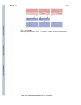
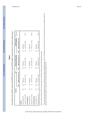
Overall, frequency distributions of the KRAS-variant genotype did not differ between cases
and controls who were genotyped from study group 1 (figure 1, table 1). However, the
KRAS variant was significantly associated with breast cancer in premenopausal patients
with ER/PR-negative tumours (table 1). This association was not observed for
postmenopausal women. Eight (33%) of 24 premenopausal women with ER/PR-negative
cancer had the KRAS variant, compared with 27 (13%) of 201 controls and four (9%) of 44
premenopausal women with cancer that was positive for ER and/or PR (webappendix p 10).
Thus, the KRAS variant might be a genetic marker of increased risk of development of
receptor-negative breast cancer for premenopausal women.
In study group 2, 478 women had luminal A breast cancer, 87 had luminal B disease, 90 had
triple-negative disease, and 35 had HER2-positive disease. 98 (14%) of 690 breast-cancer
cases from this cohort had the KRAS variant, but prevalence varied between the breast
cancer subtypes: the KRAS variant was significantly enriched in women with triple-negative
breast cancer (19 [21%] of 90 cases) compared with 64 (13%) of 478 for luminal A, 13
(15%) of 87 for luminal B, and two (6%) of 35 for HER2-positive subgroups (p=0·044;
figure 2). This association with triple-negative breast cancer was also noted in women
younger than 51 years (p=0·033, figure 2).
By comparison of cases of triple-negative breast cancer from groups 2 and 3 and controls
across all three cohorts (n=1160), we did not note a significant difference between cases or
between controls for the prevalence of the KRAS variant (webappendix p 3). However, there
were significantly more non-white women in the controls from study groups 1 and 3 than
there were in the study group 2, which allowed assessment of the association of the KRAS
variant in non-white women with triple-negative breast cancer in the multivariate analysis.
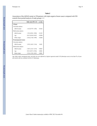
After controlling for age, ethnic origin, and study site, the KRAS variant did not predict an
increased risk of development of triple-negative breast cancer for all women in multivariate
analysis (table 2, webappendix p 4). However, the KRAS variant was associated with a
significantly increased risk of development of triple-negative breast cancer in the 361
premenopausal women in this pooled group in multivariate analysis (table 2, webappendix
pp 5–6).
Because BRCA1 coding sequence mutations are associated with risk of triple-negative
breast cancer, and because we noted an apparent enrichment of the KRAS variant in BRCA1
mutation-carriers with breast cancer,21 we aimed to establish whether the association of the
KRAS variant with premenopausal triple-negative breast cancer was due only to its
association with carriers of BRCA1 mutation. Of 36 women with triple-negative breast
cancer from cohort 2 and 3 who were BRCA tested, 25 (69%) were BRCA negative and 11
(31%) were BRCA positive. Of these patients, eight (32%) BRCA-negative women
Lancet Oncol. Author manuscript; available in PMC 2012 November 04.
$watermark-text $watermark-text $watermark-text](https://image.slidesharecdn.com/nihms-408024-141111212729-conversion-gate02/85/A-3-untranslated-region-KRAS-variant-and-triple-negative-breast-cancer-a-case-control-and-genetic-analysis-6-320.jpg)
![Paranjape et al. Page 7
harboured the KRAS variant compared with three (27%) women who were BRCA positive.
These findings suggest that the KRAS variant is associated with an independent group of
patients with triple-negative breast cancer without BRCA mutations.
Although we did not note an association between KRAS-variant status and ER or PR
negative statuses in the Rotterdam population cohort,21,23 we had not considered
menopausal status. In this study, we did not note an enrichment of the KRAS variant in 126
premenopausal BRCA1-mutation carriers who had ER/PR-negative breast cancer compared
with all 268 BRCA1-mutation-carriers from the Rotterdam cohort (21·8% vs 23·5%,
p=0·95). These findings again support the notion that association of the KRAS variant with
premenopausal triple-negative breast cancer is independent of its association with BRCA1
mutations.
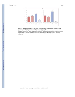
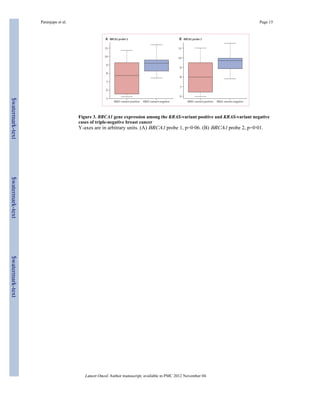
However, to further assess potential biological interaction between the KRAS variant and
altered BRCA1 expression in triple-negative disease, we appraised BRCA1 expression
levels in 74 triple-negative tumours from study group 3 (figure 1). We noted that those
patients with the KRAS variant had significantly reduced BRCA1 expression compared with
KRAS-variant-negative triple-negative tumours (p=0·06 for probe 1 [ILMN_2311089] and
p=0·01 for probe 2 [ILMN_1738027], figure 3). Furthermore, the KRAS variant was
significantly associated with a gene expression signature of decreased BRCA1 activity
(p=0·04).25 These findings suggest that, although the KRAS variant is not restricted to
patients with triple-negative breast cancer with known BRCA1 mutations, there might be
some biological interaction between the KRAS variant, altered BRCA1 expression or
functionality, and development of triple-negative breast cancer.
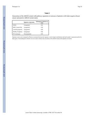
We compared signalling pathways in triple-negative breast-cancer tumours that were
KRAS-variant positive with those that were KRAS-variant negative from patients in study
group 3. Although analysis of KRAS mRNA did not vary by KRAS-variant status, this
finding agrees with the other publications about the effect of miRNA binding to the KRAS
3′-UTR.16,26 However, we noted an increase in both an NRAS mutation27 and a MAP-kinase
activation signature28 (table 3) in tumours with the KRAS variant. This supports the
notion that the KRAS variant alters gene expression of canonical RAS pathways, and is to
our knowledge the first in-vivo evidence that the KRAS variant leads to continued altered
downstream gene expression in tumours with which it is associated.
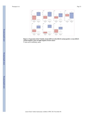
Because we had previously noted altered concentrations of let-7 miRNA in lung tumours
with the KRAS variant, we examined let-7 concentrations in triple-negative breast cancer
tumours with the KRAS variant. Consistent with our previous findings, we noted lower
concentrations of all let-7 miRNA family members in KRAS-variant-associated tumours
(figure 4).
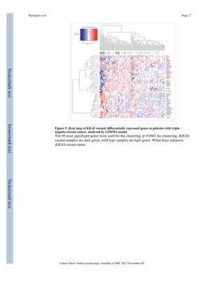
To establish how the KRAS variant integrates with known gene-expression signatures of
triple-negative breast cancer, we assessed known signatures that are differentially expressed
in such tumours. We found that KRAS-variant tumours have several features of triple-negative
and basal-like tumour biology, including decreased oestrogen signalling in a main
component derived from our expression set (p=0·04). Furthermore, KRAS-variant tumours
have a luminal progenitor signature (p=0·04), which has been suggested4 as a candidate
progenitor for basal-like breast cancer (table 3, webappendix p 11). Within the luminal
progenitor and the BRCA mutation-like signatures, markers of cell adhesion, tissue
invasion, proliferation, and angiogenesis (such as α5 integrin, DUSP6, and aurora kinase B)
were differentially regulated (webappendix p 7). This finding is in agreement with the slight
enrichment by functional annotations that we noted in three of 41 genes for wound healing
(p=0·02), three of 151 genes for glycan expression (p=0·05), and four of 148 genes for MEK
Lancet Oncol. Author manuscript; available in PMC 2012 November 04.
$watermark-text $watermark-text $watermark-text](https://image.slidesharecdn.com/nihms-408024-141111212729-conversion-gate02/85/A-3-untranslated-region-KRAS-variant-and-triple-negative-breast-cancer-a-case-control-and-genetic-analysis-7-320.jpg)
![Paranjape et al. Page 9
KRAS variant in tumorigenesis and its specific association with triple-negative breast cancer
remains to be delineated.
The KRAS variant is a biomarker of poor outcome in several cancers, including head and
neck cancer,17 and is a biomarker of poor response to targeted therapies in colon cancer.18
Our finding that patients with the KRAS variant and triple-negative breast cancer have a
luminal progenitor signature and differential expression of angiogenic and metastatic
markers within the signature suggests that tumours harbouring the KRAS variant might be
an aggressive subgroup of this cancer. Follow-up studies will be necessary to establish the
effect of the KRAS variant on outcome in patients with triple-negative breast cancer and
patients with breast cancer in general.
Our study suggests that the KRAS variant is associated with tumours that maintain unique
gene-expression patterns. Although investigations remain to be done to establish the
mechanisms of development of triple-negative breast cancer in women who are KRAS-
variant positive, our findings give insight into crucial steps and pathways required for
transformation and tumour development in these women. We believe our results are
meaningful steps towards understanding of the mechanisms of gain of function miRNA-disrupting
polymorphisms in cancer biology, which seem to be distinct in function from
previously discovered genetic markers of cancer risk.
Supplementary Material
Refer to Web version on PubMed Central for supplementary material.
Acknowledgments
We thank Neal Fischbach and the Cancer Genetic Counselling Shared Resource at the Yale Cancer Center (New
Haven, CT, USA) for contributions of samples to the study. TP was supported by a Yale Center for Clinical
Investigation (YCCI) grant made possible by Clinical and Translational Science Awards (CTSA) grant number
UL1 RR024139 from the National Centre for Research Resources (NCRR), a component of the US National
Institutes of Health (NIH), and US NIH roadmap for Medical Research. FS and JW were supported by the US
National Cancer Institute (CA131301). JW was supported by a K08 grant [CA124484]. HH was supported by a
Health Research Board Clinician Scientist Fellowship and an Irish Higher Surgical Training Group Travelling
Scholarship. JD was supported by the National Breast Cancer Research Institute in Galway, Ireland. DZ has
received royalties for books published by John Wiley and Sons, Oxford University Press, Cambridge University
Press, and the SAS institute, and payment for service on a data monitoring committee from BristolMyersSquibb.
References
1. Sørlie T, Perou CM, Tibshirani R, et al. Gene expression patterns of breast carcinomas distinguish
tumor subclasses with clinical implications. Proc Natl Acad Sci USA. 2001; 98:10869–74.
[PubMed: 11553815]
2. Haffty BG, Yang Q, Reiss M, et al. Locoregional relapse and distant metastasis in conservatively
managed triple-negative early-stage breast cancer. J Clin Oncol. 2006; 24:5652–57. [PubMed:
17116942]
3. Rakha EA, Ellis IO. Triple-negative/basal-like breast cancer: review. Pathology. 2009; 41:40–47.
[PubMed: 19089739]
4. Lim E, Vaillant F, Wu D, et al. Aberrant luminal progenitors as the candidate target population for
basal tumor development in BRCA1 mutation carriers. Nat Med. 2009; 15:907–13. [PubMed:
19648928]
5. Bild AH, Parker JS, Gustafson AM, et al. An integration of complementary strategies for gene-expression
analysis to reveal novel therapeutic opportunities for breast cancer. Breast Cancer Res.
2009; 11:R55. [PubMed: 19638211]
Lancet Oncol. Author manuscript; available in PMC 2012 November 04.
$watermark-text $watermark-text $watermark-text](https://image.slidesharecdn.com/nihms-408024-141111212729-conversion-gate02/85/A-3-untranslated-region-KRAS-variant-and-triple-negative-breast-cancer-a-case-control-and-genetic-analysis-9-320.jpg)
![Paranjape et al. Page 10
6. Yang XR, Sherman ME, Rimm DL, et al. Differences in risk factors for breast cancer molecular
subtypes in a population-based study. Cancer Epidemiol Biomarkers Prev. 2007; 16:439–43.
[PubMed: 17372238]
7. Bauer KR, Brown M, Cress RD, Parise CA, Caggiano V. Descriptive analysis of estrogen receptor
(ER)-negative, progesterone receptor (PR)-negative, and HER2-negative invasive breast cancer, the
so-called triple-negative phenotype: a population-based study from the California cancer registry.
Cancer. 2007; 109:1721–28. [PubMed: 17387718]
8. Young SR, Pilarski RT, Donenberg T, et al. The prevalence of BRCA1 mutations among young
women with triple-negative breast cancer. BMC Cancer. 2009; 9:86. [PubMed: 19298662]
9. Nanda R, Schumm LP, Cummings S, et al. Genetic testing in an ethnically diverse cohort of high-risk
women: a comparative analysis of BRCA1 and BRCA2 mutations in American families of
European and African ancestry. JAMA. 2005; 294:1925–33. [PubMed: 16234499]
10. He L, Thomson JM, Hemann MT, et al. A microRNA polycistron as a potential human oncogene.
Nature. 2005; 435:828–33. [PubMed: 15944707]
11. Esquela-Kerscher A, Slack FJ. Oncomirs—microRNAs with a role in cancer. Nat Rev Cancer.
2006; 6:259–69. [PubMed: 16557279]
12. Yu F, Yao H, Zhu P, et al. Let-7 regulates self renewal and tumorigenicity of breast cancer cells.
Cell. 2007; 131:1109–23. [PubMed: 18083101]
13. Hoffman A, Zheng T, Yi C, et al. MicroRNA miR-196a-2 and breast cancer: a genetic and
epigenetic association study and functional analysis. Cancer Res. 2009; 69:5970–77. [PubMed:
19567675]
14. Pongsavee M, Yamkamon V, Dakeng S, et al. The BRCA1 3′UTR: 5711+421T/T_5711+1286T/T
genotype is a possible breast and ovarian cancer risk factor. Genet Test Mol Biomarkers. 2009;
13:307–17. [PubMed: 19405875]
15. Tchatchou S, Jung A, Hemminki K, et al. A variant affecting a putative miRNA target site in
estrogen receptor (ESR) 1 is associated with breast cancer risk in premenopausal women.
Carcinogenesis. 2009; 30:59–64. [PubMed: 19028706]
16. Chin L, Ratner E, Leng S, et al. A SNP in a let-7 microRNA complementary site in the KRAS
3′UTR increases non-small cell cancer risk. Cancer Res. 2008; 68:8535–40. [PubMed: 18922928]
17. Christensen BC, Moyer BJ, Avissar M, et al. A let-7 microRNA binding site polymorphism in the
KRAS 3′UTR is associatied with reduced survival in oral cancers. Carcinogenesis. 2009;
30:1003–07. [PubMed: 19380522]
18. Graziano F, Canestrari E, Loupakis F, et al. Genetic modulation of the Let-7 microRNA binding to
KRAS 3′-untranslated region and survival of metastatic colorectal cancer patients treated with
salvage cetuximab-irinotecan. Pharmacogenomics J. 2010; 10:458–64. [PubMed: 20177422]
19. Zhang W, Winder T, Ning Y, et al. A let-7 microRNA-binding site polymorphism in 3′-
untranslated region of KRAS gene predicts response in wild-type KRAS patients with metastatic
colorectal cancer treated with cetuximab monotherapy. Ann Oncol. 2011; 22:104–09. [PubMed:
20603437]
20. Ratner E, Lu L, Boeke M, et al. A KRAS-variant in ovarian cancer acts as a genetic marker of
cancer risk. Cancer Res. 2010; 70:6509–15. [PubMed: 20647319]
21. Hollestelle A, Pelletier C, Hooning M, et al. Prevalence of the variant allele rs61764370 T>G in
the 3′UTR of KRAS among Dutch BRCA1, BRCA2 and non-BRCA1/BRCA2 breast cancer
famlies. Breast Cancer Res Treat. 2010 published online July 30. DOI:10.1007/s10549-010-1080-
z.
22. [accessed Jan 1, 2008] miRNA profiling. http://www.appliedbiosystems.com/absite/us/en/home/
applications-technologies/real-time-pcr/mirna-profiling.html
23. Kibriya M, Jasmine F, Roy S, Paul-Brutus R, Argos M, Ahsan H. Analyses and interpretation of
whole-genome gene expression from formalin-fixed paraffin-embedded tissue: an illustration with
breast cancer tissues. BMC Genomics. 2010; 11:622. [PubMed: 21059268]
24. Smyth, GK. Limma: linear models for microarray data. In: Gentleman, R.; Carey, V.; Huber, W.;
Irizarry, R.; Dudoit, S., editors. Bioinformatics and computational biology solutions using R and
bioconductor. Springer; New York, USA: 2005. p. 397-420.
Lancet Oncol. Author manuscript; available in PMC 2012 November 04.
$watermark-text $watermark-text $watermark-text](https://image.slidesharecdn.com/nihms-408024-141111212729-conversion-gate02/85/A-3-untranslated-region-KRAS-variant-and-triple-negative-breast-cancer-a-case-control-and-genetic-analysis-10-320.jpg)
![Paranjape et al. Page 11
25. van’t Veer LJ, Dai H, van de Vijver MJ, et al. Gene expression profiling predicts clinical outcome
of breast cancer. Nature. 2002; 415:530–36. [PubMed: 11823860]
26. Johnson SM, Grosshans H, Shingara J, et al. RAS is regulated by the let-7 microRNA family. Cell.
2005; 120:635–47. [PubMed: 15766527]
27. Croonquist PA, Linden MA, Zhao F, Van Ness BG. Gene profiling of a myeloma cell line reveals
similarities and unique signatures among IL-6 response, N-ras-activating mutations, and coculture
with bone marrow stromal cells. Blood. 2003; 102:2581–92. [PubMed: 12791645]
28. Creighton CJ, Hilger AM, Murthy S, Rae JM, Chinnaiyan AM, El-Ashry D. Activation of
mitogen-activated protein kinase in estrogen receptor α-positive breast cancer cells in vitro
induces an in vivo molecular phenotype of estrogen receptor α-negative human breast tumors.
Cancer Res. 2006; 66:3903–11. [PubMed: 16585219]
29. Iliopoulos D, Hirsch H, Struhl K. An epigenetic switch involving NF-κB Lin28, let-7 microRNA,
and IL6 links inflammation to cell transformation. Cell. 2009; 139:1–14.
30. Meylan E, Dooley A, Feldser D, et al. Requirement for NF-κB signalling in a mouse model of lung
adenocarcinoma. Nature. 2009; 462:104–08. [PubMed: 19847165]
31. Barbie D, Tamayo P, Boehm J, et al. Systematic RNA interverence reveals that oncogenic KRAS-driven
cancers require TBK1. Nature. 2009; 462:108–12. [PubMed: 19847166]
32. Atchley DP, Albarracin CT, Lopez A, et al. Clinical and pathologic characteristics of patients with
BRCA-positive and BRCA-negative breast cancer. J Clin Oncol. 2008; 26:4282–88. [PubMed:
18779615]
33. Oh AS, Lorant LA, Holloway JN, Miller DL, Kern FG, El-Ashry D. Hyperactivation of MAPK
induces loss of ERα expression in breast cancer cells. Mol Endocrinol. 2001; 15:1344–59.
[PubMed: 11463858]
34. Santen RJ, Song RX, McPherson R, et al. The role of mitogen-activated protein (MAP) kinase in
breast cancer. J Steroid Biochem Mol Biol. 2002; 80:239–56. [PubMed: 11897507]
Lancet Oncol. Author manuscript; available in PMC 2012 November 04.
$watermark-text $watermark-text $watermark-text](https://image.slidesharecdn.com/nihms-408024-141111212729-conversion-gate02/85/A-3-untranslated-region-KRAS-variant-and-triple-negative-breast-cancer-a-case-control-and-genetic-analysis-11-320.jpg)

This study investigated the association of a variant in the 3′-untranslated region of the kras oncogene with triple-negative breast cancer (TNBC) risk in premenopausal women. Results showed that the kras variant was significantly enriched in TNBC cases compared to other breast cancer subtypes, suggesting its potential as a genetic marker for TNBC. Furthermore, the study indicated that tumours with the kras variant exhibited altered gene expression and reduced let-7 microRNA levels.

![Molecular biology of breast cancer [autosaved]](https://cdn.slidesharecdn.com/ss_thumbnails/molecularbiologyofbreastcancerautosaved-191226181803-thumbnail.jpg?width=640&height=640&fit=bounds)


![HEREDITARY BREAST and OVARY CANCER [HBOC] SYNDROME, Dr BUI DAC CHI.](https://cdn.slidesharecdn.com/ss_thumbnails/hboc1-150312034459-conversion-gate01-thumbnail.jpg?width=640&height=640&fit=bounds)


