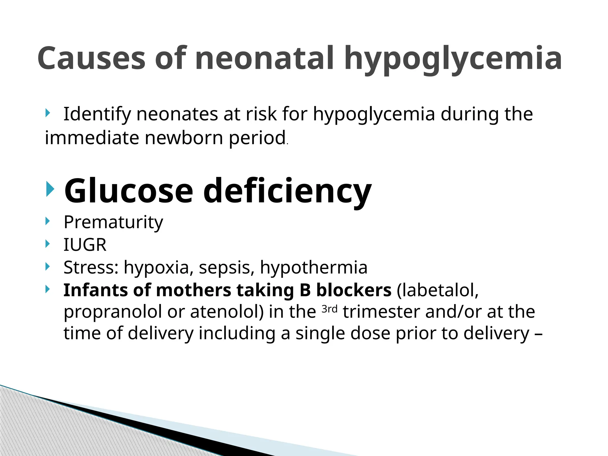
Neonatal hypoglycemia
By Marwa elsayed
Lecturer of pediatrics
•By the end of this presentation, the participant will:
 Gain an understanding of hypoglycemia in the newborn.
 Identify neonates at risk for hypoglycemia(causes) during the
immediate newborn period.
 Describe the signs and symptoms of hypoglycemia in the
neonate.
 Identify the treatment for asymptomatic and symptomatic
hypoglycemia in the neonate.
Objectives
Quiz
 Which of the following best describes
hypoglycemia?
 An inability to maintain blood sugar due to
lowered insulin efficiency
 A higher than normal blood glucose level
 Excessive fetal growth
 A lower than normal blood glucose level
 What is the diagnosis?
 what are risk factors?
 How to treat?
 In neonate, a blood glucose value of less
than40 mg/dL (2.2 mmol/L) represents
hypoglycemia. A plasma glucose level of less
than 30 mg/dL (1.65 mmol/L) in the first 24
hours of life and less than 45 mg/dL (2.5
mmol/L) thereafter constitutes hypoglycemia in
the newborn.(Apr 30, 2020)
defintion
 Identify neonates at risk for hypoglycemia during the
immediate newborn period.
 Glucose deficiency
 Prematurity
 IUGR
 Stress: hypoxia, sepsis, hypothermia
 Infants of mothers taking B blockers (labetalol,
propranolol or atenolol) in the 3rd
trimester and/or at the
time of delivery including a single dose prior to delivery –
Causes of neonatal hypoglycemia
Hyperinsulinemia
 IDM
 Exchange transfusion
 Macrosomic babies –Babies who weigh
4 kg
≥
Inborn error of
metabolism
 Galactosemia,
 Glycogen storage disease
 Aminoacids disorders
 Fatty acid oxidation defect
Endocrine
 Hypopitutrism
 Growth hormone deficiency,
 Cortisol deficiency
Pathophysiology
Hypoglycaemia may present in a number of ways within the first 48 hours of
life. These include;
 Hypotonia
 Lethargy (excessive sleepiness with or without
abnormal tone)
 Poor feeding
 Hypothermia
 Apnoea
 Irritability
 Pallor
Signs and Symptoms of Hypoglycaemia in the Neonate
 Tachypnoea
 Tachycardia or bradycardia
 Seizures
 Abnormal feeding behaviour (not waking for feeds,
not sucking effectively, appearing unsettled and
demanding very frequent feeds especially after a
period of feeding well)
 This list is not exhaustive. Medical review should be
obtained for any generally unwell infant as these
symptoms may warrant further investigation and
consideration of other causes including sepsis
•Identified infants at risk for
hypoglycemia
 -
 Breast or bottle feed within 1st
hour of life
 Check glucose level 30 minutes after initial feeding
 Continue to follow glucose levels for next 12 hours prior
to
feedings for IDM/LGA and next 24 hours for SGA/preterm
Neonates
Treatment
 When to Treat
•Borderline hypoglycemia
(asymptomatic)
 EBM or formula
 Buccal glucose gel
 Moderate hypoglycemia (20 – 25 mg/dl)
 IV glucose 2 ml/kg/dose
•Profound hypoglycemia (< 20 mg/dl) or
Symptomatic hypoglycemia
IV glucose
 - D10w 2 ml/kg/dose
Then
 IV fluids –D10w at 80 ml/kg/day
 Blood Glucose >2.5mmol/l
 If 2 consecutive values, at 3hly intervals, fall in this
zone, monitoring may cease provided there are no
clinical signs of hypoglycaemia.
 Observe feeding in hospital for further 24 hours after
monitoring ceased.
 If breastfeeding ensure at least 1 recorded breast
feeding assessment prior to transfer home.
 Remaining vigilant for the signs of hypoglycaemia. If
signs of hypoglycaemia develop or there are concerns
about feeding discuss with medical staff and consider
taking a further blood glucose.
 Green Zone/Flowchart A - Pre-Feed Glucose
2.0-2.5mmol and NO abnormal clinical signs
 Offer an additional feed if willing and continue
frequent feeds at least 3hly thereafter.
 Observe a breastfeed and ensure good
attachment and effective feeding. Encourage
skin contact and biological nurturing.
Proactively encourage hand expressing.
 If two consecutive measurements fall within
the Green Zone - - Treat as Amber Pathway
→
 Amber Zone/Flowchart B – Glucose 1.0 - 1.9 mmol/l -
Supplement and Paediatric review
 Inform Neonatal Doctor/ANP
 Administer a dose of 40% buccal glucose 200mg/kg
this must be given in conjunction with making and
documenting a detailed feeding plan.
◦ Check blood glucose 30-60 minutes after
administration of buccal gel.
◦ If glucose remains 1.0-1.9mmol on post gel blood
glucose check administer second dose of buccal
gel. The baby be reviewed by a member of the
neonatal team as soon as is practical.
◦ Repeat blood glucose after 30-60 mins.
 All doses of buccal gel MUST be administered in
conjunction with a careful review of feeding
including a clear feeding plan which ensures
adequate volumes are being administered:
 If breast feeding support breast feeding
◦ Encourage skin to skin
◦ Offer breast feed and if not feeding effectively teach
mother to hand express and use breast pump
◦ Give colostrum obtained
◦ Continue to encourage hand expressing at least 8-10 X/24
hrs and support feeding on the breast until the infant is
feeding effectively.
◦ Ensure families are aware that donor breast milk is
available as an option for supplementation.
 If formula fed ensure adequate feed volumes are
being given (at least 10ml/kg) in 3 hourly volumes.
 Check blood glucose before next feed, no later than
3 hours after feed irrespective of the blood glucose
result 30-60 minutes obtained post gel as
theoretically a delayed drop in blood glucose may
occur.
 If hypoglycaemia of <2.0mmol/L persists after two
doses of dextrose gel or the infant becomes
symptomatic manage as per red zone/Flowchart C
 If baby is not feeding adequately consider admission
to SCBU/TC for NG feeding.
 Red Zone/Flowchart C - Pre-Feed Glucose <1.0mmol/l OR
Clinical Signs Consistent with Hypoglycaemia at a higher
blood glucose concentration
 Inform Neonatal Team
 Management will include appropriate investigations
(see Investigations for Persistent Hypoglycaemia bellow) at
time of hypoglycaemia for persistent hypoglycaemia as
described below
 Continue to establish breastfeeding unless the baby is too
unwell to feed
 Obtain IV access
◦ Give 2.5ml/kg 10% glucose bolus IV
◦ If unable to obtain IV access immediately give
 40% glucose gel 200mg/kg
 For investigation of a second episode of true blood
sugar less than 2.2 mmol/L (glucose test strips
 should always be confirmed with serum sample):
 1. Obtain 3 mL blood in a serum separator tube for
 insulin, cortisol, GH and ketone bodies.
 2. Obtain 5 to 10 mL urine for a metabolic screen to
include ketones, amino acids, organic acids and
acylcarnitine profile.
 Rationale: It is important to determine whether the
hypoglycemia is ketotic or nonketotic. Nonketotic
 hypoglycemia is associated with disorders of fruc-
tose or galactose metabolism, hyperinsulinism, fatty
acid oxidation and GH deficiency. Ketotic hypoglyce-
mia is associated with organic acidurias, maple syrup
 urine disease, glycogen storage disease and adrenal
insufficiencies of central or peripheral origin (7).
 B. Consider involvement of a consultant with
expertise in inborn errors of metabolism
 OBJECTIVES
 Discuss briefly the effects of maternal Diabetes
on the Fetus
 Clinical presentation of IDM
 Review the complications encountered in an IDM
 Discuss the management of an IDM in the nursery
 Discuss the prognosis for an IDM and review the
follow up needed after hospital discharge
Infant of diabetic mother
 Morbidities in Infants of Diabetic Mothers
 Macrosomia
 Hypoglycemia
 RDS
 IUGR
 Hypocalcemia
 Hyperbilirubinemia
 Congenital Anomalies
 Polycythemia
 Hyper viscosity
 Cardiovascular
 Increased fetal death
 Postnatal problems
 Birth Injury , Macrosomia puts infant at risk for injuries
during delivery
 Shoulder dystocia can lead to
 Clavicular and/or humeral fractures
 Brachial plexus injuries
 Traumatic delivery or need for vacuum/forceps
assistance can lead to
 Cephalohematomas
 Facial bruising
 Facial nerve injuries
 Hypoglycemia is caused by hyperinsulinemia
due to hyperplasia of fetal pancreatic beta cells
consequent to maternal-fetal hyperglycemia.
Because the continuous supply of glucose is
stopped after birth, the neonate develops
hypoglycemia because of insufficient substrate.
Stimulation of fetal insulin release by maternal
hyperglycemia during labor significantly
increases the risk of early hypoglycemia in
these infants.
 Cardiovascular anomalies
 Cardiomyopathy with ventricular hypertrophy and outflow
tract obstruction may occur in as many as 30% of
IDMs.[7]
The cardiomyopathy may be associated with
congestive failure with a weakly functioning myocardium or
may be related to a hypertrophic myocardium with
significant septal hypertrophy and outflow tract
obstruction. When cardiomegaly or poor perfusion and
hypotension are present, performing echocardiography to
differentiate between these processes is important.
 These infants are also at an increased risk of congenital
heart defects, including (most commonly) ventricular septal
defect (VSD) and transposition of the great arteries (TGA).
 Congenital malformations
 Central nervous system (CNS) malformations
are 16 times more likely in IDMs. In particular,
the risk of anencephaly is 13 times higher,
whereas the risk of spina bifida is 20 times
higher. The risk of caudal dysplasia is up to 600
times higher in these infants.
 Renal (eg, hydronephrosis, renal agenesis,
ureteral duplication), ear, gastrointestinal (eg,
duodenal or anorectal atresia, small left colon
syndrome), and, as mentioned earlier,
cardiovascular (eg, single umbilical artery,
VSDs, atrial septal defects, TGA, coarctation of
the aorta, cardiomegaly) anomalies are more
frequent in these infants
 Pulmonary disease
 These infants are at an increased risk of respiratory
distress syndrome and may present within the first few
hours after birth with tachypnea, nasal flaring, intercostal
retractions, and hypoxia. Operative delivery due to
macrosomia also increases the risk for transient
tachypnea of the newborn, whereas polycythemia
predisposes the infant to persistent pulmonary
hypertension of the newborn.
 Initially, the differential diagnosis includes transient
tachypnea of the newborn, respiratory distress syndrome,
pneumonia, and persistent pulmonary hypertension.
 Metabolic and electrolyte abnormalities
 Hypoglycemia may present within the first few
hours of life. Although the infant is generally
asymptomatic, symptoms may include
jitteriness, irritability, apathy, poor feeding,
high-pitched or weak cry, hypotonia, or frank
seizure activity. Hypoglycemia that requires
intervention may persist for as long as 1 week.
 Perinatal stress may have an additive effect on
hypoglycemia due to catecholamine release and
glycogen depletion. The overall risk of
hypoglycemia is anywhere from 25-40%, with LGA
and preterm infants at highest risk.
 Hypocalcemia or hypomagnesemia may also be
apparent in the first few hours after birth.
Symptoms may include jitteriness or seizure
activity. Hypocalcemia (levels < 7 mg/dL) is believed
to be associated with a delay in parathyroid
hormone synthesis after birth.
 Hematologic problems
 Polycythemia, caused by increased erythropoiesis triggered
by chronic fetal hypoxia, may present as a clinically "ruddy"
appearance, sluggish capillary refill, or respiratory distress.
Hyperviscosity due to polycythemia increases the IDM’s risk
for stroke, seizure, necrotizing enterocolitis, and renal vein
thrombosis.
 Thrombocytopenia
 Thrombopoiesis may be inhibited because of an excess of
RBC precursors within the bone marrow as a result of
chronic in utero hypoxia and increased erythropoietin
concentration.
 Hyperbilirubinemia
 This is common, especially in association with
polycythemia. The increased red cell mass results in
increased number of RBCs that are taken out of
circulation each day and increase the bilirubin
burden presented to the liver.
 Workup
 CBC count
 Polycythemia, commonly defined as a central
hematocrit level higher than 65%, is a potential
concern. Maternal-fetal hyperglycemia and fetal
hypoxia is a strong stimulus for fetal erythropoietin
production and subsequent increase in fetal
hemoglobin concentration. Thrombocytopenia may
occur because of impaired thrombopoiesis due to
"crowding-out" of thrombocytes by the excess of
erythroid precursors in the bone marrow.
 Glucose concentration (serum or whole-blood)
 Seizures, coma, and long-term brain damage may
occur if neonatal hypoglycemia is unrecognized and
untreated. Most centers recognize levels lower than 40
mg/dL within the first 24 hours after birth as abnormal
 A policy to screen infants of diabetic mothers (IDMs)
for hypoglycemia should be in place in every hospital.
They suggested that an infant with compromised
metabolic adaptation (ie, IDM) undergo blood glucose
measurements (1) as soon as possible after birth, (2)
within 2-3 hours after birth and before feeding, and (3)
at any time abnormal clinical signs are observed
 Magnesium concentration (serum)
 Hypomagnesemia is related to younger maternal age,
severity of maternal diabetes, and prematurity. Neonatal
magnesium levels are also related to maternal serum
magnesium, neonatal calcium and phosphorus levels, and
neonatal parathyroid function. The clinical significance of low
magnesium levels in these infants remains controversial and
uncertain.
 Calcium concentration (serum, ionized or total levels)
 Low serum calcium levels in IDMs are common. They are
speculated to be caused by a functional hypoparathyroidism;
however, their clinical relevance remains uncertain and
controversial.
 Bilirubin level (serum, total and unconjugated)
 Hyperbilirubinemia (see bilirubin) is notably more common
in IDMs than in the general population of neonates.
Causative factors include prematurity, hepatic enzyme
immaturity, polycythemia, and reduced RBC half-life.
 Arterial blood gas
 Assessing oxygenation and ventilation is essential in
infants with clinical evidence of respiratory distress.
Although noninvasive methods (eg, transcutaneous
oxygen and carbon dioxide electrodes, oximeters) have
gained wide acceptance at many centers, comparison of
results with those from arterial blood is intermittently
required.
 Chest radiography
 Clinical evidence of cardiopulmonary distress requires a
detailed evaluation, which should always include a chest
radiograph. The image should be evaluated for adequacy of
lung expansion, evidence of focal or diffuse atelectasis,
presence of interstitial fluid, signs of free air in pleural or
interstitial spaces, and findings of respiratory distress
syndrome or pneumonia. The possibility of pulmonary
malformations should also be considered.
 Cardiac size, shape, and great vessel/outflow tract should be
carefully examined. In the infant with macrosomia who has a
history of shoulder dystocia, the clavicles should be evaluated
on the film as well on physical examination.
 Abdominal, pelvic, or lower extremity radiography
 When caudal dysplasia is present, other orthopedic anomalies
should be investigated, including fusion of the legs, hypoplastic
femur, defects of the tibia and the fibula, flexion contractures of the
knee and hip, or clubfoot. Caudal dysplasia or sacral agenesis is the
most common orthopedic anomaly in the IDM.
 Lower extremity congenital malformations require radiographic
evaluation to determine the exact skeletal defect or defects present.
 Cardiac echocardiography
 A thickened myocardium and significant septal hypertrophy may be
present in as many as 1 in 3 IDMs. Evidence of a hypercontractile,
thickened myocardium, often with septal hypertrophy
disproportionate to the size of the ventricular free walls, may be
noted on examination. Myocardial contractility should also be
evaluated, because the myocardium is overstretched and poorly
contractile with congenital cardiomyopathies.
 Evidence of anatomical malformation must be searched for carefully
because cardiac malformations, including VSDs and TGAs, are
significantly more common in IDMs.
 Barium enema
 Infants with feeding intolerance, abdominal distention, nonbilious
emesis, or poor passage of meconium may require a barium enema.
Congenital anomalies of the gastrointestinal tract are more common
in IDMs. These infants may have small left colon syndrome, also
known as "lazy colon."
 Clinical features of the small left colon syndrome may mimic those
of Hirschsprung disease, and distal tapering of the colon is a
radiologic feature of both disorders. The 2 disorders can be
distinguished using a biopsy because normal ganglionic cells are
present in lazy colon and absent in Hirschsprung disease.
 Post-delivery Observe / Evaluate for
 Asphyxia
 Birth injury
 Malformations
 Macrosomia
 Hypoglycemia
 Respiratory Distress
Management
 Hypoglycemic Management
 Improved maternal glucose control during the pregnancy and
labor improves postnatal glucose adaptation and a decreases
the need for IV glucose treatment in the infant. A screening
policy for hypoglycemia during the hours after birth is
necessary to detect hypoglycemia.
 Serum or whole blood glucose levels of less than 20-40 mg/dL
within the first 24 hours after birth are generally agreed to be
abnormal and to require intervention. Cornblath et al
recommended critical values of glucose that require
intervention. [12]
Determination of plasma or whole blood
glucose should be made at the following points:
 As soon as possible after birth
 Repeat determinations at 30 minutes, 1 hour, 2 hours, 4 hours,
8 hours, and 12 hours after birth
 At any time abnormal clinical signs are observed
 Guidelines for maintaining euglycemia
 If the plasma value is less than 40 mg/dL (2.5 mmol/L),
intervention is needed (1) if plasma glucose remains
below this level, (2) if it does not increase after a
feeding, or (3) if the infant develops symptoms of
hypoglycemia.
 If the plasma value is less than 20-25 mg/dL (1.1-1.4
mmol/L), IV glucose should be administered, with the
target glucose level being more than 45 mg/dL (2.5
mmol/L). This goal of 45 mg/dL is accentuated as a
margin of safety. This should include a bolus of dextrose
followed by a constant infusion of dextrose. Profound
hypoglycemia may require therapy with hydrocortisone.
 Determining which infants require the highest
dextrose administration to maintain euglycemia is
difficult. The following suggestions represent a
guideline for glucose administration to an infant
with hypoglycemia.
 Immediate IV therapy with 2-mL/kg infusion of
dextrose 10% is required in any symptomatic infant
with hypoglycemia;. Administration over 5-10
minutes is usually recommended because of the
high osmolarity. This is especially true for immature
infants younger than 32 weeks' gestational age
who are at some risk for intracranial hemorrhage.
 Maintenance of a continuous infusion of dextrose at an infusion
rate of 6-8 mg/kg/min of dextrose is necessary once bolus
therapy is complete. Failure to do so may result in rebound
hypoglycemia as a result of heightened pancreatic insulin
release triggered by the glucose infusion.
 Frequent serum or whole blood glucose analyses are important
to properly titrate the dextrose infusion. Should follow-up
glucose levels remain less than 40 mg/dL, the dextrose infusion
may be increased by 2 mg/kg/min until euglycemia is achieved.
 If the infant requires a dextrose concentration of more than
D12.5 through a peripheral vein at 80-100 mL/kg/day,
placement of a central venous catheter may be considered to
avoid venous sclerosis. Continued enteral feedings hasten
improvement in glucose control because of the presence of
protein and fat in the formula. Hydrocortisone therapy may be
required for ongoing hypoglycemia
 Once the infant's glucose levels have been stable for 12 hours, IV
glucose may be tapered by 1-2 mg/kg/min, depending on
maintenance of preprandial glucose levels higher than 40 mg/dL.
 Electrolyte management
 Hypocalcemia and hypomagnesemia may complicate the clinical
course. Because low serum calcium levels cannot be corrected in
the presence of hypomagnesemia, correction of low magnesium
levels is an initial step in the treatment of hypocalcemia.
 In infants of diabetic mothers (IDMs), calcium and magnesium
levels are commonly measured within the first hours after birth.
Ideally, ionized levels of these electrolytes should be obtained
and used to properly manage these electrolyte disturbances.
 True symptomatic hypocalcemia is extremely rare in these infants. In
most cases, symptoms interpreted to be caused by low calcium or
magnesium levels are due to low glucose levels associated with
perinatal asphyxia or associated with various CNS problems.
 Low levels may be treated by adding calcium gluconate to the IV
solution to deliver 600-800 mg/kg/day of calcium gluconate. Bolus
therapy should be avoided unless cardiac arrhythmia is present. Bolus
therapy may result in bradycardia.
 Respiratory management
 Pulmonary management is tailored to the individual infant's signs and
symptoms. Increased ambient oxygen concentrations may be
required to maintain oxygen saturations higher than 90%,
transcutaneous oxygen tensions at 40-70 mm Hg, or arterial oxygen
tensions at 50-90 mm Hg.
 When an inspired oxygen concentration (FiO2) higher than 40%
is required, the most important task is to determine a precise
diagnosis of the cause for the hypoxemia and to administer
therapy appropriate for the underlying pathophysiology.
 Assisted ventilation
 Nasal continuous positive airway pressure (NCPAP) or
endotracheal intubation with intermittent mandatory ventilation
(IMV) or synchronized positive pressure ventilation (SIMV) may
be used for the management of severe respiratory distress.
 Common criteria for such interventions include inspired oxygen
requirements (FiO2) of 60-100% to maintain arterial PO2 of 50-
80mm Hg, arterial PCO2 levels higher than 60 mm Hg or rising
10 mm Hg, and apnea. The specific criteria for using these
modes of assisted ventilation may vary depending on the
underlying respiratory pathology and clinical condition of the
infant
 Cardiac management
 If signs of congestive heart failure or cardiomyopathy with
cardiomegaly, hypotension, or significant cardiac murmur
are observed, echocardiographic evaluation is essential to
distinguish among cardiac anomalies, septal hypertrophy,
and/or cardiomyopathy.
 Once a precise diagnosis is available, management of the
cardiac disorder is no different for the IDM than for any
other newborn with a similar cardiac condition. Extreme care
in the use of cardiotonic agents is important in the presence
of any hypertrophic cardiomyopathy or significant septal
hypertrophy. These infants are at risk for actual decreased
left ventricular output resulting from this form of therapy.
Beta blockers, such as propranolol, may be used to relieve
the outflow obstruction that is seen with septal hypertrophy
 Transfer, Consultations, and Follow-Up
 Infants of diabetic mothers (IDMs) having congenital
anomalies, heart disease, or significant respiratory
illness may require transfer to a tertiary care neonatal
intensive care unit (NICU) for continued care and access
to subspecialists. Because of the frequency with which
cardiac problems occur in IDMs, early consultation with
a pediatric cardiologist often is necessary. Other
consultations depend on which other congenital
malformations or complications are present.
 Basic outpatient care should consist of routine well-
baby care provided by the infant's general pediatrician.
Additional follow-up by consultant subspecialists
depends on the neonatal clinical problems and their
resolution.
 Case study
 Term baby 3.8 kg, IDM presents on day 3 with
 1. Resp Distress -80/min
 H.R. 190/min, poor pulses, sPo2 94
 features of CHF , Systolic murmur
 Cardiac complicationsH O C M - Lasix,
Propranolol
 Resolves by 4-6 mos
 Deposition of fat and glucose along the septum
 Others-- TOGV, ASD, Coarctation
 Case 54- day old preterm IDM 36wks- 1.8 kg
has feed intolerance, vomiting, abd distension
bilious aspirates
 answer
Small Left Colon Syndrome
Questions?

Neonatal hypoglycemia and management .pptx

  • 1.
    Neonatal hypoglycemia By Marwaelsayed Lecturer of pediatrics
  • 2.
    •By the endof this presentation, the participant will:  Gain an understanding of hypoglycemia in the newborn.  Identify neonates at risk for hypoglycemia(causes) during the immediate newborn period.  Describe the signs and symptoms of hypoglycemia in the neonate.  Identify the treatment for asymptomatic and symptomatic hypoglycemia in the neonate. Objectives
  • 3.
  • 4.
     Which ofthe following best describes hypoglycemia?  An inability to maintain blood sugar due to lowered insulin efficiency  A higher than normal blood glucose level  Excessive fetal growth  A lower than normal blood glucose level
  • 8.
     What isthe diagnosis?  what are risk factors?  How to treat?
  • 9.
     In neonate,a blood glucose value of less than40 mg/dL (2.2 mmol/L) represents hypoglycemia. A plasma glucose level of less than 30 mg/dL (1.65 mmol/L) in the first 24 hours of life and less than 45 mg/dL (2.5 mmol/L) thereafter constitutes hypoglycemia in the newborn.(Apr 30, 2020) defintion
  • 10.
     Identify neonatesat risk for hypoglycemia during the immediate newborn period.  Glucose deficiency  Prematurity  IUGR  Stress: hypoxia, sepsis, hypothermia  Infants of mothers taking B blockers (labetalol, propranolol or atenolol) in the 3rd trimester and/or at the time of delivery including a single dose prior to delivery – Causes of neonatal hypoglycemia
  • 11.
    Hyperinsulinemia  IDM  Exchangetransfusion  Macrosomic babies –Babies who weigh 4 kg ≥ Inborn error of metabolism  Galactosemia,  Glycogen storage disease  Aminoacids disorders  Fatty acid oxidation defect
  • 12.
    Endocrine  Hypopitutrism  Growthhormone deficiency,  Cortisol deficiency
  • 13.
  • 14.
    Hypoglycaemia may presentin a number of ways within the first 48 hours of life. These include;  Hypotonia  Lethargy (excessive sleepiness with or without abnormal tone)  Poor feeding  Hypothermia  Apnoea  Irritability  Pallor Signs and Symptoms of Hypoglycaemia in the Neonate
  • 15.
     Tachypnoea  Tachycardiaor bradycardia  Seizures  Abnormal feeding behaviour (not waking for feeds, not sucking effectively, appearing unsettled and demanding very frequent feeds especially after a period of feeding well)  This list is not exhaustive. Medical review should be obtained for any generally unwell infant as these symptoms may warrant further investigation and consideration of other causes including sepsis
  • 16.
    •Identified infants atrisk for hypoglycemia  -  Breast or bottle feed within 1st hour of life  Check glucose level 30 minutes after initial feeding  Continue to follow glucose levels for next 12 hours prior to feedings for IDM/LGA and next 24 hours for SGA/preterm Neonates Treatment
  • 17.
     When toTreat •Borderline hypoglycemia (asymptomatic)  EBM or formula  Buccal glucose gel  Moderate hypoglycemia (20 – 25 mg/dl)  IV glucose 2 ml/kg/dose
  • 18.
    •Profound hypoglycemia (<20 mg/dl) or Symptomatic hypoglycemia IV glucose  - D10w 2 ml/kg/dose Then  IV fluids –D10w at 80 ml/kg/day
  • 19.
     Blood Glucose>2.5mmol/l  If 2 consecutive values, at 3hly intervals, fall in this zone, monitoring may cease provided there are no clinical signs of hypoglycaemia.  Observe feeding in hospital for further 24 hours after monitoring ceased.  If breastfeeding ensure at least 1 recorded breast feeding assessment prior to transfer home.  Remaining vigilant for the signs of hypoglycaemia. If signs of hypoglycaemia develop or there are concerns about feeding discuss with medical staff and consider taking a further blood glucose.
  • 20.
     Green Zone/FlowchartA - Pre-Feed Glucose 2.0-2.5mmol and NO abnormal clinical signs  Offer an additional feed if willing and continue frequent feeds at least 3hly thereafter.  Observe a breastfeed and ensure good attachment and effective feeding. Encourage skin contact and biological nurturing. Proactively encourage hand expressing.  If two consecutive measurements fall within the Green Zone - - Treat as Amber Pathway →
  • 21.
     Amber Zone/FlowchartB – Glucose 1.0 - 1.9 mmol/l - Supplement and Paediatric review  Inform Neonatal Doctor/ANP  Administer a dose of 40% buccal glucose 200mg/kg this must be given in conjunction with making and documenting a detailed feeding plan. ◦ Check blood glucose 30-60 minutes after administration of buccal gel. ◦ If glucose remains 1.0-1.9mmol on post gel blood glucose check administer second dose of buccal gel. The baby be reviewed by a member of the neonatal team as soon as is practical. ◦ Repeat blood glucose after 30-60 mins.
  • 22.
     All dosesof buccal gel MUST be administered in conjunction with a careful review of feeding including a clear feeding plan which ensures adequate volumes are being administered:  If breast feeding support breast feeding ◦ Encourage skin to skin ◦ Offer breast feed and if not feeding effectively teach mother to hand express and use breast pump ◦ Give colostrum obtained ◦ Continue to encourage hand expressing at least 8-10 X/24 hrs and support feeding on the breast until the infant is feeding effectively. ◦ Ensure families are aware that donor breast milk is available as an option for supplementation.
  • 23.
     If formulafed ensure adequate feed volumes are being given (at least 10ml/kg) in 3 hourly volumes.  Check blood glucose before next feed, no later than 3 hours after feed irrespective of the blood glucose result 30-60 minutes obtained post gel as theoretically a delayed drop in blood glucose may occur.  If hypoglycaemia of <2.0mmol/L persists after two doses of dextrose gel or the infant becomes symptomatic manage as per red zone/Flowchart C  If baby is not feeding adequately consider admission to SCBU/TC for NG feeding.
  • 24.
     Red Zone/FlowchartC - Pre-Feed Glucose <1.0mmol/l OR Clinical Signs Consistent with Hypoglycaemia at a higher blood glucose concentration  Inform Neonatal Team  Management will include appropriate investigations (see Investigations for Persistent Hypoglycaemia bellow) at time of hypoglycaemia for persistent hypoglycaemia as described below  Continue to establish breastfeeding unless the baby is too unwell to feed  Obtain IV access ◦ Give 2.5ml/kg 10% glucose bolus IV ◦ If unable to obtain IV access immediately give  40% glucose gel 200mg/kg
  • 25.
     For investigationof a second episode of true blood sugar less than 2.2 mmol/L (glucose test strips  should always be confirmed with serum sample):  1. Obtain 3 mL blood in a serum separator tube for  insulin, cortisol, GH and ketone bodies.  2. Obtain 5 to 10 mL urine for a metabolic screen to include ketones, amino acids, organic acids and acylcarnitine profile.
  • 26.
     Rationale: Itis important to determine whether the hypoglycemia is ketotic or nonketotic. Nonketotic  hypoglycemia is associated with disorders of fruc- tose or galactose metabolism, hyperinsulinism, fatty acid oxidation and GH deficiency. Ketotic hypoglyce- mia is associated with organic acidurias, maple syrup  urine disease, glycogen storage disease and adrenal insufficiencies of central or peripheral origin (7).  B. Consider involvement of a consultant with expertise in inborn errors of metabolism
  • 27.
     OBJECTIVES  Discussbriefly the effects of maternal Diabetes on the Fetus  Clinical presentation of IDM  Review the complications encountered in an IDM  Discuss the management of an IDM in the nursery  Discuss the prognosis for an IDM and review the follow up needed after hospital discharge Infant of diabetic mother
  • 29.
     Morbidities inInfants of Diabetic Mothers  Macrosomia  Hypoglycemia  RDS  IUGR  Hypocalcemia  Hyperbilirubinemia
  • 30.
     Congenital Anomalies Polycythemia  Hyper viscosity  Cardiovascular  Increased fetal death  Postnatal problems
  • 31.
     Birth Injury, Macrosomia puts infant at risk for injuries during delivery  Shoulder dystocia can lead to  Clavicular and/or humeral fractures  Brachial plexus injuries  Traumatic delivery or need for vacuum/forceps assistance can lead to  Cephalohematomas  Facial bruising  Facial nerve injuries
  • 32.
     Hypoglycemia iscaused by hyperinsulinemia due to hyperplasia of fetal pancreatic beta cells consequent to maternal-fetal hyperglycemia. Because the continuous supply of glucose is stopped after birth, the neonate develops hypoglycemia because of insufficient substrate. Stimulation of fetal insulin release by maternal hyperglycemia during labor significantly increases the risk of early hypoglycemia in these infants.
  • 33.
     Cardiovascular anomalies Cardiomyopathy with ventricular hypertrophy and outflow tract obstruction may occur in as many as 30% of IDMs.[7] The cardiomyopathy may be associated with congestive failure with a weakly functioning myocardium or may be related to a hypertrophic myocardium with significant septal hypertrophy and outflow tract obstruction. When cardiomegaly or poor perfusion and hypotension are present, performing echocardiography to differentiate between these processes is important.  These infants are also at an increased risk of congenital heart defects, including (most commonly) ventricular septal defect (VSD) and transposition of the great arteries (TGA).
  • 34.
     Congenital malformations Central nervous system (CNS) malformations are 16 times more likely in IDMs. In particular, the risk of anencephaly is 13 times higher, whereas the risk of spina bifida is 20 times higher. The risk of caudal dysplasia is up to 600 times higher in these infants.
  • 35.
     Renal (eg,hydronephrosis, renal agenesis, ureteral duplication), ear, gastrointestinal (eg, duodenal or anorectal atresia, small left colon syndrome), and, as mentioned earlier, cardiovascular (eg, single umbilical artery, VSDs, atrial septal defects, TGA, coarctation of the aorta, cardiomegaly) anomalies are more frequent in these infants
  • 36.
     Pulmonary disease These infants are at an increased risk of respiratory distress syndrome and may present within the first few hours after birth with tachypnea, nasal flaring, intercostal retractions, and hypoxia. Operative delivery due to macrosomia also increases the risk for transient tachypnea of the newborn, whereas polycythemia predisposes the infant to persistent pulmonary hypertension of the newborn.  Initially, the differential diagnosis includes transient tachypnea of the newborn, respiratory distress syndrome, pneumonia, and persistent pulmonary hypertension.
  • 37.
     Metabolic andelectrolyte abnormalities  Hypoglycemia may present within the first few hours of life. Although the infant is generally asymptomatic, symptoms may include jitteriness, irritability, apathy, poor feeding, high-pitched or weak cry, hypotonia, or frank seizure activity. Hypoglycemia that requires intervention may persist for as long as 1 week.
  • 38.
     Perinatal stressmay have an additive effect on hypoglycemia due to catecholamine release and glycogen depletion. The overall risk of hypoglycemia is anywhere from 25-40%, with LGA and preterm infants at highest risk.  Hypocalcemia or hypomagnesemia may also be apparent in the first few hours after birth. Symptoms may include jitteriness or seizure activity. Hypocalcemia (levels < 7 mg/dL) is believed to be associated with a delay in parathyroid hormone synthesis after birth.
  • 39.
     Hematologic problems Polycythemia, caused by increased erythropoiesis triggered by chronic fetal hypoxia, may present as a clinically "ruddy" appearance, sluggish capillary refill, or respiratory distress. Hyperviscosity due to polycythemia increases the IDM’s risk for stroke, seizure, necrotizing enterocolitis, and renal vein thrombosis.  Thrombocytopenia  Thrombopoiesis may be inhibited because of an excess of RBC precursors within the bone marrow as a result of chronic in utero hypoxia and increased erythropoietin concentration.
  • 40.
     Hyperbilirubinemia  Thisis common, especially in association with polycythemia. The increased red cell mass results in increased number of RBCs that are taken out of circulation each day and increase the bilirubin burden presented to the liver.
  • 41.
     Workup  CBCcount  Polycythemia, commonly defined as a central hematocrit level higher than 65%, is a potential concern. Maternal-fetal hyperglycemia and fetal hypoxia is a strong stimulus for fetal erythropoietin production and subsequent increase in fetal hemoglobin concentration. Thrombocytopenia may occur because of impaired thrombopoiesis due to "crowding-out" of thrombocytes by the excess of erythroid precursors in the bone marrow.
  • 42.
     Glucose concentration(serum or whole-blood)  Seizures, coma, and long-term brain damage may occur if neonatal hypoglycemia is unrecognized and untreated. Most centers recognize levels lower than 40 mg/dL within the first 24 hours after birth as abnormal  A policy to screen infants of diabetic mothers (IDMs) for hypoglycemia should be in place in every hospital. They suggested that an infant with compromised metabolic adaptation (ie, IDM) undergo blood glucose measurements (1) as soon as possible after birth, (2) within 2-3 hours after birth and before feeding, and (3) at any time abnormal clinical signs are observed
  • 43.
     Magnesium concentration(serum)  Hypomagnesemia is related to younger maternal age, severity of maternal diabetes, and prematurity. Neonatal magnesium levels are also related to maternal serum magnesium, neonatal calcium and phosphorus levels, and neonatal parathyroid function. The clinical significance of low magnesium levels in these infants remains controversial and uncertain.  Calcium concentration (serum, ionized or total levels)  Low serum calcium levels in IDMs are common. They are speculated to be caused by a functional hypoparathyroidism; however, their clinical relevance remains uncertain and controversial.
  • 44.
     Bilirubin level(serum, total and unconjugated)  Hyperbilirubinemia (see bilirubin) is notably more common in IDMs than in the general population of neonates. Causative factors include prematurity, hepatic enzyme immaturity, polycythemia, and reduced RBC half-life.  Arterial blood gas  Assessing oxygenation and ventilation is essential in infants with clinical evidence of respiratory distress. Although noninvasive methods (eg, transcutaneous oxygen and carbon dioxide electrodes, oximeters) have gained wide acceptance at many centers, comparison of results with those from arterial blood is intermittently required.
  • 45.
     Chest radiography Clinical evidence of cardiopulmonary distress requires a detailed evaluation, which should always include a chest radiograph. The image should be evaluated for adequacy of lung expansion, evidence of focal or diffuse atelectasis, presence of interstitial fluid, signs of free air in pleural or interstitial spaces, and findings of respiratory distress syndrome or pneumonia. The possibility of pulmonary malformations should also be considered.  Cardiac size, shape, and great vessel/outflow tract should be carefully examined. In the infant with macrosomia who has a history of shoulder dystocia, the clavicles should be evaluated on the film as well on physical examination.
  • 46.
     Abdominal, pelvic,or lower extremity radiography  When caudal dysplasia is present, other orthopedic anomalies should be investigated, including fusion of the legs, hypoplastic femur, defects of the tibia and the fibula, flexion contractures of the knee and hip, or clubfoot. Caudal dysplasia or sacral agenesis is the most common orthopedic anomaly in the IDM.  Lower extremity congenital malformations require radiographic evaluation to determine the exact skeletal defect or defects present.  Cardiac echocardiography  A thickened myocardium and significant septal hypertrophy may be present in as many as 1 in 3 IDMs. Evidence of a hypercontractile, thickened myocardium, often with septal hypertrophy disproportionate to the size of the ventricular free walls, may be noted on examination. Myocardial contractility should also be evaluated, because the myocardium is overstretched and poorly contractile with congenital cardiomyopathies.
  • 47.
     Evidence ofanatomical malformation must be searched for carefully because cardiac malformations, including VSDs and TGAs, are significantly more common in IDMs.  Barium enema  Infants with feeding intolerance, abdominal distention, nonbilious emesis, or poor passage of meconium may require a barium enema. Congenital anomalies of the gastrointestinal tract are more common in IDMs. These infants may have small left colon syndrome, also known as "lazy colon."  Clinical features of the small left colon syndrome may mimic those of Hirschsprung disease, and distal tapering of the colon is a radiologic feature of both disorders. The 2 disorders can be distinguished using a biopsy because normal ganglionic cells are present in lazy colon and absent in Hirschsprung disease.
  • 48.
     Post-delivery Observe/ Evaluate for  Asphyxia  Birth injury  Malformations  Macrosomia  Hypoglycemia  Respiratory Distress Management
  • 49.
     Hypoglycemic Management Improved maternal glucose control during the pregnancy and labor improves postnatal glucose adaptation and a decreases the need for IV glucose treatment in the infant. A screening policy for hypoglycemia during the hours after birth is necessary to detect hypoglycemia.  Serum or whole blood glucose levels of less than 20-40 mg/dL within the first 24 hours after birth are generally agreed to be abnormal and to require intervention. Cornblath et al recommended critical values of glucose that require intervention. [12] Determination of plasma or whole blood glucose should be made at the following points:  As soon as possible after birth  Repeat determinations at 30 minutes, 1 hour, 2 hours, 4 hours, 8 hours, and 12 hours after birth  At any time abnormal clinical signs are observed
  • 50.
     Guidelines formaintaining euglycemia  If the plasma value is less than 40 mg/dL (2.5 mmol/L), intervention is needed (1) if plasma glucose remains below this level, (2) if it does not increase after a feeding, or (3) if the infant develops symptoms of hypoglycemia.  If the plasma value is less than 20-25 mg/dL (1.1-1.4 mmol/L), IV glucose should be administered, with the target glucose level being more than 45 mg/dL (2.5 mmol/L). This goal of 45 mg/dL is accentuated as a margin of safety. This should include a bolus of dextrose followed by a constant infusion of dextrose. Profound hypoglycemia may require therapy with hydrocortisone.
  • 51.
     Determining whichinfants require the highest dextrose administration to maintain euglycemia is difficult. The following suggestions represent a guideline for glucose administration to an infant with hypoglycemia.  Immediate IV therapy with 2-mL/kg infusion of dextrose 10% is required in any symptomatic infant with hypoglycemia;. Administration over 5-10 minutes is usually recommended because of the high osmolarity. This is especially true for immature infants younger than 32 weeks' gestational age who are at some risk for intracranial hemorrhage.
  • 52.
     Maintenance ofa continuous infusion of dextrose at an infusion rate of 6-8 mg/kg/min of dextrose is necessary once bolus therapy is complete. Failure to do so may result in rebound hypoglycemia as a result of heightened pancreatic insulin release triggered by the glucose infusion.  Frequent serum or whole blood glucose analyses are important to properly titrate the dextrose infusion. Should follow-up glucose levels remain less than 40 mg/dL, the dextrose infusion may be increased by 2 mg/kg/min until euglycemia is achieved.  If the infant requires a dextrose concentration of more than D12.5 through a peripheral vein at 80-100 mL/kg/day, placement of a central venous catheter may be considered to avoid venous sclerosis. Continued enteral feedings hasten improvement in glucose control because of the presence of protein and fat in the formula. Hydrocortisone therapy may be required for ongoing hypoglycemia
  • 53.
     Once theinfant's glucose levels have been stable for 12 hours, IV glucose may be tapered by 1-2 mg/kg/min, depending on maintenance of preprandial glucose levels higher than 40 mg/dL.  Electrolyte management  Hypocalcemia and hypomagnesemia may complicate the clinical course. Because low serum calcium levels cannot be corrected in the presence of hypomagnesemia, correction of low magnesium levels is an initial step in the treatment of hypocalcemia.  In infants of diabetic mothers (IDMs), calcium and magnesium levels are commonly measured within the first hours after birth. Ideally, ionized levels of these electrolytes should be obtained and used to properly manage these electrolyte disturbances.
  • 54.
     True symptomatichypocalcemia is extremely rare in these infants. In most cases, symptoms interpreted to be caused by low calcium or magnesium levels are due to low glucose levels associated with perinatal asphyxia or associated with various CNS problems.  Low levels may be treated by adding calcium gluconate to the IV solution to deliver 600-800 mg/kg/day of calcium gluconate. Bolus therapy should be avoided unless cardiac arrhythmia is present. Bolus therapy may result in bradycardia.  Respiratory management  Pulmonary management is tailored to the individual infant's signs and symptoms. Increased ambient oxygen concentrations may be required to maintain oxygen saturations higher than 90%, transcutaneous oxygen tensions at 40-70 mm Hg, or arterial oxygen tensions at 50-90 mm Hg.
  • 55.
     When aninspired oxygen concentration (FiO2) higher than 40% is required, the most important task is to determine a precise diagnosis of the cause for the hypoxemia and to administer therapy appropriate for the underlying pathophysiology.  Assisted ventilation  Nasal continuous positive airway pressure (NCPAP) or endotracheal intubation with intermittent mandatory ventilation (IMV) or synchronized positive pressure ventilation (SIMV) may be used for the management of severe respiratory distress.  Common criteria for such interventions include inspired oxygen requirements (FiO2) of 60-100% to maintain arterial PO2 of 50- 80mm Hg, arterial PCO2 levels higher than 60 mm Hg or rising 10 mm Hg, and apnea. The specific criteria for using these modes of assisted ventilation may vary depending on the underlying respiratory pathology and clinical condition of the infant
  • 56.
     Cardiac management If signs of congestive heart failure or cardiomyopathy with cardiomegaly, hypotension, or significant cardiac murmur are observed, echocardiographic evaluation is essential to distinguish among cardiac anomalies, septal hypertrophy, and/or cardiomyopathy.  Once a precise diagnosis is available, management of the cardiac disorder is no different for the IDM than for any other newborn with a similar cardiac condition. Extreme care in the use of cardiotonic agents is important in the presence of any hypertrophic cardiomyopathy or significant septal hypertrophy. These infants are at risk for actual decreased left ventricular output resulting from this form of therapy. Beta blockers, such as propranolol, may be used to relieve the outflow obstruction that is seen with septal hypertrophy
  • 57.
     Transfer, Consultations,and Follow-Up  Infants of diabetic mothers (IDMs) having congenital anomalies, heart disease, or significant respiratory illness may require transfer to a tertiary care neonatal intensive care unit (NICU) for continued care and access to subspecialists. Because of the frequency with which cardiac problems occur in IDMs, early consultation with a pediatric cardiologist often is necessary. Other consultations depend on which other congenital malformations or complications are present.  Basic outpatient care should consist of routine well- baby care provided by the infant's general pediatrician. Additional follow-up by consultant subspecialists depends on the neonatal clinical problems and their resolution.
  • 58.
     Case study Term baby 3.8 kg, IDM presents on day 3 with  1. Resp Distress -80/min  H.R. 190/min, poor pulses, sPo2 94  features of CHF , Systolic murmur
  • 59.
     Cardiac complicationsHO C M - Lasix, Propranolol  Resolves by 4-6 mos  Deposition of fat and glucose along the septum  Others-- TOGV, ASD, Coarctation
  • 60.
     Case 54-day old preterm IDM 36wks- 1.8 kg has feed intolerance, vomiting, abd distension bilious aspirates  answer Small Left Colon Syndrome
  • 61.