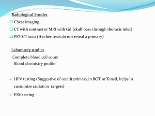
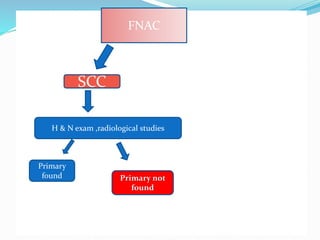
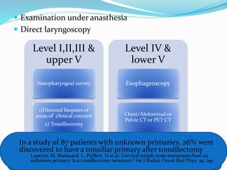
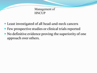
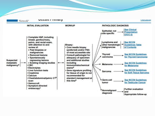
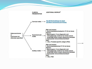
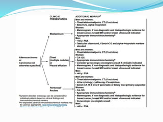
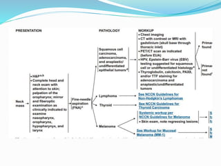
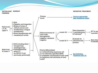
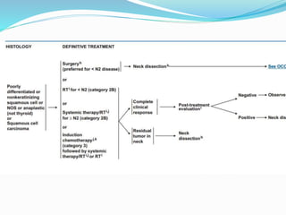
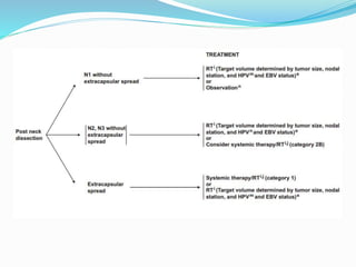
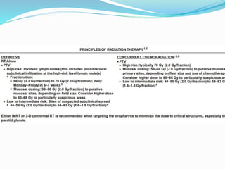
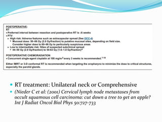
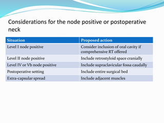
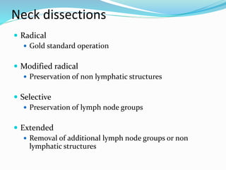
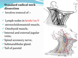
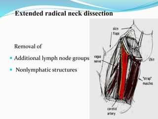
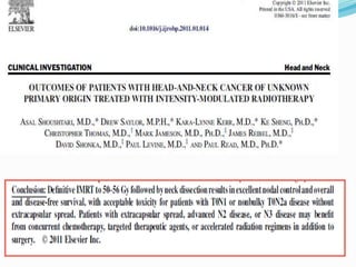
Dr Dipalee Borade
DNB student
Radiotherapy Department, Jupiter Hospital
Introduction
• Biopsy proven cancer of the neck, which even after a
complete clinical & radiological workup (that includes
physical examination, CT scan, esophgeoscopy,
laryngoscopy, bronchoscopy & multiple survillence
biopsies) reveals or yields no primary demonstrable
lesion.
Epidemology
 Exact incidence is unknown.
 Head-and-neck carcinoma of unknown primary
(HNCUP) is the final diagnosis in 3–7% of patients
with head-and-neck cancer initially presenting with
metastatic squamous cell carcinoma (SCC) to the
cervical lymph nodes
risk of lymph node metastases depends
upon:-
1) Density of capillary lymphatics
2) Location of the primary tumor
3) Histologic differentiation,
4) Size of the lesion
5) Recurrent v/s untreated lesions
Density of capillary lymphatics
Profuse capillary lymphatic network present in
Nasopharynx & Pyriform sinus
Paranasal sinuses, middle ear and true vocal
cords have sparse capillary lymphatics
Risk Groups based on location of primary tumor
Group
Estimated Risk of
Subclinical Neck
Disease % Stage Site
Low risk <20 T1 FOM, RMT, gingiva, hard palate,
buccal mucosa
Intermediate
risk
20-30 T1 Oral tongue, soft palate,
pharyngeal wall, supraglottic
larynx, tonsil
T2 FOM, oral tongue, RMT, gingiva,
hard palate, BM
High risk >30 T1-4 Nasopharynx, Pyriform sinus,
BOT
T2-4 Soft palate, pharyngeal wall,
supraglottic larynx, tonsil
T3-4 FOM, oral tongue, RMT, gingiva,
hard palate, BM
Histological differentiation
The majority of patients have either
squamous cell or poorly differentiated carcinoma.
Adenocarcinoma
High chances of primary lesion below the clavicles
If nodes are located in the upper neck
 Salivary gland
 Thyroid
 Parathyroid primary tumor.
Diagnostic workup
 History
 Physical examination
 Careful examination of the neck and supraclavicular
regions with attention to skin
 Examination of oral cavity, pharynx, and larynx
 Mirror & fiberoptic examination to visualise
nasopharynx, oropharynx, hypopharynx, larynx
STAGING OF THE NECK
FNAC
Anaplastic
epithelial &
Adenoca
FNAC
Lymphoma
Thyroid
Melanoma
Thyroglobulin
& calcitonin
SCC
Open biopsy should be avoided unless the patient is prepared for
definitive surgical managment
Radiological Studies
 Chest imaging
 CT with contrast or MRI with Gd (skull base through thoracic inlet)
 PET CT scan (If other tests do not reveal a primary)
Laboratory studies
Complete blood cell count
Blood chemistry profile
 HPV testing (Suggestive of occult primary in BOT or Tonsil, helps in
customize radiation targets)
 EBV testing
Evidence on role of PET CT
 In a meta-analysis of 16 studies looking at the role of
PET in 302 patients with cervical node metastases where
a primary has yet to be discovered through the work up,
25% of primaries are identified through PET. Previously
unrecognized regional or distant metastases were
identified in 27% of patients
 Rusthoven, KE, Koshy, M, Paulino, AC, The role of fluorodeoxyglucose PET in
cervical lymph node metastases from an unknown primary tumor. Cancer 2004;
101:2461
FNACFNAC
SCC
H & N exam ,radiological studies
Primary
found Primary not
found
 Examination under anasthesia
 Direct laryngoscopy
Level I,II,III &
upper V
Nasopharyngeal survey
1)Directed biopsies of
areas of clinical concern
2) Tonsillectomy
Level IV &
lower V
Esophageoscopy
Chest/Abdominal or
Pelvic CT or PET CT
Biopsy to be taken from
(Nasopharynx, tonsils, BOT, Pyriform sinuses & any suspicious mucosal areas)
In a study of 87 patients with unknown primaries, 26% were
discovered to have a tonsillar primary after tonsillectomy
Lapeyre, M, Malissard, L, Peiffert, D et al. Cervical lymph node metastasis from an
unknown primary: Is a tonsillectomy necessary? Int J Radiat Oncol Biol Phys; 39: 291
 Least investigated of all head-and-neck cancers
 Few prospective studies or clinical trials reported
 No definitive evidence proving the superiority of one
approach over others.
Management of
HNCUP
 RT treatment: Unilateral neck or Comprehensive
 (Nieder C et al: (2001) Cervical lymph node metastases from
occult squamous cell carcinoma: cut down a tree to get an apple?
Int J Radiat Oncol Biol Phys 50:727-733
Considerations for the node positive or postoperative
neck
Situation Proposed action
Level I node positive Consider inclusion of oral cavity if
comprehensive RT offered
Level II node positive Include retrostyloid space cranially
Level IV or Vb node positive Include supraclavicular fossa caudally
Postoperative setting Include entire surgical bed
Extra-capsular spread Include adjacent muscles
Neck dissections
 Radical
 Gold standard operation
 Modified radical
 Preservation of non lymphatic structures
 Selective
 Preservation of lymph node groups
 Extended
 Removal of additional lymph node groups or non
lymphatic structures
Standard radical neck
dissection
 Involves removal of :-
 Lymph nodes in levels I to V
 sternocleidomastoid muscle,
 Omohyoid muscle,
Internal and external jugular
veins,
Spinal accessory nerve,
Submandibular gland.
Tail of parotid
BIGGEST CONCERN
MAXIMISE CONTROL
MINIMIZE MORBIDITY
MODIFICATIONS OF RND
Modified Radical Neck Dissection
Removes
Nodal groups I-V
Preserves one or more of
the nonlymphatic
structures
 XI (I)
 IJV(II)
 SCM(III)
Selective Neck Dissection
 Remove high risk lymph node groups based on
tumor site.
 Supraomohyoid
 Levels I-III
 Lateral
 Levels II-IV
 Posterolateral
 Levels II-V
small oral cavity cancers and a
clinically negative neck.
laryngeal, oropharyngeal, and
hypopharyngeal
Post surgery management depends upon:-
1)Stage
 N1/N2-N3
2) Level of LN
 I/II-III-upper V/IV/lower level V
3)Presence of extracapsular extension
 If present chemotherapy to be added
Removal of
 Additional lymph node groups
 Nonlymphatic structures
Extended radical neck dissection
PART I: THE BASICS
 Introduction
 IHC methods
 Procedure
 IHC involves localization of antigen in tissues and
cells and is based on the principle of antigen-
antibody reaction which is detected by a tagged
visible label
 Antibodies raised specifically against the substance
of interest, and they are detected by
immunochemical methods
 Once antigen antibody reaction has occurred, an
enzyme capable of reacting with chromogen is
brought into close proximity of the antigen.
 Albert Coons used fluorescence technique in 1945
 1966 Nakane and Pierce developed enzyme labeling
method
 1977 Taylor - IHC on paraffin sections.
Uses
 Diagnose and classify undifferentiated tumors.
 Differentiate benign from malignant lesions
 Detection of small group of cancer cells
 Prognostic information on cancers
 Infections in the sections can also be detected
Methods
 Immunoenzymatic method : peroxidase, alkaline
phosphatase, biotin-avidin method
 Immunofluorescent technique: antibody labelled with
fluorochrome like Rhodamine, auramine.
 Direct conjugate labeled antibody method:
 Rapid, short and easy
 Insensitive
 Labeled antibody against every antigen
Indirect method
Unlabeled antibody method:
Secondary Ab links primary Ab to
an antiperoxidase antibody bound
to peroxidase
Avidin Biotin Conjugate method:
Secondary antibodies conjugated to
biotin, links the tissue bound primary
antibodies and avidin biotin-peroxidase
complex
 SLAB: labeled streptavidin-biotin:
Biotinylated secondary antibody links primary
antibodies to streptavidin
peroxidase conjugate
Polymer labeling 2 step method
 Polymer back bone to which
multiple enzyme molecules
are attached along with
secondary antibodies
MARKERS DIAGNOSTIC APPLICATION
CYTOKERATINS Epithelial tumors, mesothelioma
Epithelial membrane antigen Epithelial tumors, mesothelioma
Vimentin Mesenchymal tumors
Desmin Muscle lesions
S-100 Nerve, schwann cells, chondrocytes,
melanocytes
PSA Prostate
AFP Yolk sac tumor, HCC.
First tier Second tier
Epithelial tumors Pankeratin HMWK,LMWK,EMA
Mesenchymal tumors Vimentin Vimentin , Desmin,VIII
Melanomas Vimentin,S100 HMB45, NSE
Germ cell tumors Pankeratin, vimentin PLAP, AFP, hCG
Neural/neuroendocrine
tumors
NSE/NF Chromogranin,
Leu7,synaptophysin,
GFAP
Lymphomas LCA Pan-B, pan-T, kappa and
lambda light chains
The procedure…
 Formalin-fixed, paraffin-embeded blocks
 Sections are picked on slides coated with
3-aminopropyltriethoxysilane
 Deparaffinise in xylene and alcohol
 Wash in tris buffer, peroxidase block 30 minutes
 Wash, Microwave(antigen retrieval)
 Wash, Power block 30 minutes
 Primary antibody for 45-60 minutes
 Wash, super enhancer 30 minutes
 Wash, S S label (secondary antibody with poly HRP)
 Wash, DAB (diaminobenzidine) for 5 minutes
 Wash, haematoxylin 20 seconds
 Wash, mount (DPX)
Precautions
 Do not allow the slides to dry, incubate in moist
chamber
 Handle the reagents carefully
 Do not stop the procedure midway
 Cover whole sections
Quality control
 Consistency of performance and reproducibility
 Reagent control
 Tissue controls
 Positive control
 Negative control
 Internal or “built in” control
Neck node with unknown primary  dipalee

Neck node with unknown primary dipalee

  • 1.
    Dr Dipalee Borade DNBstudent Radiotherapy Department, Jupiter Hospital
  • 2.
    Introduction • Biopsy provencancer of the neck, which even after a complete clinical & radiological workup (that includes physical examination, CT scan, esophgeoscopy, laryngoscopy, bronchoscopy & multiple survillence biopsies) reveals or yields no primary demonstrable lesion.
  • 3.
    Epidemology  Exact incidenceis unknown.  Head-and-neck carcinoma of unknown primary (HNCUP) is the final diagnosis in 3–7% of patients with head-and-neck cancer initially presenting with metastatic squamous cell carcinoma (SCC) to the cervical lymph nodes
  • 4.
    risk of lymphnode metastases depends upon:- 1) Density of capillary lymphatics 2) Location of the primary tumor 3) Histologic differentiation, 4) Size of the lesion 5) Recurrent v/s untreated lesions
  • 5.
    Density of capillarylymphatics Profuse capillary lymphatic network present in Nasopharynx & Pyriform sinus Paranasal sinuses, middle ear and true vocal cords have sparse capillary lymphatics
  • 6.
    Risk Groups basedon location of primary tumor Group Estimated Risk of Subclinical Neck Disease % Stage Site Low risk <20 T1 FOM, RMT, gingiva, hard palate, buccal mucosa Intermediate risk 20-30 T1 Oral tongue, soft palate, pharyngeal wall, supraglottic larynx, tonsil T2 FOM, oral tongue, RMT, gingiva, hard palate, BM High risk >30 T1-4 Nasopharynx, Pyriform sinus, BOT T2-4 Soft palate, pharyngeal wall, supraglottic larynx, tonsil T3-4 FOM, oral tongue, RMT, gingiva, hard palate, BM
  • 7.
    Histological differentiation The majorityof patients have either squamous cell or poorly differentiated carcinoma. Adenocarcinoma High chances of primary lesion below the clavicles If nodes are located in the upper neck  Salivary gland  Thyroid  Parathyroid primary tumor.
  • 8.
    Diagnostic workup  History Physical examination  Careful examination of the neck and supraclavicular regions with attention to skin  Examination of oral cavity, pharynx, and larynx  Mirror & fiberoptic examination to visualise nasopharynx, oropharynx, hypopharynx, larynx
  • 9.
  • 10.
    FNAC Anaplastic epithelial & Adenoca FNAC Lymphoma Thyroid Melanoma Thyroglobulin & calcitonin SCC Openbiopsy should be avoided unless the patient is prepared for definitive surgical managment
  • 11.
    Radiological Studies  Chestimaging  CT with contrast or MRI with Gd (skull base through thoracic inlet)  PET CT scan (If other tests do not reveal a primary) Laboratory studies Complete blood cell count Blood chemistry profile  HPV testing (Suggestive of occult primary in BOT or Tonsil, helps in customize radiation targets)  EBV testing
  • 12.
    Evidence on roleof PET CT  In a meta-analysis of 16 studies looking at the role of PET in 302 patients with cervical node metastases where a primary has yet to be discovered through the work up, 25% of primaries are identified through PET. Previously unrecognized regional or distant metastases were identified in 27% of patients  Rusthoven, KE, Koshy, M, Paulino, AC, The role of fluorodeoxyglucose PET in cervical lymph node metastases from an unknown primary tumor. Cancer 2004; 101:2461
  • 13.
    FNACFNAC SCC H & Nexam ,radiological studies Primary found Primary not found
  • 14.
     Examination underanasthesia  Direct laryngoscopy Level I,II,III & upper V Nasopharyngeal survey 1)Directed biopsies of areas of clinical concern 2) Tonsillectomy Level IV & lower V Esophageoscopy Chest/Abdominal or Pelvic CT or PET CT Biopsy to be taken from (Nasopharynx, tonsils, BOT, Pyriform sinuses & any suspicious mucosal areas) In a study of 87 patients with unknown primaries, 26% were discovered to have a tonsillar primary after tonsillectomy Lapeyre, M, Malissard, L, Peiffert, D et al. Cervical lymph node metastasis from an unknown primary: Is a tonsillectomy necessary? Int J Radiat Oncol Biol Phys; 39: 291
  • 15.
     Least investigatedof all head-and-neck cancers  Few prospective studies or clinical trials reported  No definitive evidence proving the superiority of one approach over others. Management of HNCUP
  • 24.
     RT treatment:Unilateral neck or Comprehensive  (Nieder C et al: (2001) Cervical lymph node metastases from occult squamous cell carcinoma: cut down a tree to get an apple? Int J Radiat Oncol Biol Phys 50:727-733
  • 25.
    Considerations for thenode positive or postoperative neck Situation Proposed action Level I node positive Consider inclusion of oral cavity if comprehensive RT offered Level II node positive Include retrostyloid space cranially Level IV or Vb node positive Include supraclavicular fossa caudally Postoperative setting Include entire surgical bed Extra-capsular spread Include adjacent muscles
  • 26.
    Neck dissections  Radical Gold standard operation  Modified radical  Preservation of non lymphatic structures  Selective  Preservation of lymph node groups  Extended  Removal of additional lymph node groups or non lymphatic structures
  • 27.
    Standard radical neck dissection Involves removal of :-  Lymph nodes in levels I to V  sternocleidomastoid muscle,  Omohyoid muscle, Internal and external jugular veins, Spinal accessory nerve, Submandibular gland. Tail of parotid
  • 28.
    BIGGEST CONCERN MAXIMISE CONTROL MINIMIZEMORBIDITY MODIFICATIONS OF RND
  • 29.
    Modified Radical NeckDissection Removes Nodal groups I-V Preserves one or more of the nonlymphatic structures  XI (I)  IJV(II)  SCM(III)
  • 30.
    Selective Neck Dissection Remove high risk lymph node groups based on tumor site.  Supraomohyoid  Levels I-III  Lateral  Levels II-IV  Posterolateral  Levels II-V small oral cavity cancers and a clinically negative neck. laryngeal, oropharyngeal, and hypopharyngeal
  • 31.
    Post surgery managementdepends upon:- 1)Stage  N1/N2-N3 2) Level of LN  I/II-III-upper V/IV/lower level V 3)Presence of extracapsular extension  If present chemotherapy to be added
  • 32.
    Removal of  Additionallymph node groups  Nonlymphatic structures Extended radical neck dissection
  • 34.
    PART I: THEBASICS  Introduction  IHC methods  Procedure
  • 35.
     IHC involveslocalization of antigen in tissues and cells and is based on the principle of antigen- antibody reaction which is detected by a tagged visible label  Antibodies raised specifically against the substance of interest, and they are detected by immunochemical methods  Once antigen antibody reaction has occurred, an enzyme capable of reacting with chromogen is brought into close proximity of the antigen.
  • 37.
     Albert Coonsused fluorescence technique in 1945  1966 Nakane and Pierce developed enzyme labeling method  1977 Taylor - IHC on paraffin sections.
  • 38.
    Uses  Diagnose andclassify undifferentiated tumors.  Differentiate benign from malignant lesions  Detection of small group of cancer cells  Prognostic information on cancers  Infections in the sections can also be detected
  • 39.
    Methods  Immunoenzymatic method: peroxidase, alkaline phosphatase, biotin-avidin method  Immunofluorescent technique: antibody labelled with fluorochrome like Rhodamine, auramine.
  • 40.
     Direct conjugatelabeled antibody method:  Rapid, short and easy  Insensitive  Labeled antibody against every antigen
  • 41.
  • 42.
    Unlabeled antibody method: SecondaryAb links primary Ab to an antiperoxidase antibody bound to peroxidase
  • 44.
    Avidin Biotin Conjugatemethod: Secondary antibodies conjugated to biotin, links the tissue bound primary antibodies and avidin biotin-peroxidase complex
  • 45.
     SLAB: labeledstreptavidin-biotin: Biotinylated secondary antibody links primary antibodies to streptavidin peroxidase conjugate
  • 46.
    Polymer labeling 2step method  Polymer back bone to which multiple enzyme molecules are attached along with secondary antibodies
  • 47.
    MARKERS DIAGNOSTIC APPLICATION CYTOKERATINSEpithelial tumors, mesothelioma Epithelial membrane antigen Epithelial tumors, mesothelioma Vimentin Mesenchymal tumors Desmin Muscle lesions S-100 Nerve, schwann cells, chondrocytes, melanocytes PSA Prostate AFP Yolk sac tumor, HCC.
  • 48.
    First tier Secondtier Epithelial tumors Pankeratin HMWK,LMWK,EMA Mesenchymal tumors Vimentin Vimentin , Desmin,VIII Melanomas Vimentin,S100 HMB45, NSE Germ cell tumors Pankeratin, vimentin PLAP, AFP, hCG Neural/neuroendocrine tumors NSE/NF Chromogranin, Leu7,synaptophysin, GFAP Lymphomas LCA Pan-B, pan-T, kappa and lambda light chains
  • 51.
    The procedure…  Formalin-fixed,paraffin-embeded blocks  Sections are picked on slides coated with 3-aminopropyltriethoxysilane  Deparaffinise in xylene and alcohol  Wash in tris buffer, peroxidase block 30 minutes  Wash, Microwave(antigen retrieval)  Wash, Power block 30 minutes
  • 53.
     Primary antibodyfor 45-60 minutes  Wash, super enhancer 30 minutes  Wash, S S label (secondary antibody with poly HRP)  Wash, DAB (diaminobenzidine) for 5 minutes  Wash, haematoxylin 20 seconds  Wash, mount (DPX)
  • 54.
    Precautions  Do notallow the slides to dry, incubate in moist chamber  Handle the reagents carefully  Do not stop the procedure midway  Cover whole sections
  • 55.
    Quality control  Consistencyof performance and reproducibility  Reagent control  Tissue controls  Positive control  Negative control  Internal or “built in” control