More Related Content
Similar to NCUR CD133 Poster (20)
NCUR CD133 Poster
- 1. RESEARCH POSTER PRESENTATION DESIGN © 2011
www.PosterPresentations.com
Highly specific “markers” that distinguish stem cells from
other cell types are critical tools for isolating stem cells,
assaying their purity and functional status, and potentially
targeting cancer stem cells for destruction. Currently one
of the most widely used stem cell markers is a plasma
membrane protein of unknown function, CD133. However,
most antibodies to CD133 also recognize some non-stem
cell types, including some mature differentiated cells. By
contrast, the monoclonal antibody, AC133, seems to only
recognize a subset of CD133 uniquely expressed on stem
cells. Our aim has been to identify what variation of
CD133 makes AC133 so precise in identifying cancer stem
cells: variation in the CD133 protein sequence, post-
translational modification, or alternative conformations.
Suspecting that tyrosine sulfation, a post-translational
modification increasingly recognized on cell surface
receptors and extracellular proteins, might be involved,
we tested the effect of culturing cells with or without
sodium chlorate to competitively inhibit sulfation. 30mM
chlorate blocked CD133 recognition by both AC133 and a
specific anti-sulfotyrosine antibody, indicating that
tyrosine sulfation is a critical feature of the epitope
recognized by AC133. We are now seeking other features
of this epitope, its location on CD133, and its significance
for stem cell physiology. We will be working to structurally
define AC133 modification using cell and biochemical
approaches with the long term goal of creating a more
refined antibody for identifying stem cells.
ABSTRACT
The cell surface protein CD133 has become one of the
premier markers for identifying and isolating stem cells,
but conflicting and puzzling results have hindered its
reliability and acceptance. One major problem arises
from the use of multiple distinct anti-CD133 antibodies
and immunostaining methods by different investigators, in
part because the best antibodies to human CD133 do not
react with most other species, e.g. mouse or rat. This is
the case for the AC133 monoclonal antibody originally
used to identify CD1331. While AC133 seems to be highly
specific for human CD133 expressed on stem cells, many
other anti-CD133 antibodies also recognize the protein on
some mature cell types as well. This presents a mystery:
What is it that AC133 recognizes for CD133 as expressed
on stem cells but not for CD133 expressed on other cell
types? What is special about the epitope recognized by
AC133?
Possible explanations for AC133 epitope specificity are
variation in: the CD133 protein sequence (e.g. alternative
splicing), post-translational modification (e.g.
glycosylation or sulfation), or conformation (e.g. due to
specific association with another protein or other ligand or
due to an intracellular modification such as
phosphorylation ). Since CD133’s first discovery,
glycosylation has been implicated as important for
expression of the epitope, but this has also been
controverted and recent publications argue against it2.
Other possibilities have not yet been directly assessed.
Therefore, the objective of this study was to test the
hypothesis, that the AC133 epitope depends on sulfation
of one or more tyrosine residues. This hypothesis is
intriguing, because CD133 has been shown to post-
translationally incorporate sulfate in some form1, and
because tyrosine sulfation is increasing recognized as an
important modification for protein-ligand recognition3.
BACKGROUND and OBJECTIVES
Cell culture: A human retinoblastoma cell line (WERI-RB-1), known to express high levels of the AC133 epitope, was allowed to
proliferate in complete culture medium for three days with 0 or 30mM sodium chlorate, a competitive inhibitor of sulfate
binding to sulfurylase, so blocking cell synthesis of the critical intermediate for all cellular sulfation . Treatment of most
cultured cells with chlorate at 10-30mM blocks tyrosine sulfation by more than 90%. (Every other form of sulfation is also
inhibited to some extent.)
SDS-PAGE and immunoblotting: Cell extracts from equal numbers of cells were serially diluted (1:1, 3, 10, and 30) and analyzed
by SDS-PAGE immunoblotting. A control sample (C) of 293T cells transfected to express CD133 (H3 form) and molecular weight
markers (Amersham ECL DualVue) were included. After, SDS-PAGE, blots were immunostained with AC133 (Miltenyi-Biotec) or
anti-sulfotyrosine (clone Sulfo-1C-A2, Millipore). Bound antibodies were detected using peroxidase-conjugated second
antibodies and chemiluminescence. A matching gel stained for total protein using Coomassie Blue revealed that the chlorate
treated sample was more concentrated than the control. This was confirmed by immunostaining a matching blot with antibody
to the N-terminus of CD133.
MATERIAL AND METHODS
RESULTS
KEY REFERENCES
1. Miraglia et al. (1997) Blood 90: 5013-21
Corbeil et al. (2000) J. Biol Chem 275: 5512-20
2. Kemper et al. (2010) Cancer Res 70: 719–29
3. Moore (2009) PNAS 106: 14741-2
4. Sherry et al. (2010) Eur J Neurosci 32: 1461-72
5. Kanan et al. (2009) Exp Eye Res 89: 559-67
ACKNOWLEDGEMENTS
(Douglas N.W. Cooper, Ph.D.) Department of Natural Sciences
and Mathematics, Dominican University of California, 50
Acacia Ave, San Rafael, CA 94901
Magic Marker Mystery: Why Does Antibody AC133 So
Specifically Recognize Stem Cells?
Monica Buselli, Elizabeth Castellanos, Shiena T. Enerio, Akikta D. Murti, Kimberli D. Reed, and Alyssa M. Wong
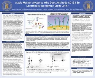
Expression Of The AC133 Epitope vs Sulfotyrosine Assessed By SDS-PAGE Immunoblot
AC133+
AC133+
AC133-
The AC133 Epitope Is A Marker For Stem Cells
The Nature And Location Of The AC133
Epitope On CD133 Has Been A Mystery
Sulfotyrosine ?
CONCLUSIONS
• CD133 expressed in WERI-RB-1 cells is modified
by sulfation of tyrosine and is clearly the
predominant sulfotyrosine modified protein.
• The AC133 epitope is apparently dependent on
tyrosine sulfation to at least a major extent.
Specific tyrosine sulfation of CD133 on stem cells is
likely to be functionally important. This modification
has been found to critically influence ligand-binding
for other proteins, e.g. tyrosine sulfation of
chemokine receptors greatly increases their affinity
for chemokines. While the function of CD133 is still
not established, its transmembrane structure suggests
receptor or transporter function.
It is intriguing that the AC133 epitope is expressed at
especially high levels in retinal photoreceptors, since
blocking tyrosine sulfation disrupts photoreceptor
outer segment morphogenesis4, and again the
predominant tyrosine sulfated protein detected by
anti-sulfotyrosine migrates at about 125kD5. Similarly,
human CD133 mutations or knockout of the mouse
gene have relatively minor effects, except for
photoreceptor degeneration!
The results reported here still require refinement with
better quantitation, confirmation with other methods
of eliminating tyrosine sulfation, and examination of
other factors that also could be important in forming
the AC133 epitope. This work should allow generation
of improved antibodies and methods for identifying
stem cells. Our results should also stimulate
investigation of the tyrosine sulfation in CD133
function.
AC133 Anti-Sulfotyrosine
MW 1 3 10 30 30 10 3 1 C MW 1 3 10 30 30 10 3 1 C
Chlorate : 30 mM 0 30 mM 0
150
100
75
50
35
25
Chlorate at 30mM reduced the immunostaining intensity with both AC133 and anti-
sulfotyrosine by at least 3 fold and closer to 10 fold after accounting for unequal sample
concentration as assessed by Coomassie Blue staining.
Monoclonal antibody AC133 recognizes an epitope of unknown structure that is expressed uniquely on
CD133 on stem cells. CD133 is a 125kD membrane glycoprotein highly conserved across multicellular
species.
CONCLUSIONS