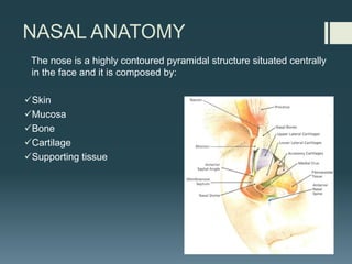
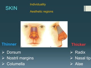
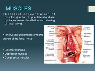
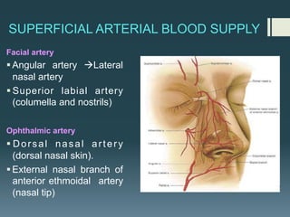
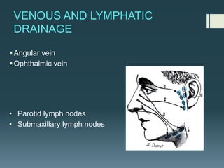
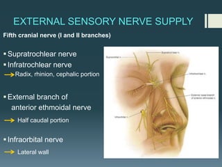
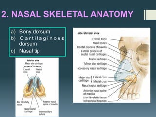
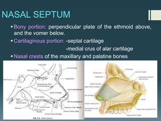
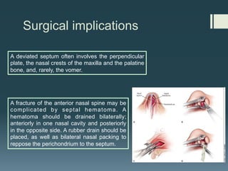
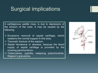
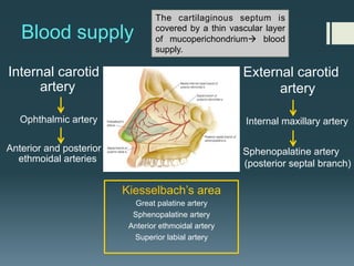
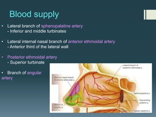
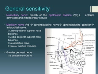
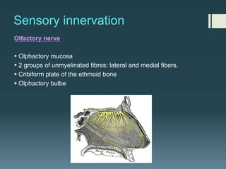
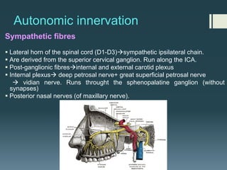
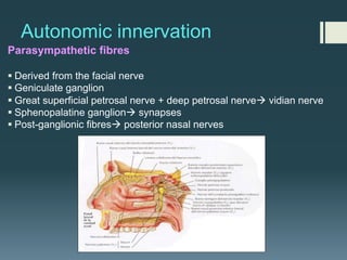
NASAL ANATOMY
Elena Rizzo Riera
R1 ORL HUSE
NASAL ANATOMY
The nose is a highly contoured pyramidal structure situated centrally
in the face and it is composed by:
üSkin
üMucosa
üBone
üCartilage
üSupporting tissue
Topographic analysis
1. EXTERNAL NASAL ANATOMY
§ Skin
§ Soft tissue
§ Muscles
§ Blood vessels
§ Nerves
² Understanding variations in
skin thickness is an essential
aspect of reconstructive
nasal surgery.
² Familiarity with blood
supplyà local flaps.
SKIN
Ø Dorsum
Ø Nostril margins
Ø Columella
Ø Radix
Ø Nasal tip
Ø Alae
Thinner Thicker
Individuality
Aesthetic regions
Surgical implications
Surgical elevation of the nasal skin should be done in the
plane just superficial to the underlying bony and cartilaginous
nasal skeleton to prevent injury to the blood supply and to the
nasal muscles. Excessive damage to the nasal muscles
causes unwanted immobility of the nose during facial
expression, so called mummified nose.
SUBCUTANEOUS LAYER
§Superficial fatty panniculus
Adipose tissue and vertical fibres between deep dermis and
fibromuscular layer.
§Fibromuscular layer
Nasal musculature and nasal SMAS
§Deep fatty layer
Contains the major superficial blood vessels and nerves. No fibrous
fibres.
§Periosteum/ perichondrium
Provide nutrient blood flow to the nasal bones and cartilage
MUSCLES
§ G r e a t e s t c o n c e n t r a t i o n o f
musclesàjunction of upper lateral and alar
cartilages (muscular dilation and stenting
of nasal valve).
§ Innervation: zygomaticotemporal
branch of the facial nerve
§ Elevator muscles
§ Depressor muscles
§ Compressor muscles
SUPERFICIAL ARTERIAL BLOOD SUPPLY
Facial artery
§Angular artery àLateral
nasal artery
§ Superior labial artery
(columella and nostrils)
Ophthalmic artery
§ Dorsal nasal artery
(dorsal nasal skin).
§External nasal branch of
anterior ethmoidal artery
(nasal tip)
VENOUS AND LYMPHATIC
DRAINAGE
§Angular vein
§Ophthalmic vein
• Parotid lymph nodes
• Submaxillary lymph nodes
EXTERNAL SENSORY NERVE SUPPLY
Fifth cranial nerve (I and II branches)
§Supratrochlear nerve
§Infratrochlear nerve
Radix, rhinion, cephalic portion
§External branch of
anterior ethmoidal nerve
Half caudal portion
§Infraorbital nerve
Lateral wall
2. NASAL SKELETAL ANATOMY
a) Bony dorsum
b) C a r t i l a g i n o u s
dorsum
c) Nasal tip
BONY DORSUM
§ Paired nasal bones
§ Paired ascending processes of
maxillary bones
§ Crest of nasal bones
§ Intercanthal line
CARTILAGINOUS DORSUM
1. Paired upper lateral cartilages
2. Cartilaginous septum
§ External lateral triangle. It’s
important in fisiology of nasal
respiration, because its support
only consists of fibrous tissue
that can collapse during the
inspiration.
NASAL TIP
Consists of:
ØLobule
ØColumella
ØVestibules
ØAlae
Structurally supported by:
Ø Alar cartilagesà attached to the
upper lateral cart. And the septum
Ø Caudal septum
Ø Accessory cartilages
Ø Fibrous fatty connective tissue
Accessory	
  cartilage	
  
Alar cartilages
§ Medial crus: footplate and columellar
segments.
§ Intermediate crus: lobular and dogmal
segments. Bound by the interdomal
ligament.
§ Lateral crus: largest component.
The appearance and projection of the
columella are influenced by the
configuration of medial crus as well as that
of the septum. Soft tissue between the
columellar segments may fill this space
but in patients with thin skin , the
columella may have a bifid appearance.
Fibrous skeleton
§ External lateral triangle
Important in the fisiology of
nasal respiration.
§ Weak triangle
§ Importance of the distal point
of attachment of the lateral
cartilage à stability of the
nose.
§ Overlaping of the alar
cartilages to the lateral
cartilages.
Weak triangle
External lateral
triangle
Surgical implications
The angle between the
septum and the upper
lateral cartilage is important
during respiration.
Obstruction of this angle by
scar tissue or trauma may
produce symptoms of nasal
obstruction.
The upper and the lower lateral cartilages are
obviously of great importance both in
achieving aesthetic results and in maintaining
nasal physiology during rhinoplasty.
A surgeon should be conservative in trimming
the alar cartilage, especially at its inferior
extremity.
3. INTERNAL NASAL ANATOMY
§Vestible
§Nasal cavity
§Septum
§Lateral wall
VESTIBULE
§ Squamous epithelium
§ Limen nasi
§ Nasal cavity: ciliated columnar epithelium
§ Nasal valvesà controls airflow direction.
§ Septo-turbinal valve (posterior, between the septum and the
mucosa of the inferior turbinate)ànasal resistance
Septum
Inferior turbinate
NASAL SEPTUM
§Bony portion: perpendicular plate of the ethmoid above,
and the vomer below.
§Cartilaginous portion: -septal cartilage
-medial crus of alar cartilage
§Nasal crests of the maxillary and palatine bones
Surgical implications
A deviated septum often involves the perpendicular
plate, the nasal crests of the maxilla and the palatine
bone, and, rarely, the vomer.
A fracture of the anterior nasal spine may be
complicated by septal hematoma. A
hematoma should be drained bilaterally;
anteriorly in one nasal cavity and posteriorly
in the opposite side. A rubber drain should be
placed, as well as bilateral nasal packing to
reppose the perichondrium to the septum.
Surgical implications
A cartilaginous saddle nose, is due to depression of
the dorsum of the nose. It may be caused by the
following:
² Excessive removal of septal cartilage, which
weakens the normal support in this area.
² Traumatic fracture of the septum.
² Septal hematoma or abscess, because the blood
supply of septal cartilage is provided by the
covering perichondrium.
² Tuberculosis, syphilis, relapsing polychondritis,
Wegner’s granuloma.
Blood supply
Internal carotid
artery
Ophthalmic artery
Anterior and posterior
ethmoidal arteries
External carotid
artery
Internal maxillary artery
Sphenopalatine artery
(posterior septal branch)
Kiesselbach’s area
Great palatine artery
Sphenopalatine artery
Anterior ethmoidal artery
Superior labial artery
	
  
The cartilaginous septum is
covered by a thin vascular layer
of mucoperichondriumà blood
supply.
LATERAL WALL
§Three (or four) nasal turbinates: inferior, middle and
superior.
§Ostia of the nasal sinuses (exception of that for the
sphenoid sinus)
§Opening of the nasolacrimal duct
Olfactory
sulcus
LATERAL WALL
§Inferior nasal turbinate
²Is a separate bone
covered by thick mucous
membrane.
§Middle turbinate
²Portion of the ethmoid
bone.
²The concha bullosa is a
pneumatized portion of
the middle turbinate
c o n t i n u o u s w i t h
ethmoidal air cells.
LATERAL WALL
§Superior turbinate
²Is approximately one- half the length of the middle
turbinate.
²The sphenoethmoidal recess lies between the superior
turbinate and the sphenoid bone.
§Supreme turbinate and supreme meatus
²Are present unilaterally or bilaterally in 60% of
individuals.
²The ostium of the posterior ethmoidal cells opens into
the supreme meatus, when present, in about 75% of
individuals.
LATERAL WALL
§ Inferior meatus: opening of the nasolacrimal duct.
§ Middle meatus
- Frontal recess: most superior portion. Opening of the frontal sinus and some anterior
ethmoidal cells.
- Ethmoidal bulla (one or more ethmoidal cells)
- Semilunar (or ethmoidal) hiatus / ethmoidal infundibulum: anterior ethmoidal
cells and maxillary sinus.
- Uncinate process
§ Superior meatus: olfactory epithelium and the opening of the posterior ethmoidal cells.
Blood supply
• Lateral branch of sphenopalatine artery
- Inferior and middle turbinates
• Lateral internal nasal branch of anterior ethmoidal artery
- Anterior third of the lateral wall
• Posterior ethmoidal artery
- Superior turbinate
• Branch of angular
artery
Veins and lymphatic drainage
§Veins
§ Pterygoid plexus - inferior turbinate, inferior meatus and
posterior part of the septum.
§ Ophtalmic vein – ethmoidal veins
§ Facial vein – subcutaneous plexus of the skin covering the alar
region
§Lymphatics
§ Lymphatic of the skin – anterior part of the nose.
§ Deep cervical lymph nodes
§ Retropharingeal lymph nodes
General sensitivity
• Nasociliary nerve: branch of the ophthalmic division (Va)à anterior
ethmoidal and infratrochlear nerves
• Maxillary nerve (Vb)à sphenopalatine nerveà sphenopalatine ganglionà
Infraorbital nerve.
²Lateral posterior superior nasal
branches
²Medial posterior superior nasal
branches
²Nasopalatine nerve
²Greater palatine branches
• Greater petrosal nerve
²Is derived from CN VII
Sensory innervation
Olfactory nerve
§ Olphactory mucosa
§ 2 groups of unmyelinated fibres: lateral and medial fibers.
§ Cribiform plate of the ethmoid bone
§ Olphactory bulbe
Autonomic innervation
Sympathetic fibres
§ Lateral horn of the spinal cord (D1-D3)àsympathetic ipsilateral chain.
§ Are derived from the superior cervical ganglion. Run along the ICA.
§ Post-ganglionic fibresàinternal and external carotid plexus
§ Internal plexusà deep petrosal nerve+ great superficial petrosal nerve
à vidian nerve. Runs throught the sphenopalatine ganglion (without
synapses)
§ Posterior nasal nerves (of maxillary nerve).
Autonomic innervation
Parasympathetic fibres
§ Derived from the facial nerve
§ Geniculate ganglion
§ Great superficial petrosal nerve + deep petrosal nerveà vidian nerve
§ Sphenopalatine ganglionà synapses
§ Post-ganglionic fibresà posterior nasal nerves
BIBLIOGRAPHY
§ Surgical anatomy of the head and neck. P. Janfaza, J. B.
Nadol, R. J. Galla, R. L. Fabian, W. W. Montgomery.
§ Otorrinolaringología. W. Becker, H. H. Naumann, C. R. Pfaltz.
§ Otorhinolaryngology, Head & Neck Surgery. M. Anniko, M.
Bernal- Sprekelsen, V. Bonkowsky, P. Bradley, S. Iurato
Thank you J

Nasal_Anatomy.pdf

  • 1.
    NASAL ANATOMY Elena RizzoRiera R1 ORL HUSE
  • 2.
    NASAL ANATOMY The noseis a highly contoured pyramidal structure situated centrally in the face and it is composed by: üSkin üMucosa üBone üCartilage üSupporting tissue
  • 3.
  • 4.
    1. EXTERNAL NASALANATOMY § Skin § Soft tissue § Muscles § Blood vessels § Nerves ² Understanding variations in skin thickness is an essential aspect of reconstructive nasal surgery. ² Familiarity with blood supplyà local flaps.
  • 5.
    SKIN Ø Dorsum Ø Nostrilmargins Ø Columella Ø Radix Ø Nasal tip Ø Alae Thinner Thicker Individuality Aesthetic regions
  • 6.
    Surgical implications Surgical elevationof the nasal skin should be done in the plane just superficial to the underlying bony and cartilaginous nasal skeleton to prevent injury to the blood supply and to the nasal muscles. Excessive damage to the nasal muscles causes unwanted immobility of the nose during facial expression, so called mummified nose.
  • 7.
    SUBCUTANEOUS LAYER §Superficial fattypanniculus Adipose tissue and vertical fibres between deep dermis and fibromuscular layer. §Fibromuscular layer Nasal musculature and nasal SMAS §Deep fatty layer Contains the major superficial blood vessels and nerves. No fibrous fibres. §Periosteum/ perichondrium Provide nutrient blood flow to the nasal bones and cartilage
  • 8.
    MUSCLES § G re a t e s t c o n c e n t r a t i o n o f musclesàjunction of upper lateral and alar cartilages (muscular dilation and stenting of nasal valve). § Innervation: zygomaticotemporal branch of the facial nerve § Elevator muscles § Depressor muscles § Compressor muscles
  • 9.
    SUPERFICIAL ARTERIAL BLOODSUPPLY Facial artery §Angular artery àLateral nasal artery § Superior labial artery (columella and nostrils) Ophthalmic artery § Dorsal nasal artery (dorsal nasal skin). §External nasal branch of anterior ethmoidal artery (nasal tip)
  • 10.
    VENOUS AND LYMPHATIC DRAINAGE §Angularvein §Ophthalmic vein • Parotid lymph nodes • Submaxillary lymph nodes
  • 11.
    EXTERNAL SENSORY NERVESUPPLY Fifth cranial nerve (I and II branches) §Supratrochlear nerve §Infratrochlear nerve Radix, rhinion, cephalic portion §External branch of anterior ethmoidal nerve Half caudal portion §Infraorbital nerve Lateral wall
  • 12.
    2. NASAL SKELETALANATOMY a) Bony dorsum b) C a r t i l a g i n o u s dorsum c) Nasal tip
  • 13.
    BONY DORSUM § Pairednasal bones § Paired ascending processes of maxillary bones § Crest of nasal bones § Intercanthal line
  • 14.
    CARTILAGINOUS DORSUM 1. Pairedupper lateral cartilages 2. Cartilaginous septum § External lateral triangle. It’s important in fisiology of nasal respiration, because its support only consists of fibrous tissue that can collapse during the inspiration.
  • 15.
    NASAL TIP Consists of: ØLobule ØColumella ØVestibules ØAlae Structurallysupported by: Ø Alar cartilagesà attached to the upper lateral cart. And the septum Ø Caudal septum Ø Accessory cartilages Ø Fibrous fatty connective tissue Accessory  cartilage  
  • 16.
    Alar cartilages § Medialcrus: footplate and columellar segments. § Intermediate crus: lobular and dogmal segments. Bound by the interdomal ligament. § Lateral crus: largest component. The appearance and projection of the columella are influenced by the configuration of medial crus as well as that of the septum. Soft tissue between the columellar segments may fill this space but in patients with thin skin , the columella may have a bifid appearance.
  • 17.
    Fibrous skeleton § Externallateral triangle Important in the fisiology of nasal respiration. § Weak triangle § Importance of the distal point of attachment of the lateral cartilage à stability of the nose. § Overlaping of the alar cartilages to the lateral cartilages. Weak triangle External lateral triangle
  • 18.
    Surgical implications The anglebetween the septum and the upper lateral cartilage is important during respiration. Obstruction of this angle by scar tissue or trauma may produce symptoms of nasal obstruction. The upper and the lower lateral cartilages are obviously of great importance both in achieving aesthetic results and in maintaining nasal physiology during rhinoplasty. A surgeon should be conservative in trimming the alar cartilage, especially at its inferior extremity.
  • 19.
    3. INTERNAL NASALANATOMY §Vestible §Nasal cavity §Septum §Lateral wall
  • 20.
    VESTIBULE § Squamous epithelium §Limen nasi § Nasal cavity: ciliated columnar epithelium § Nasal valvesà controls airflow direction. § Septo-turbinal valve (posterior, between the septum and the mucosa of the inferior turbinate)ànasal resistance Septum Inferior turbinate
  • 21.
    NASAL SEPTUM §Bony portion:perpendicular plate of the ethmoid above, and the vomer below. §Cartilaginous portion: -septal cartilage -medial crus of alar cartilage §Nasal crests of the maxillary and palatine bones
  • 22.
    Surgical implications A deviatedseptum often involves the perpendicular plate, the nasal crests of the maxilla and the palatine bone, and, rarely, the vomer. A fracture of the anterior nasal spine may be complicated by septal hematoma. A hematoma should be drained bilaterally; anteriorly in one nasal cavity and posteriorly in the opposite side. A rubber drain should be placed, as well as bilateral nasal packing to reppose the perichondrium to the septum.
  • 23.
    Surgical implications A cartilaginoussaddle nose, is due to depression of the dorsum of the nose. It may be caused by the following: ² Excessive removal of septal cartilage, which weakens the normal support in this area. ² Traumatic fracture of the septum. ² Septal hematoma or abscess, because the blood supply of septal cartilage is provided by the covering perichondrium. ² Tuberculosis, syphilis, relapsing polychondritis, Wegner’s granuloma.
  • 24.
    Blood supply Internal carotid artery Ophthalmicartery Anterior and posterior ethmoidal arteries External carotid artery Internal maxillary artery Sphenopalatine artery (posterior septal branch) Kiesselbach’s area Great palatine artery Sphenopalatine artery Anterior ethmoidal artery Superior labial artery   The cartilaginous septum is covered by a thin vascular layer of mucoperichondriumà blood supply.
  • 25.
    LATERAL WALL §Three (orfour) nasal turbinates: inferior, middle and superior. §Ostia of the nasal sinuses (exception of that for the sphenoid sinus) §Opening of the nasolacrimal duct Olfactory sulcus
  • 26.
    LATERAL WALL §Inferior nasalturbinate ²Is a separate bone covered by thick mucous membrane. §Middle turbinate ²Portion of the ethmoid bone. ²The concha bullosa is a pneumatized portion of the middle turbinate c o n t i n u o u s w i t h ethmoidal air cells.
  • 27.
    LATERAL WALL §Superior turbinate ²Isapproximately one- half the length of the middle turbinate. ²The sphenoethmoidal recess lies between the superior turbinate and the sphenoid bone. §Supreme turbinate and supreme meatus ²Are present unilaterally or bilaterally in 60% of individuals. ²The ostium of the posterior ethmoidal cells opens into the supreme meatus, when present, in about 75% of individuals.
  • 28.
    LATERAL WALL § Inferiormeatus: opening of the nasolacrimal duct. § Middle meatus - Frontal recess: most superior portion. Opening of the frontal sinus and some anterior ethmoidal cells. - Ethmoidal bulla (one or more ethmoidal cells) - Semilunar (or ethmoidal) hiatus / ethmoidal infundibulum: anterior ethmoidal cells and maxillary sinus. - Uncinate process § Superior meatus: olfactory epithelium and the opening of the posterior ethmoidal cells.
  • 29.
    Blood supply • Lateralbranch of sphenopalatine artery - Inferior and middle turbinates • Lateral internal nasal branch of anterior ethmoidal artery - Anterior third of the lateral wall • Posterior ethmoidal artery - Superior turbinate • Branch of angular artery
  • 30.
    Veins and lymphaticdrainage §Veins § Pterygoid plexus - inferior turbinate, inferior meatus and posterior part of the septum. § Ophtalmic vein – ethmoidal veins § Facial vein – subcutaneous plexus of the skin covering the alar region §Lymphatics § Lymphatic of the skin – anterior part of the nose. § Deep cervical lymph nodes § Retropharingeal lymph nodes
  • 31.
    General sensitivity • Nasociliarynerve: branch of the ophthalmic division (Va)à anterior ethmoidal and infratrochlear nerves • Maxillary nerve (Vb)à sphenopalatine nerveà sphenopalatine ganglionà Infraorbital nerve. ²Lateral posterior superior nasal branches ²Medial posterior superior nasal branches ²Nasopalatine nerve ²Greater palatine branches • Greater petrosal nerve ²Is derived from CN VII
  • 32.
    Sensory innervation Olfactory nerve §Olphactory mucosa § 2 groups of unmyelinated fibres: lateral and medial fibers. § Cribiform plate of the ethmoid bone § Olphactory bulbe
  • 33.
    Autonomic innervation Sympathetic fibres §Lateral horn of the spinal cord (D1-D3)àsympathetic ipsilateral chain. § Are derived from the superior cervical ganglion. Run along the ICA. § Post-ganglionic fibresàinternal and external carotid plexus § Internal plexusà deep petrosal nerve+ great superficial petrosal nerve à vidian nerve. Runs throught the sphenopalatine ganglion (without synapses) § Posterior nasal nerves (of maxillary nerve).
  • 34.
    Autonomic innervation Parasympathetic fibres §Derived from the facial nerve § Geniculate ganglion § Great superficial petrosal nerve + deep petrosal nerveà vidian nerve § Sphenopalatine ganglionà synapses § Post-ganglionic fibresà posterior nasal nerves
  • 35.
    BIBLIOGRAPHY § Surgical anatomyof the head and neck. P. Janfaza, J. B. Nadol, R. J. Galla, R. L. Fabian, W. W. Montgomery. § Otorrinolaringología. W. Becker, H. H. Naumann, C. R. Pfaltz. § Otorhinolaryngology, Head & Neck Surgery. M. Anniko, M. Bernal- Sprekelsen, V. Bonkowsky, P. Bradley, S. Iurato
  • 36.