
Recommended
More Related Content
Similar to multiple pregnancy(twin) by iraqi doctor.pptx
Similar to multiple pregnancy(twin) by iraqi doctor.pptx (20)
Recently uploaded
Recently uploaded (20)
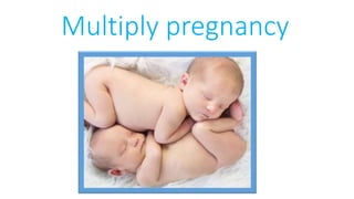
multiple pregnancy(twin) by iraqi doctor.pptx
- 2. Multiple pregnancy: overview Incidence • About 1 in 34 babies born in the UK is a twin or triplet. Incidence of multiple pregnancy was rising, but now appears to be stable at: • twins—15:1000 • triplets—1:5000 • quadruplets—1:360 000. • Higher multiples than this are extremely rare, but do occur: a surviving set of quintuplets was born in the UK in 2007.
- 3. Aetiology Multiple predisposing factors including: 1-Previous multiple pregnancy. 2- Family history. 3- Increasing parity. 4- Increasing maternal age: • <20yrs: 6:1000 • >35yrs: 22:1000 • >45yrs: 57:1000. 5- Ethnicity: • Nigeria: 40:1000 • Japan: 7:1000. 6- Assisted reproduction—incidence of multiple pregnancy: • clomiphene—10% • intrauterine insemination (IUI)—10–20% • IVF with 2-embryo transfer—20–30%.
- 4. In an attempt to decrease this complication, the Human Fertilization and Embryology Authority (HFEA) recommend that no more than two embryos should be transferred per IVF cycle.
- 5. Multiple pregnancy: types 1- Dizygotic twins Dizygotic twins result from two separate ova being fertilized by different sperm, simultaneously implanting and developing. Consequently, these fetuses will have separate amniotic membranes and placentas (dichorionic and diamniotic—DCDA). Twins may be different sexes. This mechanism of twinning accounts for two-thirds of multiple pregnancies; this type is most affected by predisposing factors, such as age and ethnicity.
- 6. 2-Monozygotic twins Monozygotic twins result from division into two of a single, already developing, embryo and will be genetically identical and, therefore, always the same sex. Whether they share the same amniotic membrane and/or chorion depends on the stage of development when the embryo divides. About two-thirds are monochorionic diamniotic.
- 7. Timing of division in monozygotic twins • <3 days DCDA 30%. • 4–7 days monochorionic, diamniotic (MCDA) 70%. • 8–12 days monochorionic, monoamniotic (MCMA) <1%. • >12 days conjoined twins (very rare). The worldwide monozygotic twining rate appears to be constant at about 3.5 per 1000. However, the rate is slightly greater than expected with IVF treatment
- 8. Diagnosis There are several signs and symptoms associated with multiple pregnancy Including 1- Hyperemesis gravidarum. 2- Uterus is larger than expected for dates. 3- Three or more fetal poles may be palpable at >24wks. 4- Two fetal hearts may be heard on auscultation. However, the vast majority are diagnosed on ultrasound in the 1st trimester (at a dating or nuchal translucency scan). As most women in the UK now have USS at some stage in their pregnancy, diagnosis is rarely missed.
- 9. Chorionicity Determining chorionicity allows risk stratification for multiple pregnancy and is best done by ultrasound in the 1st trimester or early in the 2nd. The key indicators are: • Obviously widely separated sacs or placentae—DC. • Membrane insertion showing the lambda (λ) sign—DC. • Absence of λ sign <14wks diagnostic of MC. • Fetuses of different sex—DC (dizygotic)
- 12. Multiple pregnancy: antenatal care • All multiple pregnancies are by definition ‘high risk’ and the care should be consultantled. • Establish chorionicity—most accurately diagnosed in 1st trimester (absence of sign diagnostic), so an early USS should be considered with any indications of multiple pregnancy (e.g. fundus palpable before 12wks or exaggerated symptoms of early pregnancy). • Routine use of iron and folate supplements should be considered. • A detailed anomaly scan should be undertaken. • Advise aspirin 75microgram od if additional risk factors for pre eclampsia.
- 13. • Serial growth scans at 28, 32, and 36wks for DC twins. • More frequent antenatal checks because of increase risk of pre-eclampsia. • Discuss mode, timing and place of delivery. • Establish presentation of leading twin by 34wks. • Offer delivery at 37–38wks: induction or lower segment Caesarean section (LSCS). Surveillance needs to be more intensive for MC twins particularly <24wks, or higher multiples, so referral to a specialist fetal medicine team is advisable.
- 14. Preterm delivery and multiple pregnancy • Incidence increased: principal cause of morbidity and mortality. • Predictable with transvaginal cervical scanning. • Not thought to be preventable by cervical cerclage. • Beneficial effect of progesterone limited at best.
- 15. Maternal risks associated with multiple pregnancy The risks of pregnancy appear to be heightened with twins compared with singletons, leaving mothers at increased risk of: • Hyperemesis gravidarum. • Anaemia. • Pre-eclampsia (5 ×greater risk with twins than singletons). • Gestational diabetes. • Polyhydramnios. • Placenta praevia. • Antepartum and post-partum haemorrhage. • Operative delivery.
- 16. Fetal risks associated with multiple pregnancy • All fetal risks increased with MC twins. • increase Risk of miscarriage: especially with MC twins. • Congenital abnormalities more common only in MC twins including: neural tube defects cardiac abnormalities gastrointestinal atresia.
- 17. •IUGR: up to 25% of twins . •Preterm labour: main cause of perinatal morbidity and mortality : 40 % twins deliver before 37wks 10 % twins deliver before 32wks . •I ncrease Perinatal mortality: singletons 5:1000 twins 18:1000 triplets 53:1000 .
- 18. • increase risk of intrauterine death (stillbirth): singletons 8:1000 twins 31:1000 triplets 84:1000. • increase risk of disability (mainly, but not entirely, due to prematurity and low birth weight). • increase incidence of cerebral palsy (CP): singletons 2:1000 twins 7:1000 triplets 27:1000. Vanishing twin syndrome: one twin apparently being reabsorbed at an early gestation (1st trimester).
- 19. Monochorionic, diamniotic twins The shared circulation of MC twins can lead to several problems. Twin-to-twin transfusion syndrome (TTTS) This affects about 5–25% of MC twin pregnancies and left untreated has an 80% mortality rate. It may occur acutely at any stage or more commonly take a chronic course, which, at its worst, leads to severe fetal compromise at a gestation too early to consider delivery. It is caused by aberrant vascular a nastamoses within the placenta, which redistribute the fetal blood. Effectively, blood from the ‘donor’ twin is transfused to the ‘recipient’ twin.
- 20. MC twins require intensive monitoring, usually in the form of serial USS every 2wks from 16–24wks and every 3wks until delivery. This is best performed in a specialist fetal medicine unit. The treatment options potentially available include: • Laser ablation of the placental anastamoses. This method is associated with lowest risk of neonatal handicap. • Selective feticide by cord occlusion is reserved for refractory disease. TTTS managed by laser treatment leads to survival of at least one in n 80% and both in 50%.
- 21. Selective intrauterine growth restriction • Growth discordance, even without TTTS, is more common. • A very variable pattern of umbilical artery Doppler signals (intermittent absent/reversed end diastolic flow: increase AREDF) indicates a high risk of sudden demise. • Treatment: if >28wks—delivery is safest; if <28wks, selective termination or laser ablation should be considered.
- 22. Termination of pregnancy issues • Although MC twins may be discordant for structural abnormalities, genetically they are identical. • Selective termination of pregnancy requires closure of the shared circulation so is normally performed using diathermy cord occlusion.
- 23. Twin reversed arterial perfusion (TRAP) In this rare condition, one of an MC twin pair is structurally very abnormal with no or a rudimentary heart, and receives blood from the other (umbilical artery flow direction is reversed), which is called the ‘pump twin’. This normal twin may die of cardiac failure, and unless the abnormal twin is very small or flow to it ceases, selective termination using radiofrequency ablation or cord occlusion is indicated.
- 26. Effects of twin-to-twin transfusion on the fetus Donor twin • Hypovolaemic and anaemic. • Oligohydramnios: appear ‘stuck’ to the placenta or uterine wall. • Growth restriction. Recipient twin • Hypervolaemic and polycythaemic. • Large bladder and polyhydramnios. • Cardiac overload and failure. • Evidence of fetal hydrops (ascites, pleural, and pericardial effusions). • This twin is often more at risk than the donor
- 27. Intrauterine death of a twin 1- Dichorionic: The death of one twin in the 1st trimester or early part of the 2nd does not appear to adversely affect the remaining fetus. Loss in the late part of the 2nd or 3rd trimester usually precipitates labour, with 90% having delivered within 3wks. 2- Monochorionic: Because of the shared circulation, subsequent death or neurological damage from hypovolaemia follows in up to 25%, where one of the pair dies. Delivery does not decrease the risk of brain injury.
- 28. Multiple pregnancy: labour • For all multiple pregnancies mode of delivery is debated. • The second twin is at increased risk of perinatal mortality, but it is not currently the case that all twins are delivered by CS. • For labour, the leading twin should be cephalic (80%), and there should be no absolute contraindication (e.g. placenta praevia). • Triplets and higher-order multiples are usually delivered by CS. • Some authorities advise CS for MC twins.
- 29. Intrapartum risks associated with multiple pregnancy • Malpresentation. • Fetal hypoxia in second twin after delivery of the first. • Cord prolapse. • Operative delivery. • Post-partum haemorrhage. • Rare: 1- cord entanglement (MCMA twins only) 2- head entrapment with each other: ‘locked twins’ 3- fetal exsanguination due to vasa praevia.
- 30. Management of labour and delivery for twins • Twins are usually induced at 38wks gestation, but many will have delivered spontaneously before then. • The woman should have IV access and a current Group and Save. • Fetal distress is more common in twins; continuous fetal monitoring with CTG is important throughout labour. • This becomes imperative after the first twin has delivered to avoid hypoxia in the second. • It may be helpful to monitor the leading twin with a fetal scalp electrode and the other abdominally. • An epidural may be helpful, especially if there are difficulties delivering the second twin, but is not essential. • Many units choose to deliver twins in theatre as there is more space available and it provides immediate recourse to surgical intervention if required.
- 31. • Importance of support for mother cannot be overestimated. • Leading twin should be delivered as for a singleton, but with care to ensure adequate monitoring of the second throughout. • After delivery of first baby, the lie of the second twin should be checked and gently ‘stabilized’ by abdominal palpation while a VE is performed to assess the station of the presenting part. • It may be helpful to have an ultrasound scanner available in case of concerns about malpresentation of the second twin. • Once the presenting part enters the pelvis the membranes can be broken and the second twin is usually delivered within 20min of the First.
- 32. • Judicious use of oxytocin may help if the contractions diminish after delivery of the first twin. • If fetal distress occurs in the second twin, delivery may be expedited with either forceps or ventouse. • If this is inappropriate, the choice is between CS and breech extraction (often after internal podalic version). • Breech extraction involves gentle and continuous traction on one or both feet, and must only be performed by an experienced obstetrician. It is never used to deliver singleton breeches. • As there is an increased risk of uterine atony, syntometrine and prophylactic oxytocin infusion is recommended.