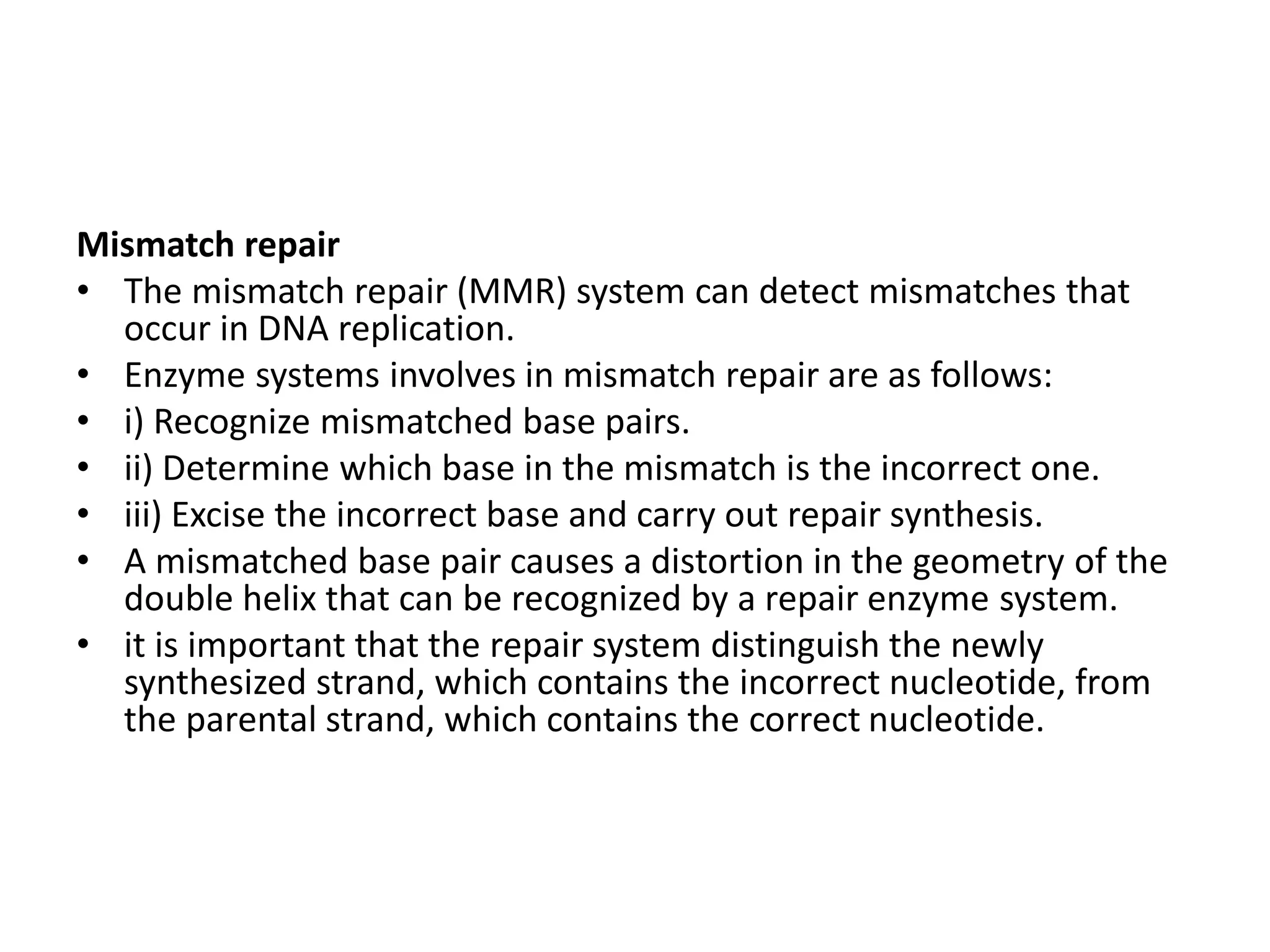
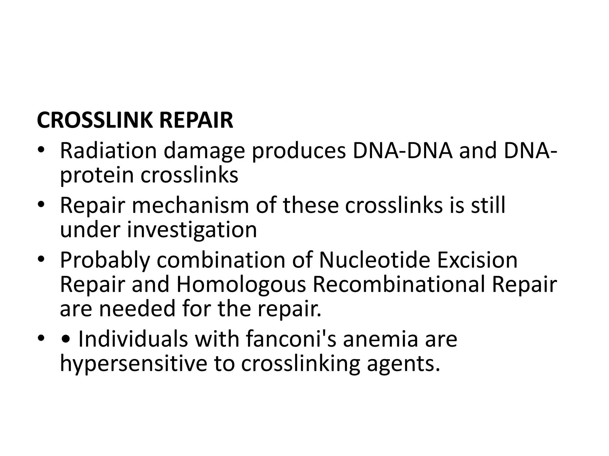
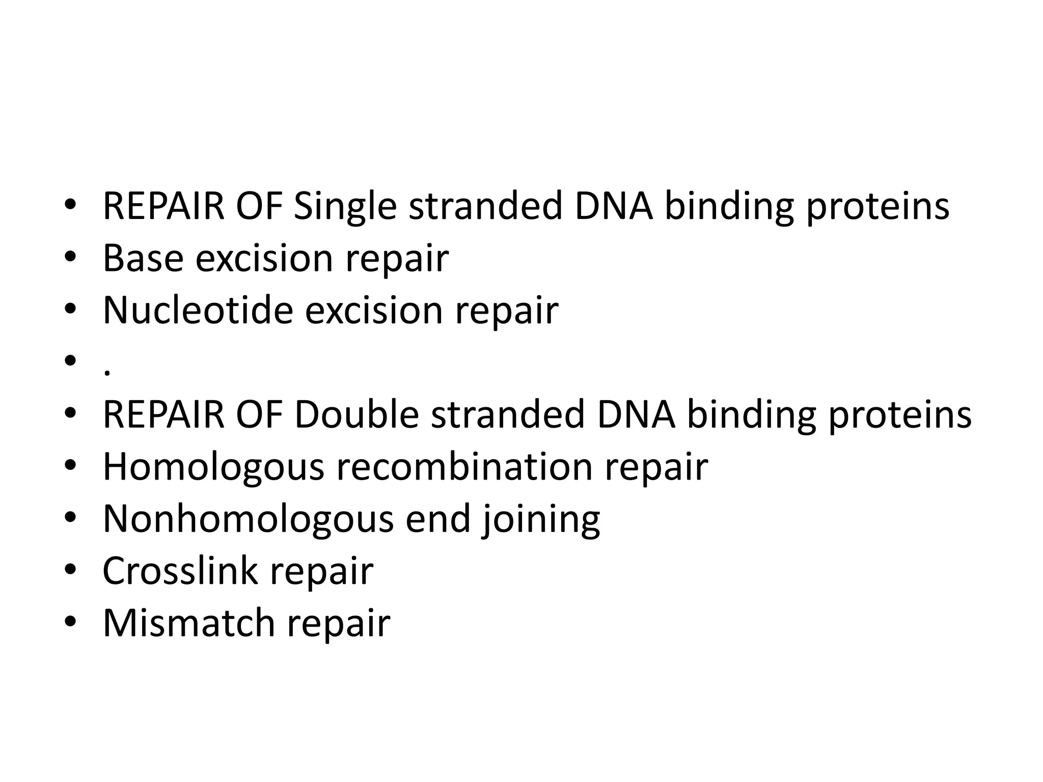
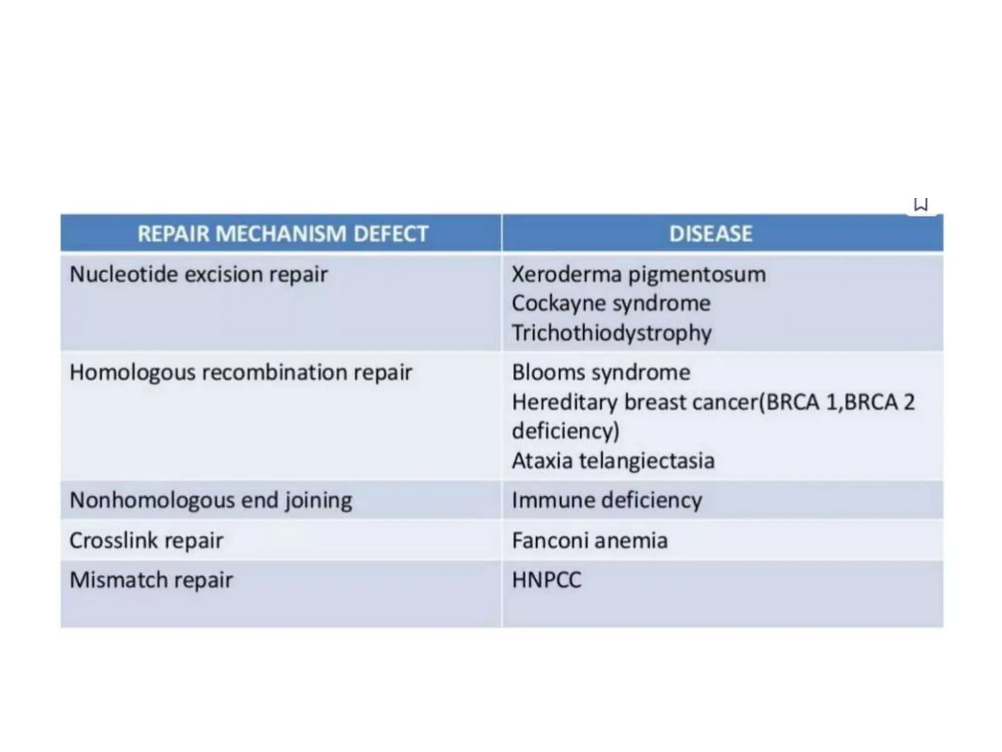
Molecular mechanism of DNA and
Chromosomal damage and repair
Molecular mechanism of DNA
• All organisms duplicate their DNA before each
cell division.DNA replication is semi
conservative because each strand in DNA
double helix act as a template for new
complementary strand and it is semi
discontinous because when one strand is
synthesized continously, the other strand is
synthesized discontinously by Okazaki
fragments.DNA replication is also
bidirectional.
DNA replication
• One of the key molecules in DNA replication is DNA
polymerase. They synthesize DNA and add nucleotide
to the DNA chain including those that are
complementary to the template.
• Replication occurs in a site called origin of replication.
Proteins recognise the site ,bind and open up the DNA.
As the DNA opens two Y shaped structures called
replication forks are formed which forms replication
bubble and moves in opposite direction as replication
proceeds.Single stranded binding proteins coat,
seperated strands of DNA, near replication fork,keeping
them from coming back together into double helix.
DNA Polymerase add the first nucleotide at a new replication
fork with the help of an enzyme called primase. Primase
makes an RNA primer. A typical primer is about five to ten
nucleotides long. The primer primes DNA synthesis, i.e., gets
it started.Once the RNA primer is in place, DNA polymerase
"extends" it, adding nucleotides one by one to make a new
DNA strand that's complementary to the template strand.
• A DNA double helix is always anti-parallel; in other
words, one strand runs in the 5' to 3' direction, while
the other runs in the 3' to 5' direction. This strand is
made continuously, because the DNA polymerase is
moving in the same direction as the replication fork.
This continuously synthesized strand is called the
leading strand.
The other new strand, which runs 5' to 3' away from
the fork, is made in fragments because, as the fork
moves forward, the DNA polymerase (which is moving
away from the fork) must come off and reattach on the
newly exposed DNA. This strand, which is made in
fragments, is called the lagging strand.
DNA Replication
• The small fragments in lagging strand are called Okazaki fragments.
The leading strand can be extended from one primer alone,
whereas the lagging strand needs a new primer for each of the
short Okazaki fragments.
• The sliding clamp protien is a ring-shaped protein and keeps the
DNA polymerase of the lagging strand from floating off when it re-
starts at a new Okazaki fragment .Topoisomerase enzyme also plays
an important maintenance role during DNA replication. It prevents
the DNA double helix ahead of the replication fork from getting too
tightly wound as the DNA is opened up. The RNA primers are
removed and replaced by DNA through the activity of DNA
polymerase I, the other polymerase involved in replication. The
nicks that remain after the primers are replaced get sealed by the
enzyme DNA ligase.
Summary
Chromosomal damage and repair
• DNA damage has been long recognized as causal factor for cancer
development. When erroneous DNA repair leads to mutations or
chromosomal aberrations affecting oncogenes and tumor suppressor
genes, cells undergo malignant transformation resulting in cancerous
growth. Genetic defects can predispose to cancer: mutations in distinct
DNA repair systems elevate the susceptibility to various cancer types.
However, DNA damage not only comprises a root cause for cancer
development but also continues to provide an important avenue for
chemo- and radiotherapy. Since the beginning of cancer therapy,
genotoxic agents that trigger DNA damage checkpoints have been applied
to halt the growth and trigger the apoptotic demise of cancer cells. We
provide an overview about the involvement of DNA repair systems in
cancer prevention and the classes of genotoxins that are commonly used
for the treatment of cancer. A better understanding of the roles and
interactions of the highly complex DNA repair machineries will lead to
important improvements in cancer therapy.
• Types of DNA Damage
• Oxidation, chemotherapy and radiation therapy can all damage
DNA. There are many ways in which this can occur.
• Base Damage: A DNA base is chemically altered.
This may cause a point mutation, or it may predispose to additional
DNA damage.
• Base Mismatch: A mistake during DNA replication leads to insertion
of the wrong base.This will cause a point mutation if it is not
repaired.
• Pyrimidine Dimers: Two adjacent pyrimidine bases are cross-linked
by ultraviolet light. This will cause a point mutation if it is not
repaired prior to replication.
• • Intercalation: This occurs when abnormal chemical groups (such
as chemotherapy drugs) are interposed in the DNA helix. This may
prevent gene function and replication.
• Crosslinking: This occurs when abnormal chemical
bonds are formed within the DNA molecule.
This may prevent gene function and replication, or
cause DNA strand breaks.
• Single strand breaks (SSBs): The sugar backbone is
broken on one strand but not the other. • This is easily
repaired as long as the other strand is still intact.
• Double strand breaks (DSBs): When the DNA is broken
on both strands, it has "sticky ends" that can react with
other DNA strands.
This causes chromatid and chromosome aberrations
that may be mutagenic or lethal.
Measuring DNA Damage
• Peripheral Blood Lymphocyte Assay
Peripheral blood lymphocytes are very sensitive to
radiation, and you can count DNA aberrations in the blood:
• Conventional light microscopy generally measures
unstable aberrations that disappear over days- months.
• Fluorescence in situ hybridization (FISH) can measure
unstable and stable aberrations. Stable aberrations may
persist for years.
• Total-body radiation doses >0.2 Gy will produce
measurable chromosome aberrations in lymphocytes.
• A linear-quadratic mathematical model can be used to
estimate absorbed dose from the number of aberrations.
DNA Repair
• DNA repair is a collection of process by which a cell
identifies and corrects damage to the DNA molecules
that encode its genome.
• The DNA repair processes is constantly active as it
responds to damage in the DNA structure.
• Most cells posses three different categories of DNA
repair systems:
• i) Direct repair
• i) Excision repair
• ii) Mismatch repair
Direct repair
• Direct repair systems act directly on damaged
nucleotides, converting each one back to its original
structure.
• One common type of UV radiation mediated damages,
pyrimidine dimers, are repaired by a light-dependent
direct system called photoreactivation.
• When stimulated by light with a wavelength between
300 and 500 nm, the enzyme binds to pyrimidine
dimers and converts them back to the original
monomeric nucleotides
Excision repair
Excision repair involves the excision of a segment of the
polynucleotide containing a damaged site, followed by
resynthesis of the correct nucleotide sequence by a DNA
polymerase.
These pathways fall into two categories:
1. Base -excision repair
• Base excision repair involves removal of a damaged
nucleotide base, excision of a short piece of the
polynucleotide and resynthesis with a DNA polymerase.
• It is used to repair many minor damage like alkylation and
deamination resulting from exposure to mutagenic agents.
• Enzyme DNA glycosylase initiates the repair process.
• A DNA glycosylase does not cleave phosphodiester bonds,
instead cleave the N- glycosidic bonds, liberating the
altered base and generating apurinic or an apyrimidinic
site, both called as AP sites.
• The resulting AP site is then repaired by an AP
endonuclease repair pathway.
• These enzymes introduce chain breaks by cleaving the
phosphodiester bonds at AP sites.
• This bond cleavage initiates an excision-repair process with
the help of three enzymes_
• Exonuclease
• DNA polymerase I
• DNA ligase
Nucleotide excision repair
• This is similar to base-excision repair, but is not
preceded by the removal of a damaged base and
can act on more substantially damaged areas of
DNA.
• This repair system includes the breaking of a
phosphodiester bond on either side of the lesion,
on the same strand, resulting in the excision of an
oligonucleotide.
• The excision leaves a gap that is filled by repair
systems, and a ligase seals the breaks.
Mismatch repair
• The mismatch repair (MMR) system can detect mismatches that
occur in DNA replication.
• Enzyme systems involves in mismatch repair are as follows:
• i) Recognize mismatched base pairs.
• ii) Determine which base in the mismatch is the incorrect one.
• iii) Excise the incorrect base and carry out repair synthesis.
• A mismatched base pair causes a distortion in the geometry of the
double helix that can be recognized by a repair enzyme system.
• it is important that the repair system distinguish the newly
synthesized strand, which contains the incorrect nucleotide, from
the parental strand, which contains the correct nucleotide.
CROSSLINK REPAIR
• Radiation damage produces DNA-DNA and DNA-
protein crosslinks
• Repair mechanism of these crosslinks is still
under investigation
• Probably combination of Nucleotide Excision
Repair and Homologous Recombinational Repair
are needed for the repair.
• • Individuals with fanconi's anemia are
hypersensitive to crosslinking agents.
• REPAIR OF Single stranded DNA binding proteins
• Base excision repair
• Nucleotide excision repair
• .
• REPAIR OF Double stranded DNA binding proteins
• Homologous recombination repair
• Nonhomologous end joining
• Crosslink repair
• Mismatch repair
CHROMOSOMAL ABERRATIONS
• Aberrations seen at metaphase are of 2 types
Chromosome aberrations
Occurs early in interphase
Before replication
Chromatid aberrations
Occurs in a single chromatid arm after
chromosome replication and leaves the opposite
arm of the same chromosome undamaged leads
to chromatid aberrations.
EXAMPLES OF RADIATION INDUCED
ABERRATIONS
• LETHAL ABERRATIONS
Rings (dicentric & ring are chromosome
aberrations)
Anaphase bridge (chromatid aberration)
• NONLETHAL ABERRATIONS
. Symmetric Translocation. e.g Burkitt lymphoma,
leukemia.
Deletion. e.g deletion of tumour suppressor genes
leads to carcinogenesis
Conclusion
• Investigating DNA damage is essential in the diagnosis and
prognosis of several cancers. The formation of some
important biomarkers in the cells such as micronuclei (MN),
nucleoplasmic bridges (NPB) and nuclear buds (NPB) can be
used as an indicator of DNA damage due to exposure to
cytotoxic or DNA damaging agents.
• Precision medicine is a new type of treatment that plays a
critical role in selecting the most appropriate therapy at the
suitable time, as it only succeeds in targeting the DNA
repair pathway in cancer. This treatment strategy is based
on a unique genetic background, environment and lifestyle
for the individual
• The innovative diagnostic methods including DNA
damage analysis are improving gradually,
especially for precision medicine, and help in
analyzing a large amount of new potential
biomarkers leading to facilitation of the detection
of early disease stages and disease prognosis
• Finally, the application of precision
medicine is the most progressively developed
field which depends on the improvement in DNA
damage analysis and the investigation of novel
markers
• DNA damage occurs on a daily basis by endogenous and exogenous
sources. Distinct DNA repair systems recognize and remove the
lesions. When the damage remains unrepaired DNA damage
checkpoints can halt the cell cycle or induce cellular senescence or
apoptosis. Erroneous repair or replicative bypass of lesions can
result in mutations and chromosomal aberrations. When mutations
affect tumor suppressor genes or oncogenes, cell might transform
into cancer cells. Therefore, DNA repair is essential for preventing
tumor development. However, once a cancer has developed, DNA
damage can be exploited to reduce cancerous growth and evoke
apoptotic demise of cancer cells. Thus, chemo- and radiotherapies
are still today, over 60 years after having been first introduced into
tumor therapy, important strategies to fight cancer.
• Given the central role of genome instability in
triggering and treating cancer, it is likely that
genotoxic treatments will remain an important
avenue of cancer therapy. Also the better
understanding of DNA repair systems will allow
therapies that specifically target selected repair
pathways. It will be of particular importance to
gain a deeper understanding how the various
DNA repair systems interact with each other in
the context of cellular homeostasis and DNA
metabolism in order to optimize targeted
approaches to cancer therapy.
Source – Radiobiology for the radiologist
Eric J Hall
Amato J Giacca
Thank You

Molecular mechanism of DNA,damage n repair.pptx

  • 1.
    Molecular mechanism ofDNA and Chromosomal damage and repair
  • 2.
    Molecular mechanism ofDNA • All organisms duplicate their DNA before each cell division.DNA replication is semi conservative because each strand in DNA double helix act as a template for new complementary strand and it is semi discontinous because when one strand is synthesized continously, the other strand is synthesized discontinously by Okazaki fragments.DNA replication is also bidirectional.
  • 3.
    DNA replication • Oneof the key molecules in DNA replication is DNA polymerase. They synthesize DNA and add nucleotide to the DNA chain including those that are complementary to the template. • Replication occurs in a site called origin of replication. Proteins recognise the site ,bind and open up the DNA. As the DNA opens two Y shaped structures called replication forks are formed which forms replication bubble and moves in opposite direction as replication proceeds.Single stranded binding proteins coat, seperated strands of DNA, near replication fork,keeping them from coming back together into double helix.
  • 5.
    DNA Polymerase addthe first nucleotide at a new replication fork with the help of an enzyme called primase. Primase makes an RNA primer. A typical primer is about five to ten nucleotides long. The primer primes DNA synthesis, i.e., gets it started.Once the RNA primer is in place, DNA polymerase "extends" it, adding nucleotides one by one to make a new DNA strand that's complementary to the template strand.
  • 6.
    • A DNAdouble helix is always anti-parallel; in other words, one strand runs in the 5' to 3' direction, while the other runs in the 3' to 5' direction. This strand is made continuously, because the DNA polymerase is moving in the same direction as the replication fork. This continuously synthesized strand is called the leading strand. The other new strand, which runs 5' to 3' away from the fork, is made in fragments because, as the fork moves forward, the DNA polymerase (which is moving away from the fork) must come off and reattach on the newly exposed DNA. This strand, which is made in fragments, is called the lagging strand.
  • 7.
  • 8.
    • The smallfragments in lagging strand are called Okazaki fragments. The leading strand can be extended from one primer alone, whereas the lagging strand needs a new primer for each of the short Okazaki fragments. • The sliding clamp protien is a ring-shaped protein and keeps the DNA polymerase of the lagging strand from floating off when it re- starts at a new Okazaki fragment .Topoisomerase enzyme also plays an important maintenance role during DNA replication. It prevents the DNA double helix ahead of the replication fork from getting too tightly wound as the DNA is opened up. The RNA primers are removed and replaced by DNA through the activity of DNA polymerase I, the other polymerase involved in replication. The nicks that remain after the primers are replaced get sealed by the enzyme DNA ligase.
  • 9.
  • 11.
    Chromosomal damage andrepair • DNA damage has been long recognized as causal factor for cancer development. When erroneous DNA repair leads to mutations or chromosomal aberrations affecting oncogenes and tumor suppressor genes, cells undergo malignant transformation resulting in cancerous growth. Genetic defects can predispose to cancer: mutations in distinct DNA repair systems elevate the susceptibility to various cancer types. However, DNA damage not only comprises a root cause for cancer development but also continues to provide an important avenue for chemo- and radiotherapy. Since the beginning of cancer therapy, genotoxic agents that trigger DNA damage checkpoints have been applied to halt the growth and trigger the apoptotic demise of cancer cells. We provide an overview about the involvement of DNA repair systems in cancer prevention and the classes of genotoxins that are commonly used for the treatment of cancer. A better understanding of the roles and interactions of the highly complex DNA repair machineries will lead to important improvements in cancer therapy.
  • 13.
    • Types ofDNA Damage • Oxidation, chemotherapy and radiation therapy can all damage DNA. There are many ways in which this can occur. • Base Damage: A DNA base is chemically altered. This may cause a point mutation, or it may predispose to additional DNA damage. • Base Mismatch: A mistake during DNA replication leads to insertion of the wrong base.This will cause a point mutation if it is not repaired. • Pyrimidine Dimers: Two adjacent pyrimidine bases are cross-linked by ultraviolet light. This will cause a point mutation if it is not repaired prior to replication. • • Intercalation: This occurs when abnormal chemical groups (such as chemotherapy drugs) are interposed in the DNA helix. This may prevent gene function and replication.
  • 14.
    • Crosslinking: Thisoccurs when abnormal chemical bonds are formed within the DNA molecule. This may prevent gene function and replication, or cause DNA strand breaks. • Single strand breaks (SSBs): The sugar backbone is broken on one strand but not the other. • This is easily repaired as long as the other strand is still intact. • Double strand breaks (DSBs): When the DNA is broken on both strands, it has "sticky ends" that can react with other DNA strands. This causes chromatid and chromosome aberrations that may be mutagenic or lethal.
  • 15.
    Measuring DNA Damage •Peripheral Blood Lymphocyte Assay Peripheral blood lymphocytes are very sensitive to radiation, and you can count DNA aberrations in the blood: • Conventional light microscopy generally measures unstable aberrations that disappear over days- months. • Fluorescence in situ hybridization (FISH) can measure unstable and stable aberrations. Stable aberrations may persist for years. • Total-body radiation doses >0.2 Gy will produce measurable chromosome aberrations in lymphocytes. • A linear-quadratic mathematical model can be used to estimate absorbed dose from the number of aberrations.
  • 16.
    DNA Repair • DNArepair is a collection of process by which a cell identifies and corrects damage to the DNA molecules that encode its genome. • The DNA repair processes is constantly active as it responds to damage in the DNA structure. • Most cells posses three different categories of DNA repair systems: • i) Direct repair • i) Excision repair • ii) Mismatch repair
  • 17.
    Direct repair • Directrepair systems act directly on damaged nucleotides, converting each one back to its original structure. • One common type of UV radiation mediated damages, pyrimidine dimers, are repaired by a light-dependent direct system called photoreactivation. • When stimulated by light with a wavelength between 300 and 500 nm, the enzyme binds to pyrimidine dimers and converts them back to the original monomeric nucleotides
  • 18.
    Excision repair Excision repairinvolves the excision of a segment of the polynucleotide containing a damaged site, followed by resynthesis of the correct nucleotide sequence by a DNA polymerase. These pathways fall into two categories: 1. Base -excision repair • Base excision repair involves removal of a damaged nucleotide base, excision of a short piece of the polynucleotide and resynthesis with a DNA polymerase. • It is used to repair many minor damage like alkylation and deamination resulting from exposure to mutagenic agents. • Enzyme DNA glycosylase initiates the repair process.
  • 19.
    • A DNAglycosylase does not cleave phosphodiester bonds, instead cleave the N- glycosidic bonds, liberating the altered base and generating apurinic or an apyrimidinic site, both called as AP sites. • The resulting AP site is then repaired by an AP endonuclease repair pathway. • These enzymes introduce chain breaks by cleaving the phosphodiester bonds at AP sites. • This bond cleavage initiates an excision-repair process with the help of three enzymes_ • Exonuclease • DNA polymerase I • DNA ligase
  • 20.
    Nucleotide excision repair •This is similar to base-excision repair, but is not preceded by the removal of a damaged base and can act on more substantially damaged areas of DNA. • This repair system includes the breaking of a phosphodiester bond on either side of the lesion, on the same strand, resulting in the excision of an oligonucleotide. • The excision leaves a gap that is filled by repair systems, and a ligase seals the breaks.
  • 21.
    Mismatch repair • Themismatch repair (MMR) system can detect mismatches that occur in DNA replication. • Enzyme systems involves in mismatch repair are as follows: • i) Recognize mismatched base pairs. • ii) Determine which base in the mismatch is the incorrect one. • iii) Excise the incorrect base and carry out repair synthesis. • A mismatched base pair causes a distortion in the geometry of the double helix that can be recognized by a repair enzyme system. • it is important that the repair system distinguish the newly synthesized strand, which contains the incorrect nucleotide, from the parental strand, which contains the correct nucleotide.
  • 22.
    CROSSLINK REPAIR • Radiationdamage produces DNA-DNA and DNA- protein crosslinks • Repair mechanism of these crosslinks is still under investigation • Probably combination of Nucleotide Excision Repair and Homologous Recombinational Repair are needed for the repair. • • Individuals with fanconi's anemia are hypersensitive to crosslinking agents.
  • 23.
    • REPAIR OFSingle stranded DNA binding proteins • Base excision repair • Nucleotide excision repair • . • REPAIR OF Double stranded DNA binding proteins • Homologous recombination repair • Nonhomologous end joining • Crosslink repair • Mismatch repair
  • 25.
    CHROMOSOMAL ABERRATIONS • Aberrationsseen at metaphase are of 2 types Chromosome aberrations Occurs early in interphase Before replication Chromatid aberrations Occurs in a single chromatid arm after chromosome replication and leaves the opposite arm of the same chromosome undamaged leads to chromatid aberrations.
  • 26.
    EXAMPLES OF RADIATIONINDUCED ABERRATIONS • LETHAL ABERRATIONS Rings (dicentric & ring are chromosome aberrations) Anaphase bridge (chromatid aberration) • NONLETHAL ABERRATIONS . Symmetric Translocation. e.g Burkitt lymphoma, leukemia. Deletion. e.g deletion of tumour suppressor genes leads to carcinogenesis
  • 27.
    Conclusion • Investigating DNAdamage is essential in the diagnosis and prognosis of several cancers. The formation of some important biomarkers in the cells such as micronuclei (MN), nucleoplasmic bridges (NPB) and nuclear buds (NPB) can be used as an indicator of DNA damage due to exposure to cytotoxic or DNA damaging agents. • Precision medicine is a new type of treatment that plays a critical role in selecting the most appropriate therapy at the suitable time, as it only succeeds in targeting the DNA repair pathway in cancer. This treatment strategy is based on a unique genetic background, environment and lifestyle for the individual
  • 28.
    • The innovativediagnostic methods including DNA damage analysis are improving gradually, especially for precision medicine, and help in analyzing a large amount of new potential biomarkers leading to facilitation of the detection of early disease stages and disease prognosis • Finally, the application of precision medicine is the most progressively developed field which depends on the improvement in DNA damage analysis and the investigation of novel markers
  • 29.
    • DNA damageoccurs on a daily basis by endogenous and exogenous sources. Distinct DNA repair systems recognize and remove the lesions. When the damage remains unrepaired DNA damage checkpoints can halt the cell cycle or induce cellular senescence or apoptosis. Erroneous repair or replicative bypass of lesions can result in mutations and chromosomal aberrations. When mutations affect tumor suppressor genes or oncogenes, cell might transform into cancer cells. Therefore, DNA repair is essential for preventing tumor development. However, once a cancer has developed, DNA damage can be exploited to reduce cancerous growth and evoke apoptotic demise of cancer cells. Thus, chemo- and radiotherapies are still today, over 60 years after having been first introduced into tumor therapy, important strategies to fight cancer.
  • 30.
    • Given thecentral role of genome instability in triggering and treating cancer, it is likely that genotoxic treatments will remain an important avenue of cancer therapy. Also the better understanding of DNA repair systems will allow therapies that specifically target selected repair pathways. It will be of particular importance to gain a deeper understanding how the various DNA repair systems interact with each other in the context of cellular homeostasis and DNA metabolism in order to optimize targeted approaches to cancer therapy.
  • 31.
    Source – Radiobiologyfor the radiologist Eric J Hall Amato J Giacca
  • 32.