The document is a revision aid titled 'Mind Maps for Medical Students' designed to assist medical students in memorizing essential clinical facts through over 100 systematically organized mind maps. It emphasizes the flexibility and adaptability of the maps for various study approaches and is intended to complement traditional textbooks. The author, Olivia Smith, is a medical student who aims to simplify complex medical knowledge for effective revision, particularly in preparation for exams.


![CRC Press
Taylor & Francis Group
6000 Broken Sound Parkway NW, Suite 300
Boca Raton, FL 33487-2742
© 2015 by Taylor & Francis Group, LLC
CRC Press is an imprint of Taylor & Francis Group, an Informa business
No claim to original U.S. Government works
Version Date: 20141104
International Standard Book Number-13: 978-1-4822-5032-9 (eBook - PDF)
This book contains information obtained from authentic and highly regarded sources. While all reasonable efforts have
been made to publish reliable data and information, neither the author[s] nor the publisher can accept any legal responsibil-
ity or liability for any errors or omissions that may be made. The publishers wish to make clear that any views or opinions
expressed in this book by individual editors, authors or contributors are personal to them and do not necessarily reflect
the views/opinions of the publishers. The information or guidance contained in this book is intended for use by medical,
scientific or health-care professionals and is provided strictly as a supplement to the medical or other professional’s own
judgement, their knowledge of the patient’s medical history, relevant manufacturer’s instructions and the appropriate best
practice guidelines. Because of the rapid advances in medical science, any information or advice on dosages, procedures or
diagnoses should be independently verified. The reader is strongly urged to consult the relevant national drug formulary and
the drug companies’ printed instructions, and their websites, before administering any of the drugs recommended in this
book. This book does not indicate whether a particular treatment is appropriate or suitable for a particular individual. Ulti-
mately it is the sole responsibility of the medical professional to make his or her own professional judgements, so as to advise
and treat patients appropriately. The authors and publishers have also attempted to trace the copyright holders of all material
reproduced in this publication and apologize to copyright holders if permission to publish in this form has not been obtained.
If any copyright material has not been acknowledged please write and let us know so we may rectify in any future reprint.
Except as permitted under U.S. Copyright Law, no part of this book may be reprinted, reproduced, transmitted, or utilized in
any form by any electronic, mechanical, or other means, now known or hereafter invented, including photocopying, micro-
filming, and recording, or in any information storage or retrieval system, without written permission from the publishers.
For permission to photocopy or use material electronically from this work, please access www.copyright.com (http://www.
copyright.com/) or contact the Copyright Clearance Center, Inc. (CCC), 222 Rosewood Drive, Danvers, MA 01923, 978-750-
8400. CCC is a not-for-profit organization that provides licenses and registration for a variety of users. For organizations that
have been granted a photocopy license by the CCC, a separate system of payment has been arranged.
Trademark Notice: Product or corporate names may be trademarks or registered trademarks, and are used only for identi-
fication and explanation without intent to infringe.
Visit the Taylor & Francis Web site at
http://www.taylorandfrancis.com
and the CRC Press Web site at
http://www.crcpress.com](https://image.slidesharecdn.com/mindmap-250108080425-ca023b05/75/Mind-Map-for-medical-students-guide-and-theory-5-2048.jpg)


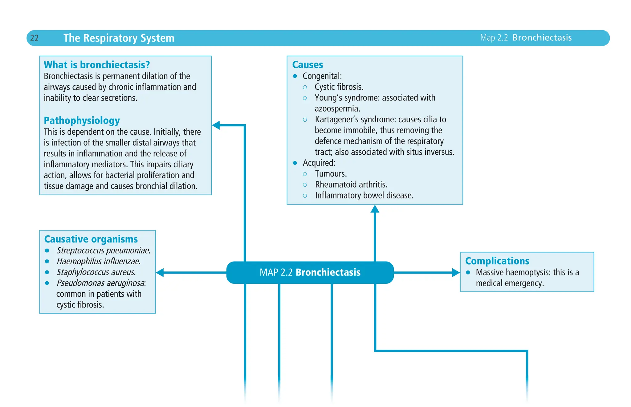
![7 The Cardiovascular System
Complications
Remember this as C PEAR DROP:
• Cardiogenic shock, Cardiac arrhythmia.
N.B.Atrial fibrillation (AF) increases a patient’s risk
of stroke.AF presents with an irregularly irregular
pulse and an ECG with absent P waves, irregular
RR intervals, an undulating baseline and narrow
QRS complexes. Start anticoagulation therapy.
• Pericarditis.
• Emboli.
• Aneurysm formation.
• Rupture of ventricle.
• Dressler’s syndrome: an autoimmune pericarditis
that develops 2–10 weeks post MI.This is a triad
of: 1) fever; 2) pleuritic pain; 3) pericardial effusion.
• Rupture of free wall.
• O
• Papillary muscle rupture.
Treatment
• Conservative: lifestyle measures such
as smoking cessation and increased excercise.
• Medical – MONA B for immediate management:
○ Morphine.
○ Oxygen (if hypoxic).
○ Nitrates (glyceryl trinitrate [GTN]).
○ Anticoagulants, e.g. aspirin and an antiemetic.
○ Beta-blockers if no contraindication.
On discharge all patients should be prescribed:
aspirin, an angiotensin converting enzyme (ACE)
inhibitor, a beta-blocker (if no contraindication; calcium
channel blockers are good alternatives) and a statin.
• Surgical: reperfusion with PCI if STEMI. PCI may
also be used in NSTEMI but if NSTEMI patients
are not having immediate PCI, fondaparinux (a
factor Xa inhibitor) or a low molecular weight
heparin (LMWH) may be given subcutaneously.
Map 1.3 Myocardial Infarction (MI)
Chapter_01.indd
7
08/12/14
5:41
PM](https://image.slidesharecdn.com/mindmap-250108080425-ca023b05/75/Mind-Map-for-medical-students-guide-and-theory-22-2048.jpg)


![67 The Renal System
Cystic diseases of the renal
medulla
Remember NAMS:
• Nephronophthisis medullary cystic
disease.
• Acquired cystic disease: usually
from dialysis.
• Medullary sponge kidney.
• Simple cysts.
Treatment
• Conservative: patient support.
• Medical:
○ Treat hypertension.
○ Antibiotic therapy for urinary trait infection
(UTI).
• Surgical: cyst decompression.
Complications
• Development of chronic kidney injury.
• Remember LAMB:
○ Liver cysts.
○ Aneurysms.
○ Mitral valve prolapse.
○ Berry aneurysm rupture leading to
subarachnoid haemorrhage.
Treatment
• Conservative: parental and patient support.
• Medical:
○ Ventilation and long-term oxygen therapy.
○ Treat hypertension (angiotensin converting
enzyme [ACE] inhibitors).
○ Antibiotics for UTI.
○ Diuretics for fluid overload.
• Surgical:
○ Nephrectomy.
○ Combined renal and liver transplant.
Complications
• Hepatic cysts.
• Congenital hepatic fibrosis.
• Proliferative bile ducts.
Map 4.6 Cystic Disease
Chapter_04.indd
67
08/12/14
5:56
PM](https://image.slidesharecdn.com/mindmap-250108080425-ca023b05/75/Mind-Map-for-medical-students-guide-and-theory-82-2048.jpg)


![Haematology
100 Map 6.4 Leukaemia
Causes
Neoplasm Cause
ALL Possibly a genetic susceptibility
coupled with an environmental
trigger
Exact cause unknown
Comment
Commonest cancer in children
Often spreads to central nervous system
Associations – DIP:
• Down’s syndrome
• Ionising radiation
• Pregnancy
Usually affects adults over 60 years old
Affects B lymphocytes
Positive ZAP-70 marker is associated with
a worse prognosis
CLL
AML Exact cause unknown
Risk factors include:
• Myeloproliferative disease
• Alkylating agents
• Ionising radiation exposure
• Down’s syndrome
Commonest leukaemia in adults
Rapidly progressing
Auer rods on microscopy are
diagnostic
CML Exact cause unknown
Risk factor: ionising radiation
exposure
Usually affects males 40–60 years old
80% associated with the Philadelphia
chromosome t[9;22], forming
bcr-abl fusion gene
Signs and symptoms
Neoplasm Clinical features
ALL Bone marrow failure
Bruising
Shortness of breath
Purpura
Malaise
Weight loss
Night sweats
Asymptomatic
Bone marrow failure
Nontender lymphadenopathy
Hepatosplenomegaly
Malaise
Weight loss
Night sweats
CLL
What is leukaemia?
This is a rare neoplasm of the blood or bone marrow.
It is classified into lymphoid and myeloid neoplasms
that may present chronically or acutely.These 4
classifications are:
1 Acute lymphoblastic leukaemia (ALL).
2 Chronic lymphocytic leukaemia (CLL).
3 Acute myeloid leukaemia (AML).
4 Chronic myeloid leukaemia (CML).
Chapter_06.indd
100
08/12/14
6:16
PM](https://image.slidesharecdn.com/mindmap-250108080425-ca023b05/75/Mind-Map-for-medical-students-guide-and-theory-115-2048.jpg)

![Haematology
102 Map 6.5 Hodgkin’s vs. Non-Hodgkin’s Lymphoma
HODGKIN’S LYMPHOMA
What is hodgkin’s lymphoma?
This is a group of uncommon malignancies; the 4 most common histological
subtypes are:
1 Lymphocyte-predominant.
2 Nodular sclerosing.
3 Mixed cellularity.
4 Lymphocyte-depleted.
Cause
Exact cause is unknown.
Risk factors include:
• Male sex.
• Infection with Epstein–Barr virus (EBV).
• Immunosuppression, e.g. HIV patients.
• Exotoxin exposure.
Signs and symptoms
• Painless lymphadenopathy.
• Unintentional weight loss.
• Fever (constitutional ‘B signs’: fever 38˚C, night sweats, weight loss).
B cell neoplasms T cell neoplasms
Burkitt’s lymphoma:
• Associated with EBV
• t[8;14]
Diffuse large B cell lymphoma
Mantle cell lymphoma: t[11;14]
Follicular lymphoma:
• t[14;18]
• bcl-2 expression
Adult T cell lymphoma; caused by human
T-lymphotrophic virus-1 (HTLV-1)
Sézary syndrome
NON-HODGKIN’S LYMPHOMA
What is non-Hodgkin’s lymphoma?
This is a group of malignancies that are either B cell or T cell in origin.
Cause
Exact cause is unknown.
Risk factors include:
• Male sex.
• Infection, e.g. EBV, Helicobacter pylori, human herpes virus (HHV)-8,
hepatitis C.
• Immunosuppression, e.g. HIV patients.
MAP 6.5
Hodgkin’s vs. Non-Hodgkin’s lymphoma
Chapter_06.indd
102
08/12/14
6:16
PM](https://image.slidesharecdn.com/mindmap-250108080425-ca023b05/75/Mind-Map-for-medical-students-guide-and-theory-117-2048.jpg)


![161 Musculoskeletal System Map 10.3 Spondyloarthropathies
Investigations
• Psoriasis is a clinical diagnosis.
• Bloods: seronegative for rheumatoid factor.
• Radiology: ‘Pencil-in-cup’ deformity on hand X-ray. X-ray of affected
joints to assess severity.
Treatment
• Conservative: patient education. Refer to physiotherapy. Explain to
patients that psoriasis does not have a cure and control of the disease
is more realistic.
• Medical: analgesia (nonsteroidal anti-inflammatory drugs [NSAIDs]) and
disease modifying antirheumatic drugs (DMARDs), e.g. methotrexate
(first line). Manage psoriasis.
• Surgery: rarely joint replacement.
Complications
• Neurological manifestations if atlanto–axial joint involvement.
• Joint destruction.
ANKYLOSING SPONDYLITIS
What is ankylosing spondylitis?
This is a chronic inflammatory disease of the spine and sacroiliac joints.There is
predominance in young males and the condition is associated with HLA B27
(positive in 95%).
Causes
The exact cause and pathophysiology of this condition are not known.
However, it is thought to be associated with HLA B27.
Signs and symptoms
• Question mark posture.
• Bamboo spine: due to calcification of ligaments.
• Pain and stiffness: symptoms improve with exercise.
Investigations
• Bloods: seronegative for rheumatoid factor.
• Radiology: CXR and MRI scan assess changes in the spine.
Treatment
• Conservative: patient education. Refer to physiotherapy.
• Medical: analgesia (NSAIDs) and DMARDs, e.g. sulphasalzine (first line).
• Surgery: corrective spinal surgery.
Complications
• Osteoprosis.
• Spinal fractures.
• Increased risk of cardiovascular disease, e.g. stroke and myocardial infarction.
Chapter_10.indd
161
08/12/14
7:02
PM](https://image.slidesharecdn.com/mindmap-250108080425-ca023b05/75/Mind-Map-for-medical-students-guide-and-theory-176-2048.jpg)


