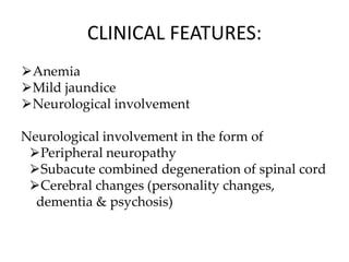
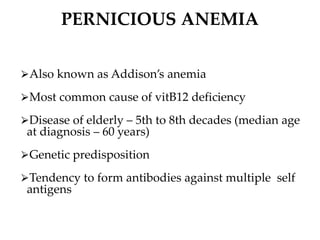
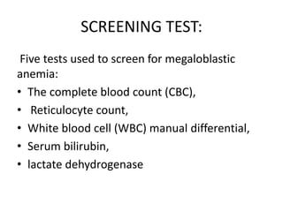
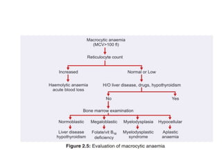
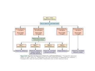
MEGALOBLASTIC ANEMIA
Dr. Pooja Chaudhary
Definition :
Megaloblastic anemias are disorders caused
by
• Impaired DNA
synthesis due to
the substances of
DNA synthesis
deficiency, such
as folic acid and
Vit B 12.
• It is characterized by
the presence of
megaloblastic cells in
the bone marrow and
macrocytic anemia
AETIOLOGY
• Vitamin B₁₂ deficiency or defective metabolism
• Folate deficiency or defective metabolism
• Drug induced ( Purine and Pyramidine
analogs)
1)Vitamin B12 deficiency
• Dietary deficiency - strict vegetarian diet
• Malabsorption - Ileal resection,
blind loop syndrome,
inflammatory bowel disease
Fish tapeworm infestation
Bacterial overgrowth in malformed small intestine
Tropical sprue
Non-tropical sprue
• Increased requirements – Pregnancy
- Disseminated cancer
• Intrinsic factor deficiency – Gastrectomy
- Pernicious anemia
2. Folic acid deficiency
• Dietary deficiency - Unbalanced diet
- Alcoholics
- Malnutrition
Impaired absorption – Sprue
- Small bowel resection
- Anticonvulsants
Increased requirements — Infancy
- Pregnancy
- Hemolytic anemias
- Myeloproliferative disorders
3)Drug-induced suppression of DNA
synthesis
• Folate antagonists
• Alkylating agents
• Metabolic inhibitors - Of purine synthesis
- Of pyrimidine synthesis:
- 0f cobalamine
metabolism :phenformin, metformin
- Of dihydrofolate
Hydroxyurea
4)Inborn errors of metabolism
• Defective folate metabolism
• Defective vitamin B12 metabolism
• Hereditary orotic aciduria
• Lesch-Nyhan syndrome
VIT. B12 METABOLISM
DIETERY SOURCE OF VITAMIN B12
DIETERY SOURCE OF FOLIC ACID
⮚Folic acid
⮚Adult men and
women-400
microgram
⮚Pregnancy
-50-100 µg
⮚Lactation
-500- 800 µg
Vitamin B12
● Daily Requirement =
2 to 3 ug/day
Other causes of megaloblastosis
Myelodysplastic syndrome
Acute erythroid leukemia
Congenital dyserythropoietic anemia
Reverse transcriptase inhibitors
Defect in Megaloblastic Anemia Caused By
Deficiency in Folate and Vitamin B12:
Conversion of Methylmalonyl
CoA to Succinyl CoA
Conversion of Methylmalonyl
CoA to Succinyl CoA
⮚Adenosylcobalamine acts as a cofactor
⮚In deficiency of B12 –
Propionyl CoA accumulates
Acts as a primer for fatty acid synthesis
⮚Production of fatty acids with odd no. of
carbon atoms
⮚Synthesis of abnormal myelin lipids
⮚Myelin degradation and neurological
abnormalities
CLINICAL FEATURES:
⮚Anemia
⮚Mild jaundice
⮚Neurological involvement
Neurological involvement in the form of
⮚Peripheral neuropathy
⮚Subacute combined degeneration of spinal cord
⮚Cerebral changes (personality changes,
dementia & psychosis)
PERNICIOUS ANEMIA
⮚Also known as Addison’s anemia
⮚Most common cause of vitB12 deficiency
⮚Disease of elderly – 5th to 8th decades (median age
at diagnosis – 60 years)
⮚Genetic predisposition
⮚Tendency to form antibodies against multiple self
antigens
PATHOGENESIS
⮚Immunologically mediated, autoimmune destruction of
gastric mucosa
⮚CHRONIC ATROPHIC GASTRITIS – marked loss of parietal
cells
⮚TYPES OF ANTIBODIES
1.Antibodies against Parietal cells -90%
2.Antibodies against Intrinsic Factor
-Blocking antibodies – prevent formation of IF-
Cobalamin complex
-Binding antibodies – React with IF-Cobalamin
binding site ,preventing its absorption
LAB DIAGNOSIS
SCREENING TEST:
Five tests used to screen for megaloblastic
anemia:
• The complete blood count (CBC),
• Reticulocyte count,
• White blood cell (WBC) manual differential,
• Serum bilirubin,
• lactate dehydrogenase
Haemoglobin : Decreased
Red cells : macrocytosis is seen.
In severe anaemia:
marked anisopoikilocytosis ,
basophilic stippling,
Howell-Jolly bodies,
Cabot’s rings.
Late or intermediate erythroblasts with fine, open
nuclear chromatin (megaloblasts) may be seen.
Retic count : Low to normal
Indices : Elevated MCV ( >120fl ),
elevated MCH ( >50pg)
MCHC normal
Leucocytes : May be reduced.
Presence of Hypersegmented Neutrophil is
characteristic.
Platelets : Moderately reduced
Specific Diagnostic Tests:
• Bone Marrow Examination
• Assays for Folate, Vitamin B12, Methylmalonic
Acid, and Homocysteine
• Gastric Analysis and Serum Gastrin
• Antibody Assays
• Holotranscobalamin Assay
• Stool Analysis for Parasites
• Shilling test
• Figlu test
• BONE MARROW EXAMINATION:
• a) Marrow cellularity : Marrow is Hypercellular
with a decreased myeloid : erythroid ratio (from
3:1 to 1:1)
• b) Erythropoiesis : Erythroid Hyperplasia is due
to characteristic megaloblastic erythropoeisis.
• c) Orthochromatic features : sieve like nucleus
and haemoglobinized cytoplasm and mitotic
figures seen
• d) Dyserythropoiesis : nuclear remnants, bi- and
trinucleated cells and dying cells
• Nuclear-cytoplasmic asynchrony.
• The nucleus lags behind.
• This asynchrony is most striking at the stage of
the polychromatic normoblast.
Other cells :
Howell Jolly bodies, Band cells, Giant
metamyelocytes, Megakaryocytes may be
increased with pseudohyperdiploidy and
agranular cytoplasm.
Marrow Iron : Increase in number and size of iron
granules in erythroid precursors. Ringed
sideroblasts are rare.
Chromosomes : Random chromosomal
abnormalities such as chromosomal breaks or
centromere spreading may be seen.
SCHILLING TEST
For evaluation of absorption of vitamin B12 in the GIT
Performed in 2 parts – part 1 and part 2
Part 1 :
0.5 to 1 µg of radiolabelled vitamin B12 is given orally
After 2 hrs IM dose (1000 µg) of unlabelled vitamin B12
is given saturates binding sites of TC I and TC II and
displaces any bound radiolabelled vitamin B12 (thus
permitting urinary excretion of absorbed radiolabelled
vitamin B12 )
Radioactivity is measured in subsequently
collected 24 hr urine sample and expressed as a
% of total oral dose
In normal persons, > 7% of the oral dose of
vitamin B12 is excreted in urine
If excretion is less than normal it indicates
impaired absorption, which may be due to
either lack of IF or small intestinal
malabsorption
Part 2 performed if part 1 of test is abnormal
Part 2 : patient is orally administered
radiolabelled vitamin B12 along with IF
while remainder of test is carried out out as
in part 1
Excretion becomes normal – lack of IF
Excretion remains below normal – defective
absorption in small intestine
Formiminoglutamate (FIGLU )
excretion test
⮚ 15 gm oral dose of histidine is given.
⮚ Urinary excretion of FIGLU is measured spectrophotometrically.
⮚Histidine in presence of adenosylcobalamine form FIGLU in
presence of terahydrofolic acid form Glutamic acid.
⮚Folic acid deficiency results in inability to degrade a
formiminoglutamic acid (FIGLU) to glutamic acid
⮚FIGLU accumulates in excessive amounts and is excreted in the
urine.
• Holotranscobalamin Assay:
• Rapid immunoassays using monoclonal
antibodies specific for holotranscobalamin
have been developed in the past several years
that are both sensitive and specific.
• Stool Analysis for Parasites:
• Eggs or proglottids of the fish tapeworm D.
latum
TREATMENT OF
MEGALOBLASTIC ANEMIA
Vitamin B12 deficiency
Initial dosage: 1000
micrograms of
hydroxycobalamin IM
injection daily for one
week
Maintenance dosage
1000μg IM once every 3
months
Folate deficiency
Folic acid 5mg daily
⮚After initiation of therapy
reticulocyte count begins to increase
around 3rd day – peak by 6th or 7th day –
gradually returns to normal by end of 3rd
week
⮚Hematocrit steadily rises and normalises
in about 1-2 months
• Within 4 to 6 hours after the initial therapy (if
parenteral), the marrow shows decreased early
megaloblasts and the appearance of
pronormoblasts.
• Within 2–4 days, the marrow is predominantly
normoblastic.
• Granulocytic abnormalities return to normal
more slowly, and hypersegmented neutrophils
disappear from the blood only after 12–14 days.
• Pernicious anemia: parenterally with 1000 µg
of cyanocobalamin daily for 1 week,
• twice weekly for the second week,
• once weekly for 4 weeks,
• then monthly for the lifetime of the patient
• Cobalamin deficiency must be excluded and
corrected if present, to avoid the occurrence
of neuropathies of cobalamin deficiency.
• Supplemental dietary folic acid during
pregnancy it reduce the incidence of neural
tube defects in the baby.
REFERENCES
• Robbins Basic Pathology, 10th edition
• Rodak’s Hematology Clinical Principles and
Applications, 6th Edition
• Henry’s Clinical Diagnosis and
Management,22nd Edition
• Dr. Tejinder Singh’s – Atlas and Text of
Hematology , 4th Edition
THANK YOU

MEGALOBLASTIC ANEMIA.pptx by pathology

  • 1.
  • 2.
    Definition : Megaloblastic anemiasare disorders caused by • Impaired DNA synthesis due to the substances of DNA synthesis deficiency, such as folic acid and Vit B 12. • It is characterized by the presence of megaloblastic cells in the bone marrow and macrocytic anemia
  • 3.
    AETIOLOGY • Vitamin B₁₂deficiency or defective metabolism • Folate deficiency or defective metabolism • Drug induced ( Purine and Pyramidine analogs)
  • 4.
    1)Vitamin B12 deficiency •Dietary deficiency - strict vegetarian diet • Malabsorption - Ileal resection, blind loop syndrome, inflammatory bowel disease Fish tapeworm infestation Bacterial overgrowth in malformed small intestine Tropical sprue Non-tropical sprue • Increased requirements – Pregnancy - Disseminated cancer • Intrinsic factor deficiency – Gastrectomy - Pernicious anemia
  • 5.
    2. Folic aciddeficiency • Dietary deficiency - Unbalanced diet - Alcoholics - Malnutrition Impaired absorption – Sprue - Small bowel resection - Anticonvulsants Increased requirements — Infancy - Pregnancy - Hemolytic anemias - Myeloproliferative disorders
  • 6.
    3)Drug-induced suppression ofDNA synthesis • Folate antagonists • Alkylating agents • Metabolic inhibitors - Of purine synthesis - Of pyrimidine synthesis: - 0f cobalamine metabolism :phenformin, metformin - Of dihydrofolate Hydroxyurea
  • 7.
    4)Inborn errors ofmetabolism • Defective folate metabolism • Defective vitamin B12 metabolism • Hereditary orotic aciduria • Lesch-Nyhan syndrome
  • 8.
  • 9.
    DIETERY SOURCE OFVITAMIN B12
  • 10.
  • 11.
    ⮚Folic acid ⮚Adult menand women-400 microgram ⮚Pregnancy -50-100 µg ⮚Lactation -500- 800 µg Vitamin B12 ● Daily Requirement = 2 to 3 ug/day
  • 12.
    Other causes ofmegaloblastosis Myelodysplastic syndrome Acute erythroid leukemia Congenital dyserythropoietic anemia Reverse transcriptase inhibitors
  • 13.
    Defect in MegaloblasticAnemia Caused By Deficiency in Folate and Vitamin B12:
  • 14.
    Conversion of Methylmalonyl CoAto Succinyl CoA Conversion of Methylmalonyl CoA to Succinyl CoA ⮚Adenosylcobalamine acts as a cofactor ⮚In deficiency of B12 – Propionyl CoA accumulates Acts as a primer for fatty acid synthesis ⮚Production of fatty acids with odd no. of carbon atoms ⮚Synthesis of abnormal myelin lipids ⮚Myelin degradation and neurological abnormalities
  • 15.
    CLINICAL FEATURES: ⮚Anemia ⮚Mild jaundice ⮚Neurologicalinvolvement Neurological involvement in the form of ⮚Peripheral neuropathy ⮚Subacute combined degeneration of spinal cord ⮚Cerebral changes (personality changes, dementia & psychosis)
  • 16.
    PERNICIOUS ANEMIA ⮚Also knownas Addison’s anemia ⮚Most common cause of vitB12 deficiency ⮚Disease of elderly – 5th to 8th decades (median age at diagnosis – 60 years) ⮚Genetic predisposition ⮚Tendency to form antibodies against multiple self antigens
  • 17.
    PATHOGENESIS ⮚Immunologically mediated, autoimmunedestruction of gastric mucosa ⮚CHRONIC ATROPHIC GASTRITIS – marked loss of parietal cells ⮚TYPES OF ANTIBODIES 1.Antibodies against Parietal cells -90% 2.Antibodies against Intrinsic Factor -Blocking antibodies – prevent formation of IF- Cobalamin complex -Binding antibodies – React with IF-Cobalamin binding site ,preventing its absorption
  • 20.
  • 21.
    SCREENING TEST: Five testsused to screen for megaloblastic anemia: • The complete blood count (CBC), • Reticulocyte count, • White blood cell (WBC) manual differential, • Serum bilirubin, • lactate dehydrogenase
  • 23.
    Haemoglobin : Decreased Redcells : macrocytosis is seen. In severe anaemia: marked anisopoikilocytosis , basophilic stippling, Howell-Jolly bodies, Cabot’s rings. Late or intermediate erythroblasts with fine, open nuclear chromatin (megaloblasts) may be seen.
  • 24.
    Retic count :Low to normal Indices : Elevated MCV ( >120fl ), elevated MCH ( >50pg) MCHC normal
  • 27.
    Leucocytes : Maybe reduced. Presence of Hypersegmented Neutrophil is characteristic. Platelets : Moderately reduced
  • 28.
    Specific Diagnostic Tests: •Bone Marrow Examination • Assays for Folate, Vitamin B12, Methylmalonic Acid, and Homocysteine • Gastric Analysis and Serum Gastrin • Antibody Assays • Holotranscobalamin Assay • Stool Analysis for Parasites • Shilling test • Figlu test
  • 29.
    • BONE MARROWEXAMINATION: • a) Marrow cellularity : Marrow is Hypercellular with a decreased myeloid : erythroid ratio (from 3:1 to 1:1) • b) Erythropoiesis : Erythroid Hyperplasia is due to characteristic megaloblastic erythropoeisis. • c) Orthochromatic features : sieve like nucleus and haemoglobinized cytoplasm and mitotic figures seen • d) Dyserythropoiesis : nuclear remnants, bi- and trinucleated cells and dying cells
  • 30.
    • Nuclear-cytoplasmic asynchrony. •The nucleus lags behind. • This asynchrony is most striking at the stage of the polychromatic normoblast.
  • 32.
    Other cells : HowellJolly bodies, Band cells, Giant metamyelocytes, Megakaryocytes may be increased with pseudohyperdiploidy and agranular cytoplasm. Marrow Iron : Increase in number and size of iron granules in erythroid precursors. Ringed sideroblasts are rare. Chromosomes : Random chromosomal abnormalities such as chromosomal breaks or centromere spreading may be seen.
  • 35.
    SCHILLING TEST For evaluationof absorption of vitamin B12 in the GIT Performed in 2 parts – part 1 and part 2 Part 1 : 0.5 to 1 µg of radiolabelled vitamin B12 is given orally After 2 hrs IM dose (1000 µg) of unlabelled vitamin B12 is given saturates binding sites of TC I and TC II and displaces any bound radiolabelled vitamin B12 (thus permitting urinary excretion of absorbed radiolabelled vitamin B12 )
  • 36.
    Radioactivity is measuredin subsequently collected 24 hr urine sample and expressed as a % of total oral dose In normal persons, > 7% of the oral dose of vitamin B12 is excreted in urine If excretion is less than normal it indicates impaired absorption, which may be due to either lack of IF or small intestinal malabsorption Part 2 performed if part 1 of test is abnormal
  • 37.
    Part 2 :patient is orally administered radiolabelled vitamin B12 along with IF while remainder of test is carried out out as in part 1 Excretion becomes normal – lack of IF Excretion remains below normal – defective absorption in small intestine
  • 39.
    Formiminoglutamate (FIGLU ) excretiontest ⮚ 15 gm oral dose of histidine is given. ⮚ Urinary excretion of FIGLU is measured spectrophotometrically. ⮚Histidine in presence of adenosylcobalamine form FIGLU in presence of terahydrofolic acid form Glutamic acid. ⮚Folic acid deficiency results in inability to degrade a formiminoglutamic acid (FIGLU) to glutamic acid ⮚FIGLU accumulates in excessive amounts and is excreted in the urine.
  • 42.
    • Holotranscobalamin Assay: •Rapid immunoassays using monoclonal antibodies specific for holotranscobalamin have been developed in the past several years that are both sensitive and specific. • Stool Analysis for Parasites: • Eggs or proglottids of the fish tapeworm D. latum
  • 43.
    TREATMENT OF MEGALOBLASTIC ANEMIA VitaminB12 deficiency Initial dosage: 1000 micrograms of hydroxycobalamin IM injection daily for one week Maintenance dosage 1000μg IM once every 3 months Folate deficiency Folic acid 5mg daily
  • 44.
    ⮚After initiation oftherapy reticulocyte count begins to increase around 3rd day – peak by 6th or 7th day – gradually returns to normal by end of 3rd week ⮚Hematocrit steadily rises and normalises in about 1-2 months
  • 45.
    • Within 4to 6 hours after the initial therapy (if parenteral), the marrow shows decreased early megaloblasts and the appearance of pronormoblasts. • Within 2–4 days, the marrow is predominantly normoblastic. • Granulocytic abnormalities return to normal more slowly, and hypersegmented neutrophils disappear from the blood only after 12–14 days.
  • 46.
    • Pernicious anemia:parenterally with 1000 µg of cyanocobalamin daily for 1 week, • twice weekly for the second week, • once weekly for 4 weeks, • then monthly for the lifetime of the patient
  • 47.
    • Cobalamin deficiencymust be excluded and corrected if present, to avoid the occurrence of neuropathies of cobalamin deficiency. • Supplemental dietary folic acid during pregnancy it reduce the incidence of neural tube defects in the baby.
  • 48.
    REFERENCES • Robbins BasicPathology, 10th edition • Rodak’s Hematology Clinical Principles and Applications, 6th Edition • Henry’s Clinical Diagnosis and Management,22nd Edition • Dr. Tejinder Singh’s – Atlas and Text of Hematology , 4th Edition
  • 49.

Editor's Notes

  • #6 Alcoholics ( bioavailability of FA and folate dependent biochemicals is affected)
  • #7  purine synthesis: hydroxyurea, acyclovir 6-mercaptopurine,,,,,, Of pyrimidine synthesis: 5-fluorouracil……so we add b12 in dm treatment
  • #8 Orotic aciduria-def of uridine monophosphate……LN SYNDROME HGPRT DEF IN THIS OVERPRODUCTION OF URIC ACID UTILISE FOLIC ACID
  • #9 Cubilin receptors…bind with transcobalamin….bone marrow pronormoblast …methyl coba-is coenzyme support homocystn methionine convrsn….hepatocyte transcob receptor bind cbl-tc and release the cbl
  • #14 DUMP-DEOXYURIDINE MONOPHOSPHATE, DTMP-DEOXYTHYMIDINE MONOPHOSPHATE, DTTP-DEOXYTHYMIDINETRIPHOSPHATE
  • #31 Nuclear-cytoplasmic asynchrony in which the cytoplasm matures as expected with increasing pinkness as hemoglobin accumulates, nucleus lags behind, appearing younger than expected for the degree of maturity of the cytoplasm. Polychro..cytoplasm..pinkgreyish…
  • #32 Proerythro-basophilic erythroblast-polychromatic erythroblast—orthochromatic eryblast—polychromatic erythrocytrs/reticulocytes-rbcs