Lymphatic endothelial cells(LECs) arises from venous endothelial
cells.
LECs organize and coalesce to form lymph sacs, the first structures
of the lymphatic system
lymph sacs in three regions: jugular (near brachiocephalic veins);
cranial abdominal (future cysterna chyla); and iliac region.
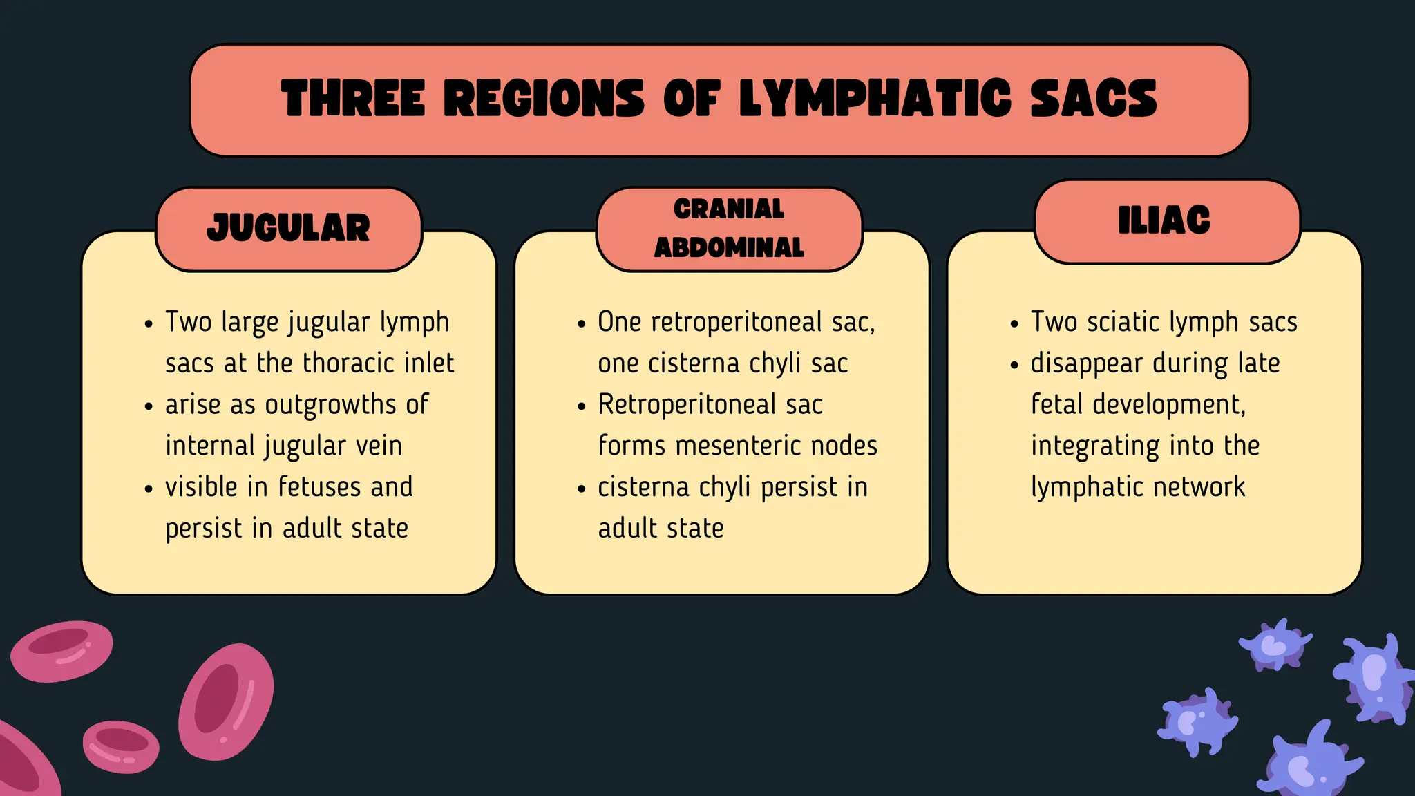
LYMPH SACS FORMATION
4.
Two large jugularlymph
sacs at the thoracic inlet
arise as outgrowths of
internal jugular vein
visible in fetuses and
persist in adult state
THREE REGIONS OF LYMPHATIC SACS
JUGULAR
One retroperitoneal sac,
one cisterna chyli sac
Retroperitoneal sac
forms mesenteric nodes
cisterna chyli persist in
adult state
CRANIAL
ABDOMINAL
Two sciatic lymph sacs
disappear during late
fetal development,
integrating into the
lymphatic network
ILIAC
5.
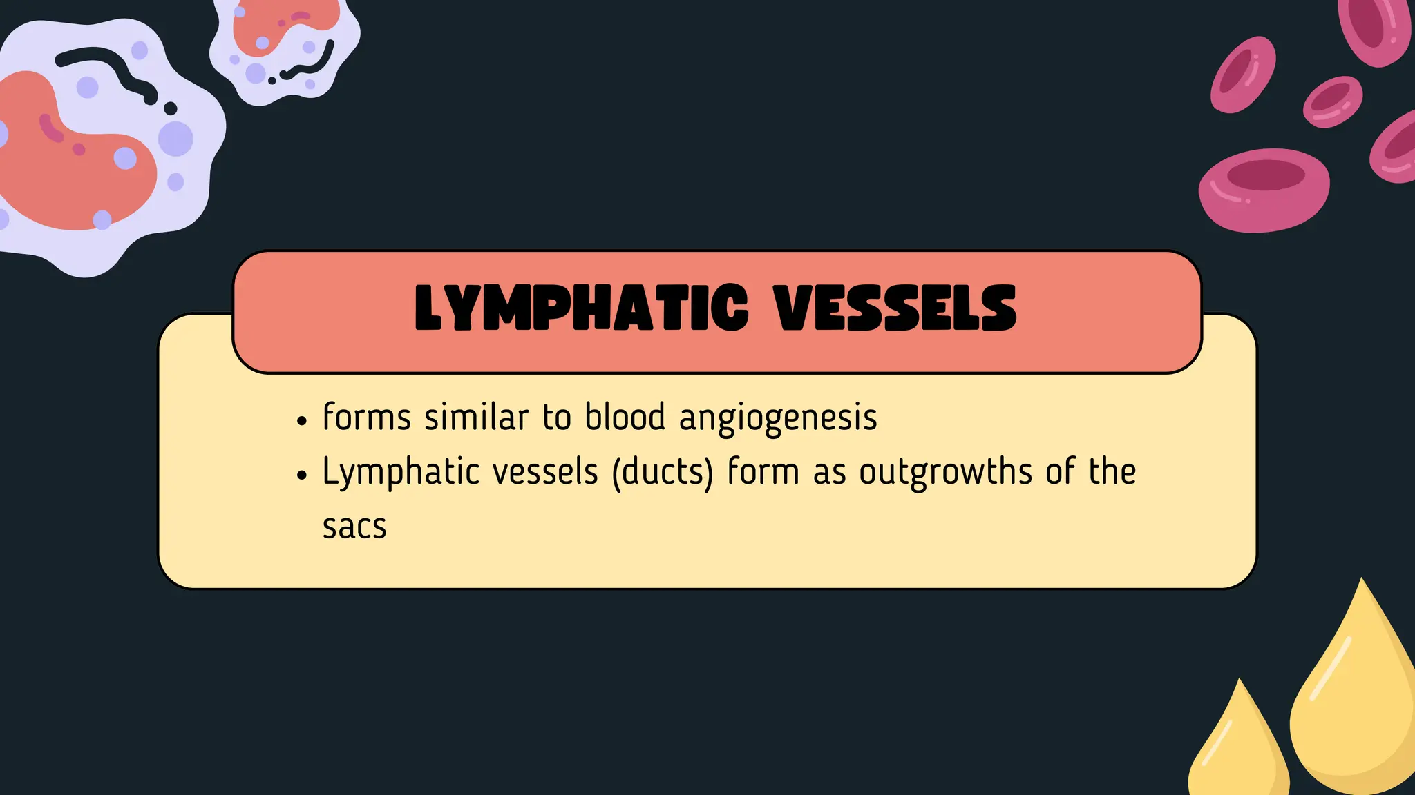
forms similar toblood angiogenesis
Lymphatic vessels (ducts) form as outgrowths of the
sacs
LYMPHATIC VESSELS
6.
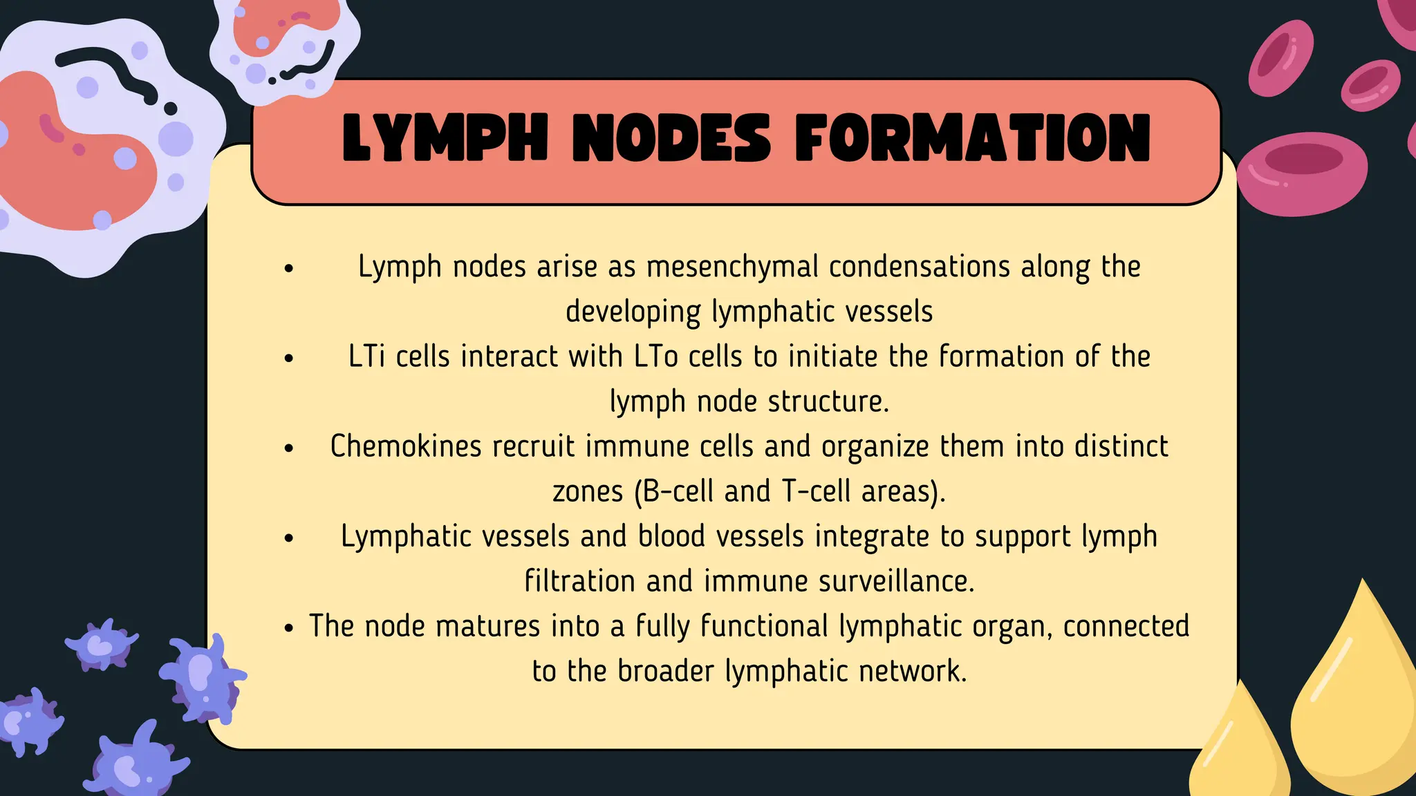
Lymph nodes ariseas mesenchymal condensations along the
developing lymphatic vessels
LTi cells interact with LTo cells to initiate the formation of the
lymph node structure.
Chemokines recruit immune cells and organize them into distinct
zones (B-cell and T-cell areas).
Lymphatic vessels and blood vessels integrate to support lymph
filtration and immune surveillance.
The node matures into a fully functional lymphatic organ, connected
to the broader lymphatic network.
LYMPH NODES FORMATION
8.
Fletcher, T. F.,& Weber, A. F. (2013). Veterinary developmental
anatomy.
Noden, D. M., & de Lahunta, A. (Eds.). (2009). The embryology of
domestic animals: Developmental mechanisms and malformations.
Elsevier.
REFERENCES


![Lymphatic system [autosaved]](https://cdn.slidesharecdn.com/ss_thumbnails/lymphaticsystemautosaved-180608144519-thumbnail.jpg?width=640&height=640&fit=bounds)


