2. corneum, the outermost layer of the skin.6,7
Researchers have tried
various approaches to either disrupt or weaken the stratum cor-
neum to improve skin delivery. The first major approach to over-
come the skin barrier is the use of chemical enhancers such as
azones, glycols, ethanol, terpenes, and so on.6,8
They facilitate drug
transport by partially fluidizing skin lipids and increasing drug
partitioning. A second approach is to use physical enhancement
methods, such as sonophoresis (ultrasound), electroporation,
magnetophoresis, microneedles, thermal ablation, micro-
dermabrasion, and iontophoresis.7,9-12
This approach bypasses the
stratum corneum and delivers the drug directly to the target skin
layer. Both of the aforementioned approaches have shown suc-
cessful delivery for variety of drugs.11,13
However, physical ap-
proaches are mostly painful, expensive, and lack patient
compliance while chemical permeation enhancers can cause skin
irritation and permanent skin damage.6
Finally, the third approach
is the use of drug delivery systems like nanoparticles, microparti-
cles, and lipid-based delivery systems. These systems can increase
skin transportation by improving drug solubilization in the
formulation, drug partitioning into the skin, and by fluidizing the
skin lipids.6
Among the various studied drug delivery systems,
lipid-based delivery systems have shown a great potential for both
topical and transdermal delivery, especially in the last few
decades.14
Lipid-based delivery systems are composed of biocompatible
and biodegradable lipids that can be utilized for controlled
release, targeted delivery, and drug protection. The first com-
mercial product utilizing lipid-based delivery system was mar-
keted in 1988 for antimycotic agent, econazole.15
Since then,
several reports are published indicating the success of these de-
livery systems.6,14,16-19
Based on the recent literature review for
skin application, majority of the lipid-based skin delivery systems
are classified into vesicular carriers and lipid particulate systems.
Vesicular carriers comprise liposomes, ethosomes, ultra-
deformable liposomes, and other specialized novel vesicular car-
riers. Due to the limited success of conventional liposomes in the
skin delivery, majority of the recent research are predominantly
focused on polymeric liposomes (PLs) and elastic liposomes like
ultradeformable liposomes and ethosomes. Lipid particulate sys-
tems have also gained popularity in the recent past. Among this
class, lipospheres, solid lipid nanoparticles (SLNs), and more
recently nanostructured lipid carriers (NLCs) have been success-
fully utilized for skin delivery. Table 1 provides a brief summary of
various lipid-based delivery systems.
The lipid-based delivery systems can be tailored to target
various skin conditions depending on the delivery system selected,
formulation composition, manufacturing processes, and process
variables. However, fabrication of these delivery systems requires
understanding of process and formulation variables, mechanism of
skin delivery, knowledge of physicochemical characteristics, recent
technological advancements, and specific limitations. To address
these needs, this review article focuses on lipid-based delivery
systems (specifically vesicular and lipid particulates) with
emphasis on recent research, advancements, and challenges. Also,
acknowledging that the literature provides only a limited review on
lipid-based delivery systems for unique areas like transcutaneous
immunization (TCI), vaccine delivery via the skin, and cosmeceut-
icals, we have attempted to encompass these areas within the
limited scope of this review article. Furthermore, it is also imper-
ative to understand the associated regulatory implications for
achieving commercial success of these delivery systems for skin
application. However, because literature review of past decade
provides little information of this subject, the regulatory aspects
and U.S. Food and Drug Administration (FDA) standpoint for lipid-
based delivery systems are also covered in this article.
Skin Anatomy and Physiology
The skin is the largest organ of the human body. The total surface
area of the skin of an average male adult is approximately 2 m2
.35
The
major functions of the skin include protection against mechanical
stresses, prevention of excessive water loss; facilitating transpirational
cooling, and preventing absorption of foreign bodies. Anatomically,
skin is composed of 3 main distinguishable layers, namely epidermis,
dermis, and subcutaneous (SC) “fat” tissues (Fig. 1).36
Epidermis
The epidermis is divided into 2 regions: the nonviable epidermis
(the stratum corneum) and the viable epidermis. It consists of 70%
water and keratinizing epithelial cells responsible for synthesis of the
stratum corneum.37
The epidermis does not contain any blood vessels
and hence molecules permeating across the epidermis must cross the
dermal-epidermal layer to enter the body’s systemic circulation.
The stratum corneum is the outermost layer of the skin and is
involved in skin homeostatic and protective functions. The stratum
corneum is the final product of epidermal differentiation with
approximately 10-20 mm thickness and is considered as metaboli-
cally inactive.37
It consists of 10-25 layers of dead, elongated, fully
keratinized corneocytes, which are embedded in a matrix of the
lipid bilayers. It typically resembles “Brick and Mortar” type
structure, where corneocyte from hydrated keratin of the skin re-
sembles Bricks embedded in a Mortar, comprising of extracellular
lipid components.38
The extracellular lipid is constituted of 2
lamellar phases with predominant crystalline phase and the sub-
population of liquid lipid phase.39
Lipids that constitute the extra-
cellular matrix of the stratum corneum have a unique composition
and are very different from the lipids that constitute most biolog-
ical membranes.
The viable epidermis is present below the stratum corneum and is
approximately 50-100 mm thick.40
It is different from the stratum
corneum because it is physiologically more closely akin to the other
living cellular tissues and contains many metabolizing enzymes. The
viable epidermis is involved in the generation of stratum corneum and
metabolism of the foreign substances. It is also involved in the immune
response of the skin due to the presence of Langerhan cells (LCs).41
Dermis
The dermis is a supportive, compressible, and elastic con-
nective tissue protecting the epidermis. It is composed of
fibrous proteins (collagen and elastin) and an interfibrillar gel of
glycosaminoglycans, salts, and water. Blood and lymphatic
vessels, nerve endings, hair follicle, sebaceous glands, and sweat
glands are embedded within the dermis. Extensive vascular
network in the dermis plays a crucial role in skin nutrition,
repair, immune responses, and thermal regulation.37
The hair
follicles and sweat ducts form a direct connecting path from
dermis to the skin surface, bypassing stratum corneum and
henceforth involved in providing appendageal route of skin
permeation.42
Subcutaneous “Fat” Tissue
The SC fat tissue located below the dermis is composed of
the cells that contain large quantities of fat, making the
cytoplasm lipoidal in character.37
The collagen between the fat
cells provides the linkage of the epidermis and the dermis
with the underlying structures of the skin. The main function
of SC fat tissue is to act as a heat insulator and shock
absorber.
S. Jain et al. / Journal of Pharmaceutical Sciences xxx (2016) 1-232
3. Table 1
Summary of Lipid-Based Delivery Systems
Lipid-Based Delivery System Definition Typical Formulation
Composition
Advantages Challenges
Vesicular carriers
Liposomes20,21
These are conventional vesicles (single or multilayers)
that are formed when biodegradable lipids
(phospholipid and cholesterol) come into contact
with the aqueous medium, wherein the hydrophilic
head group of the lipid surrounds the aqueous core
while the hydrophobic tail group is exposed to the
external medium
Phospholipid
Cholesterol
Aqueous medium
Lipids are biocompatible and biodegradable High cost of lipids in general. Synthetic lipids are even
more expensive than natural lipids
Well-studied manufacturing (conventional)
processes and its process parameters at
laboratory scale
Process scalability challenges for commercial
application along with risk of residual organic
solvent in the drug product
Suitable for both hydrophobic and hydrophilic
drug loading
Poor chemical (e.g., oxidative degradation) and
physical stability (e.g., aggregation and fusion)
Improves localized delivery Poor permeation to viable epidermis and dermis
Lack of well-established regulatory guidance for skin
delivery
Poor physicochemical characteristics (higher particle
size, higher rigidity, and low encapsulation
efficiency)
Ultradeformable liposomes
(also called as
transferosomes or
deformable liposomes)22-24
These are elastic liposomes similar to conventional
liposomes in terms of its preparation techniques
and vesicular structure but functionally they are
sufficiently deformed due to presence of edge
activator
Phospholipid
Edge activator
Aqueous medium
Lipids are biocompatible and biodegradable High cost of lipids
Manufacturing process and process parameters
are similar to that of liposomes (which are
extensively studied at laboratory scale)
Process scalability challenges for commercial
application along with risk of residual organic
solvent in the drug product
Higher elasticity and smaller vesicle size than
conventional liposomes due to the presence
of edge activator
Hydrophobic drug loading can compromise elasticity
of these vesicles
Higher skin permeation potential compared to
conventional liposomes
Limited skin permeation under occlusive condition
High membrane hydrophilicity and elasticity
facilitate these vesicles to avoid aggregation
and fusion under osmotic stress, which poses
a problem to the conventional liposomes
Lack of well-established regulatory guidance for skin
delivery
Ethosomes25,26
These are elastic liposomes similar to conventional
liposomes in terms of its preparation techniques
and vesicular structure but functionally they are
sufficiently deformed due to the presence
of ethanol
Phospholipid
Cholesterol
Water and ethanol
cosolvent medium
Lipids are biocompatible and biodegradable High cost of lipids
Manufacturing process and process parameters
are similar to that of liposomes (which are
extensively studied at laboratory scale)
Process scalability challenges for commercial
application along with risk of residual organic
solvent in the drug product
Suitable for both hydrophobic and hydrophilic
drug loading
Lack of long-term structural and chemical stability
data during storage
Higher elasticity, smaller vesicle size and higher
entrapment efficiency than conventional
liposomes.
Challenge in optimizing lipid and ethanol
concentration to achieve improved
physicochemical properties without compromising
stability of the ethosomes
Unlike ultradeformable liposomes, it enhances
skin permeation under both occlusive and
nonocclusive conditions
Lack of well-established regulatory guidance for skin
delivery.
Higher skin permeation than conventional and
ultradeformable liposomes (in most cases).
Possibility of skin irritation and toxicity due to high
ethanol content
Lipid particulate systems
Lipospheres27-29
These are microspheres, composed of solid
hydrophobic lipid core stabilized by a
monolayer of phospholipid embedded
on the surface
Fats (mainly solid
triglyceride)
Stabilizer (e.g.,
phospholipid)
Aqueous medium
Biodegradable and biocompatible Poor skin permeation compared to lipid-based
vesicles, SLNs, and NLC.
Relatively cost effective compared to lipid-based
vesicular carriers
Lack of long-term physical stability data
Ease of preparation and scale-up Higher particle size than lipid-based vesicular carriers,
SLN, and NLC.
Possibility for extended release of entrapped
drug
Poor drug loading for hydrophilic compounds
Improved stability for photo-labile drugs
Controlled particle size
High dispersability in aqueous medium
Lack of well-established regulatory guidance for skin
delivery
(continued on next page)
S.Jainetal./JournalofPharmaceuticalSciencesxxx(2016)1-233
4. Pathways for Skin Penetration
In accordance to the above-discussed Brick and Mortar model,
the process of percutaneous absorption can occur via 2 different
routes: transepidermal (intercellular and intracellular) and trans-
appendageal (hair follicles, sweat ducts, and sebaceous glands)
pathways (Fig. 2).36
Transepidermal Pathway
Transepidermal pathway consists of intercellular and intracel-
lular pathways. Intercellular pathway involves solute diffusion
through the intercellular lipid domains via tortuous pathway (via
cornified cells of stratum corneum, the viable epidermis, and the
dermis).43
Tracer studies have provided evidences that intercellular
lipids, and not the corneocyte proteins, are the main epidermal
permeability barrier.44
Intercellular pathway was initially rejected
as a dominant skin permeation mechanism due to its small volume
occupancy.43
However, later the intercellular volume fraction was
found to be much larger than originally estimated.45,46
These
studies suggest that intercellular pathway provided a major resis-
tance for skin permeation.
Intracellular (transcellular) pathway involves permeation
through the corneocytes followed by the intercellular lipids.
Compounds permeating through this route utilize the imper-
fections in the corneocytes that create openings comprised of
water. This route is therefore believed to prefer hydrophilic
compounds for delivery. It is interesting to note that the intra-
cellular pathway requires not only partitioning into and diffu-
sion through corneocytes but also into and across the
intercellular lipids.47
Transappendageal Pathway
In transappendageal pathway, the penetrant traverse the
stratum corneum via a “shunt” pathway provided by the hair
follicles or sweat glands. In particular, hair follicles play a major
contributor for this pathway due to higher follicular distribution.
Although the available surface area for the follicular route is
assumed to be limited to approximately 0.1% of total skin surface
area, it has recently been suggested that follicular number,
opening diameter, and follicular volume are important consid-
erations to define the extend of delivery.42,48
Also, the hair fol-
licles extend deep into the dermis with significant increase in
the actual surface area available for the penetration. Many
studies have indicated the relevance of this pathway in skin
permeation.49-51
Principle of Skin Permeation
Passive permeation is the most simplistic scenario for skin
permeation and is governed via Fick’s first law of diffusion,
where the rate of transfer (dQ/dt) of a solute through a mem-
brane with unit area A in one dimension (x) is directly propor-
tional to the concentration gradient (dc/dx) across the
membrane. The permeation flux (J) can be mathematically
defined as follows52
:
J ¼
dQ
dt*A
¼ D
dc
dx
(1)
As indicated in the equation, the permeation flux is directly
proportional to the concentration gradient across the membrane.
The diffusion coefficient (D) can further be represented by
Equation 2:
Table1(continued)
Lipid-BasedDeliverySystemDefinitionTypicalFormulation
Composition
AdvantagesChallenges
Solidlipidnanoparticles30-32
Thesearecolloidallipidnanoparticlescomposedof
biodegradablesolidlipophilicmatrix(attheroom
temperatureandbodytemperature)inwhichthe
drugmoleculescanbeincorporated
Solidlipid
Surfactant
Aqueousmedium
BiodegradableandbiocompatibleDuetohighwatercontent,ithastobegenerally
incorporatedintosemisolidcarrierslikeointment
andgel
Relativelycosteffectivecomparedtolipid-based
vesicularcarriers
Lackoflong-termphysicalstabilitydata.Potential
expulsionofactivecompoundsduringstorage
Manufacturingprocessesarereproducibleand
scalable
Gelationandconsequentlyparticleagglomeration
Avoiduseoforganicsolventduringthe
manufacturingprocess
Lackofwell-establishedregulatoryguidanceforskin
delivery
Smallerparticlesizethanlipospheres
Protectdrugfromchemicaldegradation
Flexibilityofmodulatingdrugrelease
Nanostructuredlipid
carriers33,34
Thesearecolloidalnanoparticlesproducedbymixing
liquidlipid(oils)withthesolidlipidinwhichthe
liquidlipidiseitherembeddedintothesolidmatrix
orlocalizedatthesurfaceofsolidparticles
Solidlipids
Liquidlipids(oils)
Surfactant
Aqueoussolution
BiodegradableandbiocompatibleLackoflong-termphysicalstabilitydata
Manufacturingprocessesarereproducibleand
scalable
Lackofwell-establishedregulatoryguidanceforskin
delivery
HigherdrugloadingcapacitycomparedtoSLNs
Smallerparticlesizethanlipospheres
Avoid/minimizepotentialexpulsionofactive
compoundsduringstorage
LowerwatercontentcomparedtoSLNs
S. Jain et al. / Journal of Pharmaceutical Sciences xxx (2016) 1-234
5. D ¼
BT
6phR
(2)
where B is the Boltzmann constant, T the temperature, h the vis-
cosity of the solute medium, and R the radius of the solute.
As indicated in Figure 3, Equation 1 can be represented as
follows:
J ¼ D
A ðC1 À C2Þ
h
(3)
In this equation, C1 and C2 are the concentrations across the
membrane while h is the thickness of the membrane. Based on
Figure 3, the partition coefficient (K) can be defined as follows:
K ¼
C1
Cd
¼
C2
Cr
(4)
where Cd and Cr represent the concentration in the donor and re-
ceptor compartment. Considering the partition coefficient,
Equation 3 can be represented as follows:
J ¼
DAKðCd À CrÞ
h
(5)
It can be inferred that the passive diffusion of drug is dependent
on the concentration gradient, temperature, viscosity of the solute
medium or delivery system, and the particle size of drug molecule
or delivery system.
Figure 2. The pathways for percutaneous absorption. Adapted with permission from Erdo et al.36
Figure 1. Schematic representation of anatomical structure of the human skin. Adapted with permission from Erdo et al.36
S. Jain et al. / Journal of Pharmaceutical Sciences xxx (2016) 1-23 5
6. Lipid-Based Delivery Systems
Vesicular Carriers
The vesicular carriers have traditionally been used for topical
and transdermal drug delivery. They are typically composed of
biocompatible lipids and aqueous phase (water, buffer solutions,
or cosolvents). Structurally, these lipids form concentric lamellae
entrapping the aqueous phase. Owing to the lipophilic nature of
the lipids, these vesicles (with entrapped drug) can supposedly
partition into the skin layers and deliver the drug across stratum
corneum. Additionally, because the vesicles are typically in nano-
size range, they can further enhance the skin delivery of drug-
loaded vesicular carriers. In general, it is suggested that vesicle
size !600 nm do not penetrate the deeper layers of the skin and
stay in/or on the stratum corneum, vesicles 300 nm can pene-
trate more deeply, but vesicles 70 nm can deliver to both the
viable epidermal and dermal layers.53
For improving skin
permeation potential, researchers have invented and modified
various vesicular carriers with unique structural and functional
properties in the last 4 decades.
The first-generation lipid-based vesicular carrier was called li-
posomes. The first reported publication in this field was from Mezei
and Gulasekharam in 1980.54,55
However, the success of liposomal
delivery was mainly limited by its vesicular size (typically 200-800
nm) and rigidity, which can impede skin permeation.6,53
In 1992,
Cevc and Blume introduced the second-generation vesicular car-
riers named Ultradeformable liposomes or Transfersomes®
, which
possess smaller vesicular size (typically <300 nm) and higher
elasticity (typically 5-8 times higher compared to conventional
liposomes).56,57
In 2000, Touitou et al.58
developed third-
generation vesicular carrier called ethosomes. Ethosomes are
ethanol-based nanosized elastic lipid vesicles. The improved skin
permeation of ethosomes is attributed to the unique physico-
chemical properties, that is, smaller vesicular size (typically <300
nm) and higher elasticity (typically 10-30 times higher than con-
ventional liposomes), as well as permeation enhancement effect of
ethanol.6,57
More recently, various modifications of these vesicular
carriers are also studied to provide specific structural or functional
attribute for skin delivery.
Each of these vesicles has its specific features, mechanism of
drug delivery, advantages, and challenges. The following section
discusses the vesicular carriers in detail.
Liposomes
Conventional liposomes are one of the most famous and
extensively studied lipid vesicles, which are typically composed of
phospholipids, cholesterol, and aqueous medium (water or buffer
solution with varying pH). These vesicles are formed when natu-
rally or synthetically occurring biodegradable lipids come into
contact with the aqueous medium, wherein the hydrophilic head
group of the lipid surrounds the aqueous core while the hydro-
phobic tail group is exposed to the external medium. Due to this
unique structural property, water-soluble drugs can be loaded in
the aqueous core while the water-insoluble drugs can be loaded in
the lipid bilayer.
Although both natural and synthetic phospholipids are avail-
able, conventional choice is often limited to naturally occurring
phosphatidylcholines (e.g., soy or egg source) due to toxicological
considerations and relative cost.59
Phosphatidylcholine is the major
component of the liposomes and act as a permeation enhancer for
skin delivery of the drugs. Due to the lower gel-liquid crystalline
phase transition temperature, these lipids are in fluid state at the
skin temperature of 32C.6
The fluid-state phospholipids disturb
the rigid bilayer structure of the skin lipids leading to increase in
drug partitioning into the lipid phase. Cholesterol is generally
added to impart rigidity and stabilization by increasing the gel
(stable) to liquid crystalline state (metastable) transition temper-
ature of the lipid bilayer.60
However, the inclusion of cholesterol in
the liposome may decrease the encapsulation efficiency of hydro-
philic drugs by reducing the volume of the aqueous phase.61
Furthermore, because the addition of cholesterol increases the ri-
gidity of the vesicles, it can negatively impact the permeation of
these vesicles through the skin.6
The most commonly used conventional techniques for liposome
preparation include thin-film hydration,6
reversed phase evapora-
tion,62
and solvent injection techniques.63
Based on the available
literature, among the aforementioned conventional techniques,
thin-film hydration is most commonly used for skin delivery
studies. In this technique, lipids (phospholipid and cholesterol) are
Figure 3. Permeation of drug molecule from donor compartment to receptor compartment across concentration gradient.
S. Jain et al. / Journal of Pharmaceutical Sciences xxx (2016) 1-236
7. dispersed in the organic solvent. Then, organic solvent is removed
by means of evaporation (using a rotary evaporator at reduced
pressure) leaving behind a dry lipid film on the wall of the flask.
Finally, the dry lipid film is hydrated by aqueous phase (while
vortexing the content) to obtain liposomes. In this technique,
processing parameters like hydration time, hydration temperature
(temperature at which the lipids are hydrated by aqueous me-
dium), and vortexing speed may affect various parameters (espe-
cially vesicle size and entrapment efficiency) of the liposome and
subsequently may modify its skin permeation.64
For example, a
study conducted on liposomes prepared with imiquimod:phos-
phatidylcholine:cholesterol weight ratio of 1:10:1 indicated that
increase in hydration time from 90 to 150 min resulted in increase
in entrapment efficiency from 36.85% to 65.32%, respectively.64
Also, hydration temperature higher than lipid phase transition
temperature is preferred for this technique.6
However, most of the
above-mentioned conventional technologies encounter severe
drawbacks. For example, thin-film hydration utilizes organic sol-
vent and renders larger vesicle size liposomes.6
In case of solvent
injection technique, a relatively dilute preparation of liposomes is
obtained which decreases the encapsulation efficiency of the
aqueous phase. Furthermore, most of these techniques exhibit
scale-up issues. For detailed discussion on these conventional
techniques and their challenges, the readers can refer to the
recently published review articles.20,65
Recently, more advanced technologies such as supercritical
fluid,66,67
dual asymmetric centrifugation,68
and microfluidic
channels69,70
have been employed in liposome preparation for skin
delivery application. Supercritical fluid technology provides a
green, nontoxic, inexpensive, and scalable alternative to the con-
ventional liposome preparation techniques.71
Briefly, phospholipid
and cholesterol are dissolved in supercritical CO2 and then allowed
to precipitate in the form of ultrafine lipid particles. Afterwards,
aqueous medium is added to consequently form liposome vesicles.
Processing parameters like operational pressure, vessel tempera-
ture, and flow rate ratio between CO2 and ethanol can affect various
properties of the liposomes (especially drug loading, entrapment
efficiency, and particle size). In a recent study, effect of process
parameters involved in supercritical fluid technology was studied
on CoQ10-loaded liposomes (phosphatidylcholine to drug weight
ratio of 10:1).66
It was observed that with decrease in the opera-
tional pressure from 16 to 8 MPa, drug loading could increase up to
4 times (2.95% and 8.92%, respectively), at constant vessel tem-
perature of 35C. Additionally, increase in temperature from 35C
to 55C can further improve drug loading from 8.92% to 10.2%,
respectively (keeping all other parameters constant). Several re-
searchers have shown promising results using supercritical fluid
technology.72,73
Dual asymmetric centrifugation is another latest
technology for liposome preparation.74
This is a unique advanced
centrifugation technique wherein 2 types of rotational forces are
applied. Conventional centrifugation rotational force moves the
sample outward, while additional rotational force is provided to
move the sample toward the center of the centrifuge. This unique
combination of 2 contra-rotational movements causes shearing of
the sample (typically a dispersion of phospholipid, cholesterol, and
aqueous medium) and consequently results in formation of the li-
posomes. For model compound calcein, dual asymmetric centri-
fugation technique was used on the concentrated blend of
hydrogenated phosphatidylcholine and cholesterol (55:45 mol%)
and 0.9% NaCl solution. After optimization of process parameters
like centrifugation speed and time, the formed liposomes exhibited
particle size of 60 ± 5 nm and entrapment efficacy of 56 ± 3.3%.74
In
another study, siRNA (short-interfering RNAs) liposome, composed
of phosphatidylcholine and cholesterol, was prepared using dual
asymmetric centrifugation. The obtained liposomes resulted in
mean particle sizes of 79-109 nm with entrapment efficiency
ranging from 43% to 81%. Additionally, based on spectral fluorim-
etry, it was concluded that all entrapped siRNA was structurally
intact with no chemical degradation. Based on these results, this
technology can be effectively utilized to load RNA (without causing
degradation problems) for skin delivery application.68
Another
recent but widely used technique is microfluidic channels where
liposomes are formed by passing the stream of alcoholic solution of
lipid through 2 aqueous streams in a microfluidic channel.70,75-77
The laminar flow in the channels enables to control the size and
size distribution of the liposomes. It was demonstrated that lipo-
some (cholesterol, dimyristoylphosphatidylcholine, and dihex-
adecyl phosphate) vesicle size could be modified from 50 to 150 nm
by adjusting alcohol-to-aqueous volumetric flow rate.75
Various studies have shown the effect of formulation variables
(e.g., lipid composition, type of lipid, drug-lipid ratio, concentration
and type of surface charge imparting compound, etc.) on the
physicochemical properties and skin permeation behavior of the
liposomes.78-80
In our earlier work, we investigated the effect of
lipid composition (phosphatidylcholine to cholesterol ratio) on the
vesicle size, entrapment efficiency, elasticity, and skin permeation
of diclofenac-loaded liposomes.6
It was observed that with increase
in the phosphatidylcholine to cholesterol ratio from 50:50 to 90:10
wt/wt, the vesicle size decreased (252-182 nm, respectively),
entrapment efficiency increased (34.6%-53.6%, respectively), elas-
ticity index increased (0.05-0.62, respectively), and in vitro cumu-
lative drug permeate increased (0e94 mg/cm2
, respectively). These
results were attributed to the presence of cholesterol that embeds
into the bilayer structure of the phosphatidylcholine, resulting in
increase in thickness (vesicle size), decrease in motion of the lipid
tails (decreases elasticity), reduction in free volume for drug
entrapment, and consequently decrease in drug permeation
through skin. The type of lipid selected for liposome preparation
also needs to be carefully evaluated. For example, because egg-
based phosphatidylcholine is more saturated than soy-based
phosphatidylcholine, liposomes prepared using the latter may
provide a better oxidative stability.81
In another study, skin
permeation behavior of natural lipid (soy phosphatidylcholine and
egg phosphatidylcholine) and synthetic lipid (hydrogenated soy
phosphatidylcholine) was compared by preparing curcumin-
loaded liposomes with phosphatidylcholine to cholesterol ratio of
8:1.82
It was observed that although the particle size, entrapment
efficiency, and zeta potential were similar, the skin permeation
behavior was significantly different for natural and synthetic
phospholipid-based liposomal formulations. Natural lipid-based
liposomal formulations exhibited higher skin permeation
(approximately 1.5 times) and skin retention (approximately 1.7
times) compared to synthetic lipid-based liposomal formulations.
This behavior was attributed to the low phase transition temper-
ature of the natural lipids (below 0C), which results in increased
fluidity of the liposome and consequently enhances skin
permeation.
Another factor that can play a critical role is the surface charge of
the lipid. Positively charged liposome has shown enhanced skin
permeation compared to neutral and negatively charged liposome,
presumably due to interaction with negatively charged skin
membrane.19,83,84
More recently, drug-loaded liposomes are con-
jugated with cationic cell penetrating peptide (CPP) to improve skin
membrane penetration of the liposomes.85
In a study, Polygonum
aviculare L. extract (antioxidative and cellular membrane protective
activity) was loaded into CPP conjugated liposome for transdermal
delivery. In vivo studies indicated that the CPP conjugated lipo-
somes were more effective in depigmentation and antiwrinkle
potential than the conventional liposomes (without CPP). This
result was attributed to the ability of cationic peptide conjugated
S. Jain et al. / Journal of Pharmaceutical Sciences xxx (2016) 1-23 7
8. liposomes to effectively interact with the intercellular lipid
lamellae of the stratum corneum compared to the conventional
liposomes.85
Similarly, for topical delivery of lidocaine (LID),
transactivation transcriptional activator (TAT), one of the CPP was
conjugated on the octadecyl-quaternized lysine-modified chitosan
PLs (TAT-PLs).86
The in vitro skin permeation results indicated
approximately 4.17 and 1.75 times higher permeation flux of
LID-loaded TAT-PLs than that of LID solution and LID-loaded con-
ventional liposome (composed of phosphatidylcholine and
cholesterol), respectively. The author attributed the cationic PL
(octadecyl-quaternized lysine-modified chitosan) and the posi-
tively charged arginine group in TAT peptide sequences to facilitate
binding to the negatively charged skin membrane.86
Several theories have been proposed with regard to the mech-
anism of skin delivery via liposomes. Some of the prominent the-
ories include intact vesicular skin penetration,54,55
adsorption
effect,87
and the penetration of liposomes through the trans-
appendageal route.88,89
However, some researchers have recently
suggested that the permeation enhancement effect of the liposome
is due to the interaction of liposome with the skin lipid causing
partial fluidization of skin lipid and consequently delivering the
drug to the deeper skin layers (below the stratum corneum
layer).87,90-92
Confocal laser scanning microscopy (CLSM) has
revealed that conventional liposome might disintegrate and fuse
with stratum corneum lipids, and consequently form a depot of the
drug on the skin surface.93
Thereafter, the extent of delivery will be
guided by the physicochemical properties (solubility and partition
coefficient) of the drug. This is evident from the limited success that
has been achieved in the field of liposomal skin delivery.94,95
Conventional liposomes are generally reported to be confined (or
disintegrates) in the upper layer of the stratum corneum and
accumulate in the skin appendages with minimal penetration to
the deeper skin layers, owing to their large size and lack of
elasticity.96
Literature review of past decade suggests that most of the
liposome research is in the area of topical drug delivery. Liposomes
have been utilized for topical delivery of variety of drugs including
curcumin,82,97
siRNA,98
loperamide,99
clotrimazole,100
resvera-
trol,101
LID,102
and so on. Solubilizing ability of the liposomes was
utilized to load curcumin, a poorly water-soluble drug.82,97
It was
found that liposomal curcumin with entrapment efficiency up to
98% was 2-fold to 6-fold more potent than corresponding curcu-
minoids. In another study, liposomes were utilized to deliver siRNA
through skin for melanoma treatment.98
It was observed that li-
posomes were able to not only penetrate into the skin layers but
were also effectively internalized into the viable cells of basal
epidermis and knock down the target protein expression.98
Topical
application of loperamide hydrochloride-encapsulated liposomal
gel (composed of phosphatidylcholine, cholesterol, and Carbopol®
940) resulted in potent and prolonged analgesic and anti-
inflammatory activity compared to controls (free loperamide gel
and empty liposomal gel) in a rodent model.99
Despite various reported research work on conventional lipo-
somes and PLs, clinical and commercial success of these vesicles are
rather limited. This is due to the fact that skin permeation of the
liposomes is mainly limited by its large vesicle size and lack of
elasticity.6
Furthermore, scalability of the manufacturing process,
chemical instability, residual organic solvent in the drug product,
liposomes aggregation, cost of lipids, and regulatory implications
also pose additional challenges in liposomes' success (Table 1).103
Ultradeformable Liposomes
To overcome some of the drawbacks of conventional liposomes,
a novel highly deformable elastic liposomes called ultradeformable
liposomes (also called as Transferosomes®
or deformable
liposomes) were introduced with the ability to penetrate the intact
skin if applied nonocclusively.104
These elastic liposomes are
similar to conventional liposomes in terms of its preparation
techniques and vesicular structure but functionally they are suffi-
ciently deformed to penetrate pores (i.e., skin pores) much smaller
than their own size. Additionally, in contrast to the conventional
liposomes, the ultradeformable liposomes are made up of phos-
pholipids, aqueous medium, and edge activators (Table 1). The edge
activators are capable of increasing the deformability of the bilayer
by affecting the interfacial tension of these vesicles. Transmission
electron microscopy has conclusively demonstrated the deforma-
tion of the vesicles into oval and irregular structures upon addition
of edge activator.57,105,106
Another major difference between the
ultradeformable liposomes and the conventional liposomes is the
higher hydrophilicity of the former, which allows the elastic
membrane to swell more in comparison to the conventional lipid
bilayer. High membrane hydrophilicity and elastic nature facilitate
these vesicles to avoid aggregation and fusion under osmotic stress,
which poses a problem to the conventional liposomes.107
Apart from the formulation variables discussed in case of lipo-
somes (e.g., type and concentration of lipid), the type and concen-
tration of edge activator can significantly affect the physicochemical
properties of these vesicles. Edge activators typically used for
ultradeformable liposome preparation include sodium cholate, so-
dium deoxycholate, Span 60, Span 65, Span 80, Tween 20, Tween 60,
Tween 80, and dipotassium glycyrrhizinate.108,109
In a study, the
effect of type of edge activators (sodium cholate, sodium deoxy-
cholate, and Tween 80) on physicochemical properties of ultra-
deformable liposomes (phosphatidylcholine to edge activator
weight ratio of 6:1) was evaluated. It was observed that sodium
cholate and sodium deoxycholate resulted in the smaller vesicle size
and higher zeta potential compared to Tween 80.108
Ultra-
deformable liposomes prepared with 95%:5% (wt/wt) (Phosphati-
dylcholine:Edge activator) ratio showed entrapment efficiency in
the following orderdSpan 85 Span 80 Na cholate Na deoxy-
cholate Tween 80dand it was attributed to the hydrophyllic
lipophyllic balance values of the respective edge activator.110
In
another study, the effect of Tween 20 was studied on vesicular
elasticityand the electron spin resonance study revealed that Tween
20 increased the fluidity at the C5 atom of the acyl chain of the
phospholipid (egg-based phophatidylcholine) bilayer.111
In addition, the concentration of edge activator plays a critical
role as well. Ultradeformable liposomes (Lipid:Phospholipon 100®
)
prepared at different molar fractions of sodium cholate revealed
that increase in the molar fraction of sodium cholate 0.2 may
cause formation of phospholipid/sodium cholate aggregates, such
as mixed vesicles, opened vesicles, mixed micelles, and rod-like
mixed micelles, which can consequently lead to lower entrap-
ment efficiency of the drug.112
Similarly, for diclofenac-loaded
ultradeformable liposomes (composed of phosphatidylcholine
and Span 80), increase in concentration of Span 80 (edge activator)
from 2% to 5% (wt/wt) resulted in increase in the entrapment effi-
ciency from 50.73% to 55.19%, respectively.110
However, with
further increase in edge activator concentration to 15% then 25%
(wt/wt), the entrapment efficiency decreased from 44.93% to
42.80%, respectively. The decrease in entrapment efficiency at
higher concentration of edge activator was attributed to the for-
mation of micelle aggregates.110
There are 2 major proposed mechanisms of skin delivery via
ultradeformable liposomes. First mechanism proposes that intact
vesicles enter the stratum corneum carrying drug molecules into
the skin.56
It is suggested that owing to the deformable nature,
these vesicles are able to squeeze through the stratum corneum to
the deeper skin layers intact, under the influence of the naturally
occurring transcutaneous hydration gradient. The skin surface is
S. Jain et al. / Journal of Pharmaceutical Sciences xxx (2016) 1-238
9. relatively dry compared to the viable epidermis. When ultra-
deformable liposomes are applied on the skin surface that is
partially dehydrated, the vesicles move toward the deeper skin
layers (e.g., viable epidermis and dermis) that are relatively hy-
drated. The stress induced during the movement to deeper skin
layers is alleviated by the deformable nature of these vesicles. In an
in vitro skin (200-300 mm thickness) permeation study, the pre-
treatment of empty deformable liposomes on the skin surface fol-
lowed by application of saturated aqueous solution of the drugs
(pergolide or rotigotine) was compared with drug-loaded
deformable liposomes. It was observed that skin permeation was
significantly higher in case of deformable liposome encapsulated
drugs compared to pretreatment of empty deformable liposomes
followed by application of drug solution. This study suggests that
ultradeformable liposomes may also act as carrier systems (rather
than acting as a permeation enhancer for free drug) to deliver drug
into deeper layers of the skin (up to 200-300 mm deep from the skin
surface).113,114
However, researchers have reported that the hydra-
tion gradient in the skin layer might not be linear. The water con-
tent in the deeper region of the stratum corneum close to the viable
epidermis is much lower than in the central regions of the stratum
corneum. Therefore, if hydration gradient is the driving force for
ultradeformable liposome delivery, it might be difficult for the
vesicles to penetrate beyond the lowest layers of the stratum cor-
neum.22
In another interesting study, CLSM indicated that fluo-
rescein sodium-loaded ultradeformable liposomes utilized
transfollicular pathway to penetrate to viable epidermis and
dermis; however, the fluorescence intensity still remained higher
in the stratum corneum region.115
Second mechanism proposes that vesicles act as the penetration
enhancer, whereby vesicles enter the stratum corneum layers and
subsequently modify the intercellular skin lipids.71
This will facil-
itate penetration of free drug molecules into and across the stratum
corneum. In a recent study, it was observed that deformable vesi-
cles actually reduced the transdermal absorption of calcein, most
probably by controlling the drug release from the formulation on
the skin surface.116
In an interesting study, skin permeation and
skin deposition of ketotifen fumarate-loaded deformable liposomes
(phosphatidylcholine to Tween 80 ratio of 84.5:15.5 wt/wt) and
conventional liposomes (without Tween 80) were studied,
respectively.117
It was observed that for deformable liposomes, skin
deposition was 5 times higher than the skin permeation. Addi-
tionally, even though skin deposition for deformable liposome was
similar to conventional liposome, skin permeation of deformable
liposome was significantly higher (2 times) than the conventional
liposomes. Based on these findings, it was suggested that deform-
able liposome acts as penetration enhancer for the drug by inter-
acting with the skin lipid.117
Despite various scientific efforts
summarized above, it is still controversial whether the ultra-
deformable liposomes act as a drug carrier or permeation enhancer
or both.
Although the mechanism of skin delivery via deformable lipo-
some is still unclear, researchers have successfully utilized ultra-
deformable liposomes to deliver various drugs.57,106,112,118-120
Deformable liposomes composed of quercetin, phosphatidylcho-
line, cholesterol, and Tween 80 showed 3.8-fold higher penetration
rate compared to it quercetin suspension.119
Similarly after 1 h of
nonocclusive incubation, the total accumulation of amphotericin in
the human skin was 40 times higher when applied as amphotericin-
loaded ultradeformable liposomes than as AmBisom (marketed
amphotericin-loaded liposome).106
Itraconazole-loaded deform-
able liposomes in the presence of hydroxypropyl-b-cyclodextrin
(HP-b-CD) exhibited improvement in itraconazole delivery in stra-
tum corneum and deeper skin layers compared to conventional
liposomes.105
In an attempt to find an alternative to the painful
penile injections for erectile dysfunction, topical deformable lipo-
somes for papaverine hydrochloride (a vasoconstrictor) was studied
in 9 patients. Compared to control, statistically significant
improvement on pharmacodynamic responses were observed in
these patients.121
Ultradeformable liposomes have also been used
for delivery of macromolecules via skin.112,122
The optimized
transferosomal gel containing insulin showed good permeation
behavior with in vitro permeation flux of 13.50 ± 0.22 mg/cm2
/h
through porcine ear skin and demonstrated prolonged hypoglyce-
mic effect in diabetic rats over 24 h.122
In another study, ultra-
deformable liposomes provided a 10-fold increase in in vitro skin
permeation of asiaticoside compared to the free drug solution and
facilitated an increase in in vivo collagen biosynthesis.112
However, despite the success of ultradeformable liposomes,
these vesicles possess some practical difficulties (Table 1). Because
their transport across the skin is driven by the hydration gradient,
occlusive application can compromise the action of the deformable
vesicles by eliminating the gradient force. Another major disad-
vantage of these vesicles corresponds to the difficulty in loading
hydrophobic drugs into the vesicles without compromising their
deformability and elastic properties.22
Ethosomes
Ethosomes are new generation elastic lipid carriers; those have
shown enhanced skin delivery for both hydrophilic and lipophilic
drugs. Although ethosomes are conceptually sophisticated, the
simplicity involved in their preparation along with improved safety
and efficacy have made these vesicles suitable for skin delivery.25
The vesicular structure is composed mainly of phosphatidylcho-
line, cholesterol, ethanol, and water. Preparation techniques are
also similar to conventional liposomes. Despite utilization of
organic solvent, thin-film hydration technique is generally
preferred for ethosomes because of its simplicity and high
entrapment efficiency.123
Furthermore, unlike ultradeformable li-
posomes, ethosomes can provide effective delivery under both
occlusive and nonocclusive conditions. Effectiveness of these ves-
icles in skin delivery is attributed to its soft and elastic nature,
instigated due to the presence of ethanol. Owing to their elastic
nature, ethosomes are able to penetrate through the small pores
and channels of the skin.58
In order to understand the elastic nature of ethosomes, it is
important to understand the interplay of phosphatidylcholine,
ethanol, and cholesterol on phase transition temperature (TP) of the
ethosome vesicles. Phosphatidylcholine has a characteristic TP, that
is, the temperature at which its gel state transitions into the liquid
crystalline state. In gel state, the molecular motion of the lipids is
severely restricted while in the liquid crystalline state the confor-
mational disorder predominates resulting in smaller sized vesicles
with high elasticity.6
Cholesterol increases the TP value of the lipid
vesicles making these vesicles more rigid and consequently more
stable. On the other hand, ethanol interacts with the hydrophilic
head group region of the phosphatidylcholine and eventually de-
creases the TP of the lipid vesicles, facilitating the transition from
gel state into the liquid crystalline state. In our earlier investigation,
we studied the effect of phosphatidylcholine (soy based) to
cholesterol ratio (PC:CH) and ethanol on TP of the lipid vesicles. It
was observed that with increase in cholesterol concentration from
10% to 50% wt/wt, the TP value increased from 8.78C to 11.26C,
while with increase in ethanol concentration from 0% to 30% vol/
vol, the TP value decreases from 14.13C to 8.66C.6
These results
indicate that a fine balance is required between cholesterol and
ethanol in order to achieve ethosome vesicles with higher elasticity
while maintaining the physical stability.
Researchers have also studied the influence of lipid and ethanol
content on physicochemical properties of ethosomes such as
S. Jain et al. / Journal of Pharmaceutical Sciences xxx (2016) 1-23 9
10. vesicle size and morphology, entrapment efficiency, zeta potential,
and elasticity. Transmission electron microscopy study on etho-
somes indicated that they are relatively imperfect round shape
vesicles owing to fluidizing effect of ethanol on lipid bilayer
compared to ultradeformable liposome.124
In our investigation on
diclofenac-loaded ethosomes, we found that with increase in
ethanol concentration from 0% to 30%, the vesicle size decreased,
elasticity increased, and zeta potential decreased. On the other
hand, entrapment efficiency increased with increase in the ethanol
concentration from 0% to 20% due to its cosolvent effect.6,125
However, entrapment efficiency decreased with further increase
in ethanol concentration, probably due to excessive vesicular
fluidization causing leakage of the drug. Similar results were
observed by other researchers.126
Additionally, among studied
properties (i.e., vesicle size, elasticity, zeta potential, and entrap-
ment efficiency), vesicle size and elasticity of the ethosomes were
identified as the only 2 dominating physicochemical properties
that affect the skin permeation of the ethosomes. These physico-
chemical properties could be suitably manipulated by modification
of formulation variables (PC:CH ratio and ethanol) to achieve
desired therapeutic permeation flux.6
Presence of ethanol also provides a net negative surface charge
that prevents aggregation of the vesicles due to electrostatic
repulsion. In a study, colloidal stability of liposomes and ethosomes
were evaluated using Turbiscan optical analyzer. It was found that
compared to liposomes (Phospholipon 100G®
, cholesterol, and
water), ethosomes (Phospholipon 100G, ethanol, and water)
showed no coalescence, sedimentation, and flocculation indicating
superior physical stability.127
In another study, the econazole
nitrate-loaded ethosomes (soy phosphatidylcholine, ethanol, and
water) was found to be physically stable for 6 months under the
tested condition 25C.128
Although the presence of ethanol in the ethosomes can enhance
skin permeation, it can also lead to skin irritation. To address this
concern, few researchers have studied the impact of ethosomal
vesicles on skin morphology. Buspirone-loaded ethosomes with
38% ethanol exhibited no change in skin morphology. The thickness
and appearance of the horny layer were found to be unchanged in
comparison to the normal untreated rat skin.129
Similar results on
human skin were also reported in other studies.130,131
In general,
ethanol concentration of approximately 30%-40% is considered
widely acceptable for skin delivery via ethosomes.
Several studies have been conducted to explore the application
of ethosomal delivery for variety of drugs.3,128,132-135
Table 2 sum-
marizes the recent patents on the application of ethosomal drug
delivery. There are several reported studies of superior skin de-
livery of ethosomes compared to liposomes, ultradeformable lipo-
somes, and marketed formulations. Psoralen-loaded ethosomes (an
antipsoriasis drug) has shown 3.50 and 2.15 times higher perme-
ation flux and skin deposition respectively, compared to that of
liposomes.136
Ethosome-mediated apigenin delivery produced a
more prominent effect on UVB-induced skin inflammation by
suppressing COX-2 levels, compared to liposomes and deformable
liposomes.137
Ethosomal formulation of 5-aminolevulinic acid also
showed better skin permeation than liposomes in photodynamic
therapy.138
Anticandidal activity against Candida albicans was found
to be highest for clotrimazole-loaded ethosomal formulation with
the highest zone of inhibition (34.6 ± 0.57 mm), in contrast to
deformable liposomal formulation (29.6 ± 0.57 mm) and marketed
cream formulation (19.0 ± 1.00 mm).124
The superior anticandidal
activity of the ethosomes was attributed not only to the obvious
permeation enhancement effect of these vesicles but also to the
presence of ethanol that has potential to kill organisms by dena-
turing their proteins. Similarly, healing time in herpes infected
patients was significantly improved by acyclovir-loaded ethosomal
cream in comparison to the market formulation.139
In another
experiment, ammonium glycyrrhizinate-loaded ethosomes elicited
an increase in in vitro percutaneous permeation (in human skin)
and anti-inflammatory activity (in human volunteers) compared to
the ethanolic or aqueous solutions of this drug.131
For diclofenac-
loaded ethosomes, the permeation flux of the optimized formula-
tion was 12.9 ± 1.0 mg/h$cm2
, which was significantly higher than
the drug-loaded conventional liposome, ethanolic or aqueous so-
lution.6
Furthermore, in vivo pharmacodynamic study indicated
that optimized ethosomal hydrogel exhibited enhanced anti-
inflammatory activity (reduction in paw edema volume)
compared with liposomal and plain drug hydrogel formulations. In
order to improve the skin permeation of ethosomes, researchers
have explored various interesting arena. For example, low fre-
quency ultrasound was utilized to deliver hydrophilic macromol-
ecules, hyaluronic acid (MW 1500 kDa). In vitro permeation study
revealed that the combination of low frequency ultrasound and
ethosomes improves the permeation enhancing effect for hyal-
uronic acid by 2.1 times and 6.4 times compared to ethosomes
alone and hyaluronic acid solution with low frequency ultrasound,
respectively.140
Furthermore, some promising results are also observed in the
hormonal therapy. In a study, the skin permeation potential of the
testosterone-loaded ethosomes was compared with the marketed
transdermal patch of testosterone. The authors observed nearly 30
times higher skin permeation of testosterone from the ethosomal
formulation compared to the marketed formulation.130
Touitou
et al. tested the effect of an ethosomal insulin formulation on the
blood glucose level that was applied to the skin. The ethosomal
formulation instigated up to 60% decrease in blood glucose levels in
both normal and diabetic rats and the level was maintained for at
least 8 h.25
Some researchers have also compared the effectiveness of
ethosomal delivery via transdermal route with that of the drug
delivery via oral route. In vivo pharmacokinetic study of ethosomal
transdermal therapeutic system showed approximately 3 times
higher bioavailability compared with oral suspension of valsar-
tan.141
Transdermal application of buspirone-loaded ethosomes for
menopausal syndromes showed similar Cmax value compared to the
oral aqueous solution (120.07 ± 86.97 and 93.44 ± 76.46 ng/mL,
respectively).129
Furthermore, buspirone delivered via transdermal
route was present for longer time period compared to the oral
administration (12 vs. 4 h, respectively), suggesting more sustained
and nonfluctuated delivery to plasma with reduced side effects via
transdermal application of buspirone-loaded ethosomes.129
Moreover, there have been various reported studies exploring
the mechanism of ethosomal delivery via skin. Study on the
amphiphilic fluorescent probe D-289 containing trihexyphenidyl
hydrochloride ethosomes has suggested that ethosomes permeate
deeper into the skin compared to classic liposomes (about 170 and
96 mm respectively from the skin surface).142
Recently, CLSM study
also indicated that “intact” ethosomes were able to reach deeper
stratum corneum layers due to higher elasticity of these vesicles.143
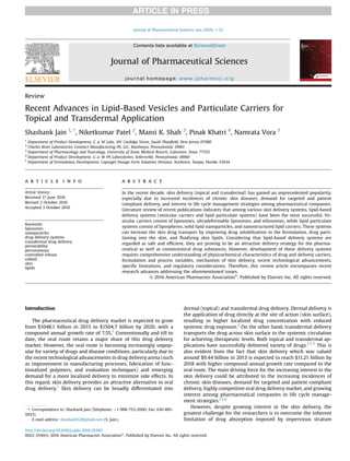
Skin permeation behavior via ethosomes is generally attributed to
the solvent properties of ethanol followed by “ethosome”
effect.26,58
Figure 4 shows the diagrammatic representation of this
mechanism. The stratum corneum lipids, at physiological temper-
ature, are densely packed and highly ordered. Ethanol in ethosomes
interacts with the polar head group region of the lipid molecule,
resulting in a reduction in the transition temperature of the stratum
corneum lipids and consequently increasing their fluidity. Ethoso-
mal vesicles containing drug, owing to the smaller vesicle size and
high elasticity, can then permeated through the partially fluidized
skin lipids to deliver the drug into deeper skin layers. In a study,
CLSM has demonstrated enhanced skin permeation of ethosomes
S. Jain et al. / Journal of Pharmaceutical Sciences xxx (2016) 1-2310
11. compared to liposomes, with permeation up to the last layer of
epidermis (to stratum basale).128,141
In another interesting study,
CLSM revealed the presence of “intact de-shaped” vesicles pene-
trating through the skin, suggesting that these vesicles squeeze
themselves to deeper skin layers owing to their high elastic
nature.143
A study using double staining technique indicated that etho-
somes entered the skin between the coreocytes through the
intercellular lipid domain.144
FTIR studies indicated that mild
swelling of corneocytes and skin lipid fluidization (penetration
pathways) were observed with ethosomal formulation.124
Howev-
er, ethosomes were found to deliver drug to deeper skin layers
Table 2
Some of the Recent Patents on Ethosome Delivery System
Application Title Inventors Filling Year Results
CN 104706571 A Preparation method of ethosome/
natural material/polyvinyl alcohol
composite hydrogel
Yang Xingxing, Lynn, Chen Mengxia,
Fanlin Peng
2015 Addition of the polyvinyl alcohol improved
the mechanical properties of the
hydrogel
CN103536700 A Chinese medicinal ethosome gel patch
for treating herpes zoster and
preparation method thereof
Bu Ping, Hu Rong, Chen Lin, Wei Rong,
Wu Huanhuan, Huang Xiaoli
2014 Easy in medication and convenient to use,
has a good therapeutic effect, quick
response, strong analgesic action but no
adverse reaction
CN103893394 (A) Ethosome gel film-coating agent with
multiple wound repair effects and
preparation method of ethosome gel
film-coating agent
Chen Jie, Huang Changping, Zheng Maoxin,
Nie Kaipin
2014 The ethosome entrapped film-coating agent
helps to promote healing and nutrition
supplying of the wound tissue. The
ethosome gel film-coating agent is
suitable for wound clinical care and
treatment
CN103800277 (A) Leflunomide ethosome composition
and its preparation method
Zhang Tao, Ding Yanji, Deng Jie, Luo Jing,
Zhong Xiaodong
2014 Improves the transdermal rate of
leflunomide, can significantly reduce side
effects of oral administration of
leflunomide, and improves curative
effects
EP 2810642 A1 Chitosan-modified ethosome structure Chin-Tung Lee, Po-Liang Chen 2013 The chitosan-modified ethosome structure
contains active substances with different
effects, such that it improves the storage
and transportation of multiactive
substances
CN103006562 (A) Daptomycin ethosome preparation Li Chong, Liu Xia, Yin Qikun, Wang Xiaoying,
Chen Zhangbao
2013 The daptomycin ethosome preparation is a
stable translucent dispersion system
with light blue opalescence, small and
uniform in particle size, high in
entrapment efficiency and excellent in
transdermal performance, drug release
and has certain slow-release effect, and
the preparation method is simple and
convenient, low in cost and good in
stability
CN102688194 B Preparation method of lidocaine
ethosome
Liang Ju, Wu Wenlan, Li Mei, Miao Juan,
Wei Xuefeng, Chen Shan, Wang Xiao taro
2012 The method obtained lidocaine ethosomes
stable, high encapsulation efficiency,
process optimization encapsulation
efficiency up to 80.93%. Lidocaine
ethosomes good compatibility with the
skin
CN102552147 (A) Bullatacin ethosome gel and
preparation method thereof
Jianping Tan, Lixin Jiang, Tanran Chang,
Zhiwen Zhou
2012 The bullatacin ethosome gel provided by
the invention can reduce irritation to the
skin and has good percutaneous
penetration effects
CN102813624 (A) Lidocaine ethosome and preparation
method thereof
Zhao Xianying, Su Yongping, Gao Jining,
Liu Yimin, Zhao Huawen, Xiao Xiang,
Zhou Xiaoxia, Zhang Dinglin, Wu Liping
2012 The lidocaine ethosome of the present
invention provides advantages of rapid
onset, prolonged drug action time,
further has advantages of small particle
size, high penetration efficiency, high
encapsulation efficiency
CN102579323 (A) Paclitaxel ethosome gel and
preparation method thereof
Jianping Tan, Lixin Jiang, Tanran Chang,
Zhiwen Zhou
2012 The action of stimulation to the skin can be
reduced, and the percutaneous
permeation effect is good
CN102397255 (A) Progesterone ethosome, and
preparation method and application
thereof
Shu Zhang, Hong Deng, Huaqing Lin,
Xiaoling Zhang
2012 The progesterone ethosome is mainly
applied to hormone replacement
therapy, secondary amenorrhea,
functional aplastic bleeding, and
premenstrual syndrome
CN102133183 (A) Acyclovir ethosome and preparation
method thereof
Xuewen Wu, Yan Xiong 2011 Acyclovir ethosome has high stability and
narrow particle size distribution
CN102144972 (A) Podophyllotoxin ethosomes and
preparation methods thereof
Nianping Feng, Yanyan Yu, Jihui Zhao,
Haiting Weng, Xiaoqin Shi
2011 The aims of increasing curative effect and
reducing relapse and toxic side effects
are fulfilled. The invention also discloses
2 preparation methods for the
podophyllotoxin ethosomes
S. Jain et al. / Journal of Pharmaceutical Sciences xxx (2016) 1-23 11
12. (dermis) via hair follicular route in another reported study.145
It is
still unclear whether ethosomes transport mechanism involves
intracellular route or follicular route or both.
Ethosomes have provided a new frontier in the field of vesicular
skin delivery. However, based on the literature review, we feel that
some key issues are not addressed in this field (Table 1). For
example, although short-term skin toxicity of ethanol (in etho-
somes) is available in the literature, but long-term effects of
repeated applications (clinically relevant dosing) of ethosomal
formulation is not studied. Also, long-term structural and chemical
stability during storage is not investigated in the systematic
manner. Finally, scalability of the manufacturing process for etho-
some is not available in the literature.
Emerging Lipid Vesicles
The utility of various lipid vesicles has encouraged researchers
to modify these vesicle to impart specific structural or application
properties.146
Table 3 lists these new emerging lipid vesicles
developed in recent past for drug delivery. Current research on
these vesicles is rather limited, especially for topical drug delivery
and not in the scope of this review article. Interested readers are
directed to the references mentioned in Table 3.
Lipid Particulate Systems
In the recent years, the lipid particulate systems have gained
huge popularity as an alternative to lipid vesicular delivery systems
such as liposomes, ultradeformable liposome, and ethosomes.160,161
Some of the currently marketed products based on lipid-based de-
livery systems are listed in Table 4. Additionally, compared to poly-
meric nanoparticles, lipid particulate systems are preferred due to
availability of biocompatible and nontoxic lipid excipients for
fabrication of these delivery systems.162
Lipid particulate systems
typically include lipospheres and lipid nanoparticles such as SLNs
and NLCs. SLNs can be considered as the first generation of lipid
nanoparticles, whereas NLCs are regarded as the second-generation
lipid nanoparticles overcoming the shortcomings of SLNs. Table 1
summarizes the information on lipid particulate delivery systems.
Lipospheres
Lipospheres are water dispersible solid microparticles with
particle size range of 0.2-500 mm.163
It consists of a solid hydro-
phobic lipid core stabilized by a monolayer of phospholipid
embedded on the surface. Some of the benefits of liposphere drug
delivery are improved drug stability, possibility for extended
release of entrapped drug, controlled particle size, high drug
loading mainly for hydrophobic drugs, high dispersability in an
aqueous medium, low cost of ingredients, ease of preparation, and
scale-up.27
Lipospheres have been reported to enhance the pene-
tration of drugs through the stratum corneum for variety of drugs
by forming an occlusive film on the skin surface.28
Several techniques such as melt dispersion technique, solvent
emulsification evaporation, solvent emulsification-diffusion tech-
nique, hot and cold homogenization, multiple microemulsion
method, ultrasonication or high-speed homogenization, and high-
pressure homogenization have been used for the production of
lipospheres.27
For hydrophobic core of the lipospheres, naturally
occurring lipids such as triglycerides, waxes, or fatty acids are used.
In addition, neutral fats and stabilizers are also used in the prepa-
ration of the hydrophobic core. Some of the phospholipids that are
used to form the surrounding layer of lipospheres include soybean
phosphatidylcholine, pure egg phosphatidylcholine, phosphati-
dylethanolamine, dimyristoyl phosphatidylglycerol, and food grade
lecithin.27
Depending on the physicochemical properties, the drug
is either dissolved or dispersed in a solid fat matrix. Hydrophilic
drugs have shown lower entrapment in such lipids. However, polar
lipids such as cetyl alcohol, stearyl alcohol, and cetostearyl alcohol
have been utilized to successfully overcome this limitation.27,164
Entrapment efficiency and particle size are considered pre-
dominant physicochemical properties that influence the skin de-
livery potential of the lipospheres. Type of lipid, amount of
Figure 4. Mechanism of skin permeation via ethosomes.
Table 3
List of Emerging Lipid Vesicles for Skin Drug Delivery
Emerging Lipid
Vesicles
Definition Reference
Archeosomes Archeosomes are vesicles consisting of
archebacteria lipids, which are chemically
distinct from eukaryotic and prokaryotic
species. They are less sensitive to oxidative
stress, high temperature, and alkaline pH
147,148
Lipoplexes Cationic lipid-DNA complexes, named lipoplexes,
are efficient carriers for cell transfection but
have certain drawbacks due to their toxicity.
These toxic effects may result from either
cationic lipids or nucleic acids
149
Proliposomes Proliposomes are defined as dry, free-flowing
particles that immediately form a liposomal
dispersion on contact with water
150-152
Cubosomes Cubosomes are discrete, submicron,
nanostructured particles of bicontinuous cubic
liquid crystalline phase
153-155
Ufasomes Ufasomes containing lipid carriers that attached
to the skin surface and allows lipid exchange
between the outermost layers of the stratum
corneum
156,157
Niosomes Niosomes are nonionic surfactant and
cholesterol-based vesicle with improved
stability than liposomes (especially oxidative
stability due to absence of phosphatidylcholine
in niosomes)
158,159
S. Jain et al. / Journal of Pharmaceutical Sciences xxx (2016) 1-2312
13. phospholipid, method of preparation, and concentration of stabi-
lizer are some of the factors that affect the entrapment efficiency of
lipospheres.29
In general, smaller lipospheres are preferred to
improve skin penetration over larger lipospheres. Small size of the
lipospheres (especially in submicron range) ensures close contact
to stratum corneum and can also increase the amount of drug
penetrating into the mucosa or skin layers.29
With an emergence of lipid nanoparticles (SLNs and NLCs), the
research involving application of lipospheres for skin drug delivery
has been limited. Major research utilizing lipospheres for skin drug
delivery focuses on the anti-inflammatory drugs such as benzo-
caine, flurbiprofen (FP), and aceclofenac165-167
; photolabile drugs
such as melatonin and UV filters168,169
; and protein and peptide.170
In one of the reported studies, aceclofenc lipospheres prepared
using tristearin to phosphatidylcholine weight ratio of 2:1 exhibi-
ted superior anti-inflammatory activity compared to the marketed
product in rat paw edema test.167
The result was attributed to the
occlusive film forming ability of the lipospheres. The solid matrix of
the lipospheres can also protect photo or thermal labile drugs
against physical and chemical degradation. In one study, the effect
of formulation components on the physicochemical properties and
shielding efficiency of the photo labile drug-loaded lipospheres was
investigated.168
Lipospheres loaded with melatonin were prepared
using tristearin or tripalmitin as the lipid core and hydrogenated
phosphatidylcholine or polysorbate 60 as the emulsifier. It was
observed that the liposphere yield was significantly affected by the
lipid/emulsifier ratio with the highest yield obtained for triglycer-
ide/emulsifier ratio of 3:1.168
In addition, the photolysis experi-
ments demonstrated that the light-induced decomposition of
melatonin was markedly decreased by encapsulation into lipid
microspheres based on tristearin and phosphatidylcholine (the
extent of degradation was 19.6% for unencapsulated melatonin
compared to 5.6% for the melatonin-loaded microparticles). These
results indicate that lipospheres can provide an effective strategy to
enhance the photostability of melatonin.168
In another study, in-
clusion complex between HP-b-CD and butyl methoxydibenzoyl-
methane (BMDBM, the sunscreen agent) was loaded into the
lipospheres to study the influence of this system on sunscreen
photostability. BMDBM/HP-b-CD complex was prepared and
loaded in melted lipids during liposphere preparation.171
Release of
BMDBM from the lipospheres was lower when it was incorporated
as inclusion complex rather than as a free molecule. The photo-
degradation studies showed that complex-loaded liposphere sys-
tem achieved a significant reduction in light-induced
decomposition of the free sunscreen agent (the BMDBM loss
decreased from 28.9% to 17.3%-15.2%).171
Although aforementioned studies indicate successful application of
lipospheres, the insufficiently reported physical stability data are a
concern. Furthermore, relatively higher particle size of lipospheres
poses challenges during skin delivery. Also, emergence and advance-
ment of lipid nanoparticles has drawn researchers away from lipo-
spheres due to obvious advantage of nanocarriers overs microcarriers.
Solid Lipid Nanoparticles
SLNs are colloidal drug delivery systems composed of physio-
logical and biodegradable lipids.172-174
These lipids form a solid
lipophilic matrix at the room temperature in which hydrophilic or
lipophilic drug molecules can be incorporated (Fig. 5). Typically, the
lipid content ranging from 1% to 30% wt/wt and surfactant con-
centration ranging from 0.5% to 5% wt/wt is used. Structurally, they
are spherical in shape with an approximate mean particle size in
the range of 50-1000 nm and usually yield narrow particle size
distribution around the mean particle size.
SLNs are widely studied for therapeutic efficacy via skin delivery
route. Compared to lipid-based vesicular carriers, SLNs provide
flexibility in modulating the drug release, higher drug loading of
lipophilic moieties, and enhance drug stability by protecting the
drugs from chemical degradation, oxidation, light degradation, and
moisture (Table 1). Due to small particle size and consequently
higher surface area, these nanoparticles achieve close contact with
superficial junction of corneocyte clusters and channels of stratum
corneum.172
This is particularly important to improve drug accu-
mulation and local drug depot formation, which can be utilized for
controlled delivery of the drug over a period of time. SLNs also
possess a distinct occlusive property, which may enhance the
penetration of drugs through stratum corneum by decreasing
transepidermal water loss. Due to higher water content of SLNs,
lipid nanoparticle dispersions are now incorporated into
commonly used dermal carriers (e.g., gels or creams) such as car-
bopol gel and hydrogel to obtain semisolid formulations.175-177
Furthermore, it has been reported that SLNs enhance the pene-
tration and transport of active substances, particularly lipophilic
agents, and therefore intensify the concentration of these agents in
the skin.30,161,172
In addition, the manufacturing processes of SLNs are cost
effective, reproducible, and scalable. Manufacturing processes
Table 4
Currently Marketed Cosmetic Products with Lipid-Based Delivery Systems
Trade Name Manufacturer
Liposome
Rovisome ACE Plus ROVI Cosmetics
International GmbH
Ageless Facelift cream I-Wen Naturals
Ameliox Mibelle Biochemistry
AstraForce
Lipobelle Glacier
Nano-Lipobelle S100/PA
PhytoCellTec™
Revitalift L’Oreal
Lanc^ome Soleil Soft-Touch Anti-Wrinkle
Sun Cream SPF 15
L’Oreal
Ethosomes
Celltight EF Hampden Health
Decorin cream Genome Cosmetics
Nanominox Sinere
Noicellex Novel Therapeutic
Technologies
SkinGenuity Physionics
Lipid nanoparticles
Cutanova Cream Nano Repair Q10 Dr. Rimpler
Intensive Serum Nanorepair Q10
Cutanova Cream Nanovital Q10
SURMER Creme Legere Nano-Protection Isabelle Lancray
SURMER Creme Riche Nano-Restructurante
SURMER Elixir du Beaute Nano-Vitalisant
SURMER Masque Creme Nano-Hydratant
NanoLipid Restore CLR Chemisches Laboratorium
Nanolipid Q10 CLR Dr. Kurt Richter (CLR)
Nanolipid Basic CLR
Nanolipid Repair CLR
IOPE SuperVital line of- AmorePacific
Cream
Serum
Eye cream
Extra moist softener
Extra moist emulsion
NLC Deep Effect Eye Serum Beate Johnen
NLC Deep Effect Repair Cream
NLC Deep Effect Reconstruction Cream
Regenerationscreme Intensiv Scholl
Swiss Cellular White Illuminating
Eye Essence
Swiss Cellular White Intensive Ampoules
La Prairie
SURMER Creme Contour Des Yeux Nano-
Remodelante
Isabelle Lancray
S. Jain et al. / Journal of Pharmaceutical Sciences xxx (2016) 1-23 13
14. utilized to prepare SLNs include high shear homogenization, high-
pressure homogenization (hot and cold homogenization), solvent
emulsification and evaporation or diffusion, microemulsion, w/o/w
double emulsion method, and high-speed stirring and sonicat-
ion.30,178
High-pressure homogenization has been accepted as a
reliable and effective technique for the preparation of SLNs with
better submicron nanoparticles than high shear mixing or ultra-
sound.179
Furthermore, high-pressure homogenization technique
has also shown a good scalability and feasibility for SLN
manufacturing.30
Most of the preparation techniques have reported
to result in solid lipid matrix type structure from which drug
release occurs by diffusion.160,172,180,181
Several reports have indicated that formulation composition can
be suitably tailored to modify the physicochemical properties that
can lead to effective drug delivery via skin.162,172
Based on literature
review, particle size and entrapment efficiency of SLNs are most
relevant parameters controlling the effectiveness of the drug de-
livery. In one of the reported studies, SLNs with different particle
size (80, 333, and 971 nm) were prepared using hot melt homog-
enization technique using Precirol as solid lipid and Rhodamine B
as fluorescent dye. Effect of particle size on skin permeation was
then studied in rat skin using fluorescent microscopy. The results
indicated size-dependent skin permeation with lowest particle size
SLNs exhibiting the highest skin permeation. Based on these find-
ings, authors suggested that sub 100 nm size range is optimal for
skin delivery of SLNs, possibly via hair follicular route.182
In this
regard, the type of lipid used for preparation of SLNs plays a crucial
role.30,172
For example, using the hot homogenization, it has been
demonstrated that lipids with higher melting point result in higher
particle size of SLNs.162,183
SLNs prepared with same concentration
(5%) of either steric acid (low melting lipid) or Compritol ATO 888
(high melting lipid) resulted in particle size of 50 and 80 nm,
respectively.183
Other than lipids, selection of surfactants and their
concentrations has also shown significant impact on the physico-
chemical properties of SLNs. It was found that the use of lipid-based
surfactants (Labrasol or Labrafil) enhances the solubility of the
lipophilic drug and thus increases its entrapment efficiency in the
SLNs.160
In another study, the effect of Poloxamer F-68, Poloxamer
F-127, and Tween 80 on the physicochemical properties of
Amphotericin B-loaded SLNs was evaluated. It was observed that
for drug:lipid ratio of 1:10, compared to Poloxamer F-68 and Tween
80-based SLNs, Poloxamer F-127-based SLNs exhibited lowest
particle size (242.0, 373.0 vs. 111.1 nm, respectively), higher zeta
potential (À12.64, À6.12 vs. À23.98, respectively) and higher
entrapment efficiency (86.4%, 81.9% vs. 93.8%, respectively).184
In an
interesting study, the effect of 3 independent factors, that is, the
concentration of lipid, surfactant, and drug on the response vari-
ables (particle size and entrapment efficiency) of fluocinolone
acetonide (FA)-loaded SLNs, was studied using 3-factor, 3-level
Box-Behnken design. It was concluded that the concentration of
lipid had positive effect while concentration of surfactant had
negative effect on the particle size of SLNs (particle size in the range
of 99.26-132.66 nm). On the other hand, all 3 independent factors,
that is, concentration of lipid, surfactant, and drug, had positive
effect on entrapment efficiency of SLNs (entrapment efficiency in
the range of 67.28%-88.79%).185
Major reported research on SLNs include topical delivery of (1)
antifungal agents such as amphotericin B,184
griseofulvin,180
and
terbinafine hydrochloride175
; (2) antioxidants such as hydroqui-
none,186
idebenone (IDB),187
and isotretinoin188,189
; (3) drugs for
skin diseases such as adapalene,190
psoralen,31
and curcumi-
noids191
; (4) drugs for treatment of chronic wounds192,193
; (5)
nonsteroidal anti-inflammatory drugs (NSAIDS) such as melox-
icam,194
dexflurbiprofen,195
and ketoprofen196
; and (6) glucocorti-
coids such as betamethasone dipropionate197
and FA.185
In in vitro cytotoxicity studies on human keratinocyte cells
(HaCaT), griseofulvin-loaded lipid nanoparticles (GF-LN) have re-
ported a better safety profile compared to pure griseofulvin sus-
pension.180
In addition, GF-LN demonstrated comparable
antifungal activity against Trichophyton rubrum (minimum inhibi-
tory concentration 0.5 mg/mL) and Trichophyton mentagrophytes
(minimum inhibitory concentration 0.25 mg/mL), indicating no loss
of antifungal activity after being incorporated in LN. Furthermore,
higher drug accumulation was reported in upper layers of the skin,
which may minimize diffusion of drug from the dermis layer into
the systemic circulation. Amphotericin B-loaded SLN dispersion gel
also demonstrated similar drug localizing effect in the skin with
improved antifungal activity.184
Similarly for glucocortioids, in vitro
skin distribution studies showed the presence of significant
amount of FA on the epidermal layer of skin when treated with FA-
loaded SLN suspension compared to plain FA suspension. Further-
more, drug release study confirmed prolonged release from the
SLNs following Higuchi release kinetics with R2
value of 0.995.185
As mentioned above, another interesting area of research is the
treatment of wound healing. In a study, Astragaloside IV-loaded
SLNs were prepared using the solvent evaporation method and
further incorporated in carbomer hydrogel to study wound healing
and antiscar effects by topical route. The wound scratch test and
Figure 5. Structural difference between SLN and NLC.
S. Jain et al. / Journal of Pharmaceutical Sciences xxx (2016) 1-2314
15. lipid nanoparticle uptake study via skin cells revealed enhanced
migration and proliferation of keratinocytes with increased drug
uptake on fibroblasts through the caveolae endocytosis pathway.
Moreover, Astragaloside IV-based SLN gel strengthened wound
healing and inhibited scar formation in vivo by increasing wound
closure rate and by contributing to angiogenesis and collagen
regular organization.198
SLNs are also reported to be an excellent carrier for loading of
antioxidants as well. It is well known that the photochemical
instability of these compounds has been a limiting factor for their
skin applications. In a study, stearyl ferulate-based solid lipid
nanoparticles (SF-SLNs), as vehicles for b-carotene and a-tocoph-
erol, were formulated to improve the stability of these compounds.
Ferulic acid (by-product of stearyl ferulate) is a potent antioxidant
having synergistic effects with other antioxidants (e.g., b-carotene
and a-tocopherol) and it is able to protect and stabilize them from
degradation. SF-SLNs were demonstrated to provide a good vehicle
for b-carotene and a-tocopherol by preventing oxidation and
degradation of both compounds.199
Despite the aforementioned success in skin drug delivery, low
drug-loading capacity (especially hydrophilic drugs) and drug
expulsion during storage have caused major challenges for this
delivery system. Additionally, SLNs can undergo a rapid, unpre-
dictable, and irreversible gelation phenomenon, where low-
viscosity SLN dispersion can transform into viscous gel during the
cooling of dispersion.32
Gelation can also result in increase in par-
ticle size and particle agglomeration.32
Furthermore, SLNs are
manufactured mostly in dispersion form and therefore are required
to be incorporated in semisolid carriers like gels or ointments.
Physical stability of SLNs can also cause issues during shelf life.
However, recent studies have suggested that SLNs can be stored at
4C without affecting its physicochemical properties.184,185
Simi-
larly, FA-loaded SLNs showed no significant changes in particle size,
zeta potential, and entrapment efficiency when stored at 4C for 3
months.185
Also, amphotericin B-loaded SLNs exhibited a good
stability over the period of 3 months storage (at 2C-8C and 25C)
with no significant change in clarity and any phase separation
through visual examination.184
Nanostructured Lipid Carriers
As discussed above, SLNs’ success in topical drug delivery is
mainly limited by poor drug loading, risk of gelation, particle
agglomeration, and drug leakage during storage. In order to over-
come the potential challenges of SLNs, the second generation of
lipid nanoparticles, namely NLCs, is now widely studied.31,33,200
NLCs have been introduced for pharmaceutical and cosmetic
application with more than 40 products currently available in the
cosmetic market.200
Its commercial success is mainly attributed to
high drug loading, biodegradable components, prevention or
minimization of active ingredients expulsion, avoidance of organic
solvents during the preparation, and suitability for large-scale
production by using existing technologies.
Preparation techniques and components for NLCs are similar to
SLNs. However, unlike SLNs, NLCs are produced by mixing at least one
liquid lipid (oils) with the solid lipid(s) to form nanocapsule in which
the liquid lipid phase can be embedded into the solid matrix or to be
localized at the surface of solid particles.34,201
In literature, the typical
weight ratio of solid lipid to liquid lipid ranges from 70:30 to 90:10.
Mixing of liquid and solid lipids induces a melting point depression
comparedtothe pure solid lipid.The resultingstructure remainssolid
at room temperature with API-loaded liquid pocket.34,202,203
Incor-
porating oil in the solid lipid matrix distorts the lipid crystals by
creating imperfections in the lattice, which facilitate higher drug
loading (Fig. 3). NLCs also minimize drug expulsion during storage
and possess less water content unlike SLN dispersions.204
Table 5
provides the comparison between SLNs and NLCs.
The impact of formulation variables on the particle size and
entrapment efficiency of the NLCs has also been studied. For
minoxidil-loaded NLCs, it was demonstrated that the ratio of solid
lipid (tristearin) to liquid lipid (oleic acid) could be suitably modi-
fied to achieve smaller particle size, higher entrapment efficiency,
and improved physical stability.205
In another study, the effect of
concentration of lipid, concentration of surfactant, and concentra-
tion of drug was studied on the particle size and entrapment effi-
ciency using 3-factor, 3-level Box-Behnken design. It was observed
that the ratio of liquid lipid to total lipid and concentration of drug
has positive effect while concentration of surfactant has negative
effect on the particle size of NLCs. On the other hand, the ratio of
liquid lipid to total lipid, concentration of surfactant, and concen-
tration of drug has positive effect on the entrapment efficiency of
NLCs. Furthermore, release study indicated prolonged drug release
from the NLCs following Higuchi release kinetics and zero-order
release kinetics.206
Mechanism of action of NLCs in the topical drug delivery is
similar to that of the SLNs. Smaller size of NLCs improves surface
contact to the stratum corneum and consequently increases the
amount of active compound penetrated through the skin. In addi-
tion, nano-sized particles can tightly adhere to the skin surface and
allow the delivery of drug in a more controlled fashion. Further-
more, because NLCs provide higher drug loading than SLNs, it can
achieve high drug concentration gradient on the skin surface to
facilitate drug permeation.4,207
NLCs have been successfully utilized to deliver the drugs via
topical route for improving drug permeation, skin hydration,
controlled drug release, and drug stability.150,208,209
In recent past,
topical delivery of clotrimazole,210
psoralen,31
enoxaparin,211
lutein,212
and CoQ10-loaded NLCs213
has been reported. In a
study, the effect of NLCs, nanoemulsion (NE), or oil solution on the
Table 5
Comparison of Solid Lipid Nanoparticles and Nanostructured Lipid Carriers
Parameters Solid Lipid Nanoparticles Nanostructured Lipid Carriers
Composition SLNs are composed of 1% (wt/wt) to 30% (wt/wt) solid lipid
dispersed in an aqueous medium and if necessary stabilized
with preferably 0.5% (wt/wt) to 5% (wt/wt) surfactant
Blends of solid lipids and liquid lipids (oils), preferably in a weight ratio
of 70:30 up to a ratio of 90:10
Microstructure Highly ordered lipid matrix Less ordered lipid matrix
Preferred method of
preparation
High-pressure homogenization High-pressure homogenization
Drug loading Due to its high degree of order, the number of imperfections in
the crystal lattice is reduced leading to drug expulsion
By blending solid and liquid lipids, a less ordered lipid matrix is created
with higher drug load potential
Occlusivity Higher Lower
Mechanism of enhancing
skin permeation
Due to small particle size and consequently higher surface area,
these nanoparticles achieve close contact with superficial
junction of corneocyte clusters and channels of stratum
corneum
Similar to SLNs. Due to small particle size and consequently higher
surface area, these nanoparticles achieve close contact with
superficial junction of corneocyte clusters and channels of stratum
corneum
S. Jain et al. / Journal of Pharmaceutical Sciences xxx (2016) 1-23 15
16. permeation of IDB (a synthetic antioxidant) was evaluated using
ex vivo guinea pig skins. It was observed that for NLC formulation,
the cumulative amount of IDB in the epidermis, dermis, and
acceptor medium of diffusion cells was approximately 3-fold
higher than the NE or oil solution at the end of 24 h experiment.
Furthermore, the stability of NLCs and NE was also evaluated by
measuring their diameter, zeta potential, and entrapment effi-
ciency after 30, 90, and 180 days of storage at 25C in dark, 40C in
dark, and 25C under daylight, respectively. NLCs exhibited supe-
rior physical stability at all storage conditions compared to NE.214
Thus, NLCs not only showed better permeation profile but also
improved physical stability during storage. Researchers have also
reported NLC-loaded gel as a ready to use topical delivery systems
with no adverse effect on the properties and behavior of NLCs.4,215
In one of the reported study for delivery of NSAIDs, FP (for treat-
ment of arthritis)-loaded NLCs were prepared by the optimized o/w
emulsification homogenization-sonication technique. Differential
scanning calorimetry of the treated skin indicated that the NLCs
penetrate into follicles of the skin and accumulate in the dermis
and consequently improved bioavailability.216
The in vivo evalua-
tion revealed 1.7-fold improved bioavailability to that of commer-
cial gel. Similarly, FP-loaded NLCs were prepared by hot high-
pressure homogenization method with Compritol®
ATO 888,
Miglyol®
812, lecithin, FP, and aqueous surfactant solutions of
Poloxamer 188 and sodium deoxycholate. NLCs were then loaded in
Carbopol (FP-NLC-gel) and compared with FP-loaded gel through
rat skin. NLC gel exhibited pseudoplastic flow with thixotropy
behavior, which is essential for topical drug delivery.217
Further-
more, FP-NLC-gel showed a more pronounced permeation profile
compared to FP-loaded common gel through rat skin. The
maximum concentration in plasma was 29.44 and 2.49 g/ml after
oral (FP methylcellulose suspension) and transdermal (FP-NLC-gel)
administration, respectively. Lower plasma level exposure of FP via
transdermal delivery suggests lower systemic (especially gastro-
intestinal) side effects, which is commonly associated with oral
delivery of NSAIDs like FP.5
In addition to improved dermal delivery, NLCs were explored to
provide protection for photolabile drugs. In one of such studies,
photolabile alpha-lipoic acid (ALA-antioxidant)-loaded NLCs were
prepared with hot high-pressure homogenization technique. Free
ALA and ALA-loaded NLC aqueous dispersion (composed of glycerin
monostearate, glyceryl triacetate, and glyceride) was exposed to
natural daylight and improvement in ALA photostability was vali-
dated by evaluating the percentage of retained ALA under the natural
lightexposure. Free ALA (dissipated inmethanol) degraded more than
99% of its original concentration under natural daylight in 4 months,
while ALA-NLCs allowed ALA retention up to about 88.5% under the
same conditions. This finding suggests potential use of NLCs as an
effective alternative to improve the photostability of various com-
pounds utilized in nutrition, dermal and cosmetic applications.218
Modified NLCs have also been studied to further exploit and
expand the utility of NLCs for topical drug delivery, especially for
skin disorders including psoriasis, atopic dermatitis, and allergic
contact dermatitis.219,220
In an interesting study, NLCs were pre-
pared by modified hot melt homogenization technique to load
2-model anti-inflammatory drugsdspantide II and ketoprofen,
respectively. NLCs were further tagged with polyarginine peptide to
improve skin permeation of the actives. Surface modified NLCs
showed enhanced skin permeation of spantide II and ketoprofen to
the deeper skin layers (viable epidermis and dermis) and conse-
quently reduced ear swelling associated with allergic contact
dermatitis. Authors also claimed that these results could be appli-
cable to various other skin disorders like psoriasis, fungal, bacterial,
viral infections, and skin cancers like melanoma.220
In another
study, Tacrolimus (poorly soluble drug)-loaded modified NLCs were
prepared using lipophilic solubilizer in place of liquid lipids.221
Tacrolimus was dissolved in a minimum amount of lipophilic
solubilizer (propylene glycol monocaprylate) before preparing
NLCs with high-pressure homogenization technique with glyceryl
trimyristate as solid lipid. Delivery of Tacrolimus-NLC-enriched gels
showed significantly higher in vitro drug release, skin permeation,
and in vivo bioavailability compared to commercial ointment.
Furthermore, in vivo gamma scintigraphy also revealed that
radioactivity remained localized in skin at the application site
avoiding unnecessary biodisposition to other organs with pro-
spective minimization of toxic effects.
In recent years, researchers have compared NLCs to the tradi-
tional SLNs and other lipid-based delivery systems. In one such
study, dibucaine (DBC)-loaded SLNs and NLCs were prepared by the
high-pressure homogenization technique.222
Although DBC-loaded
NLCs exhibited higher encapsulation efficiency (90.54 ± 0.95%)
compared to its SLN counterpart (76.58 ± 7.88%), both nanocarriers
showed comparative significant decrease in its intrinsic cytotoxic
effect of DBC compared to control (free DBC solution).222
In another
study, SLNs and NLCs of sildenafil (for treatment of erectile
dysfunction) were prepared using a modified high-shear homog-
enization method. Both nanocarriers exhibited small particle size
(180 and 100 nm, respectively) and high entrapment efficiency
(96.7% and 97.5%, respectively).223
Furthermore, permeation study
across stratum corneum exhibited higher initial release from both
SLN and NLC formulations followed by controlled release, sug-
gesting promising implications for faster onset and longer duration
of action. It is worth noting that although both SLNs and NLCs
demonstrated almost similar initial permeation profile, however,
after 5 h NLCs achieved higher skin permeation compared to
SLNs.223
Similarly, other researchers have also compared SLN- and
NLC-loaded clotrimazole and psoralen as well.31,224
Although NLCs overcome some of the major limitations of SLNs,
however, lack of long-term stability data and regulatory challenge
associated with lipid particulate systems also impedes the success
of NLCs in skin application area (Table 1).
Special Interest Areas
Transcutaneous Immunization
Vaccination triggers specific immune response and induces
long-lasting immunologic memory to protect against subsequent
infections. Almost all vaccines are administered by intramuscular
(IM) or SC injection currently, which could be painful and requires
aseptic technique and trained personnel.225
Consequently, this
technique is associated with poor patient compliance especially in
children. In this regard, TCI provides a feasible cost-effective
alternative to the invasive routes of administration (IM and SC),
along with providing uniform blood levels, reduced systemic side
effects, and increased compliance.
Due to the presence of abundant LCs (dermal dendritic cells)
beneath the epidermis, potent immune response could be seen
with TCI. The epidermal antigen presenting cells and migratory
T-lymphocytes are also present along with LCs. In fact, skin tissue
contains more antigen presenting cells than muscles and SC tis-
sue.226
All these cells are collectively known as skin-associated
lymphoid tissue (SALT), which constitutes for the skin immune
system. Cellular and humoral-mediated immune response can be
seen because of SALT in association with lymph nodes.225,226
However, impervious nature of stratum corneum poses a signifi-
cant challenge to deliver vaccine via skin route. The advances in
elastic liposomes (especially ethosomes and ultradeformable li-
posomes) for other disease areas have provided a unique oppor-
tunity for TCI. Promising results for TCI has been recently reported
S. Jain et al. / Journal of Pharmaceutical Sciences xxx (2016) 1-2316