How to do quick user assign in kanban in Odoo 17 ERP
IP del Ductus venoso como predictor de cardiopatía congénita
1. Ultrasound Obstet Gynecol 2010; 36: 668–675
Published online in Wiley Online Library (wileyonlinelibrary.com). DOI: 10.1002/uog.7742
First-trimester measurement of the ductus venosus pulsatility
index and the prediction of congenital heart defects
E. TIMMERMAN*, S. A. CLUR†‡, E. PAJKRT* and C. M. BILARDO*§
*Fetal Medicine Unit, Department of Obstetrics & Gynecology and †Department of Pediatric Cardiology, Emma Children’s Hospital,
Academic Medical Centre, Amsterdam, ‡Center for Congenital Heart Defects Amsterdam-Leiden (CAHAL), Leiden and §Fetal Medicine
Unit, Department of Obstetrics & Gynecology, University Medical Centre, Groningen, The Netherlands
KEYWORDS: cardiac function; congenital heart defect; ductus venosus; first-trimester screening; nuchal translucency
ABSTRACT
Objective This study was carried out to evaluate the
additional predictive value of ductus venosus pulsatility
index for veins (DV-PIV) in the identification of
congenital heart defects (CHDs) in fetuses with an
enlarged nuchal translucency (NT) and a normal
karyotype.
Methods All chromosomally normal fetuses referred to
our Fetal Medicine Unit between September 1996 and
December 2008 with known NT, DV-PIV and ductus
venosus (DV) a-wave measurements were included.
Intrafetus variation in DV-PIV was overcome by
averaging three recordings. Follow-up included special
focus on CHD. The odds of CHD at any NT and DV-PIV
value were evaluated using logistic regression analysis.
Results Of 792 fetuses included, the NT was enlarged
(equal to or above the 95th
percentile (P95)) in 318
(40.2%). The DV-PIV was abnormal (≥ P95) in 41.8%
of the fetuses with an enlarged NT and the a-wave was
abnormal (negative or reversed) in 29.9%. CHD was
diagnosed in 35 fetuses, 33 of which had an enlarged NT.
Amongst the fetuses with an enlarged NT, the sensitivities
for CHD of abnormal DV-PIV and DV a-wave were
73% and 55%, with specificities of 62% and 73%,
respectively. Logistic regression analysis showed that in
this risk group the DV-PIV multiple of the median (MoM)
(as a continuous variable) was significantly associated
with the risk of CHD (odds ratio = 2.4), independent of
the degree of NT enlargement, whereas the DV a-wave
did not significantly add to the prediction of CHD.
Conclusion Two-thirds of fetuses with an enlarged NT,
a normal karyotype and CHD have an increased DV-
PIV. DV-PIV can be used as continuous variable in
combination with NT to increase specificity in the
identification of CHD and to refine the individual risk
assessment. Copyright 2010 ISUOG. Published by
John Wiley & Sons, Ltd.
INTRODUCTION
Although cardiac defects are among the most common
congenital defects, prenatal detection rates are still
disappointing, varying from 27 to 60%1,2. The experience
of, and the equipment used by, the sonographer are
essential to achieve high detection rates and an accurate
diagnosis3
. A two-step screening and diagnosis policy,
whereby patients with risk factors are referred to
specialized units where congenital heart defects (CHD)
can be diagnosed or excluded by expert echocardiography,
is effective. Recently it has been suggested that early
diagnosis of CHD, in late first/early second trimester, has a
high sensitivity in expert hands4,5. It is therefore important
to define specific and cost-effective screening protocols in
order to detect the genuinely high-risk fetuses. Although
different nuchal translucency (NT) cut-offs have been
used in the literature, enlarged NT has been confirmed
as a marker for CHD3,6–8 and there is general consensus
that specialized fetal echocardiography is indicated in
this situation. If the 95th percentile is used as selection
criterion, 5% of fetuses will be referred for specialized
echocardiography, which is a burden for specialized
fetal medicine units. Interestingly, the number of fetuses
referred for early echocardiography can be reduced by
the addition of other selection criteria for CHD to the
enlarged NT.
The finding of abnormal ductus venosus (DV) flow
patterns, (negative/reversed a-wave, increased pulsatility
index for the veins (PIV)), in euploid first-trimester fetuses
Correspondence to: Dr E. Timmerman, Fetal Medicine Unit, Department of Obstetrics & Gynecology, Academic Medical Centre, H4-205,
Meibergdreef 9, 1105 AZ Amsterdam, The Netherlands (e-mail: e.timmerman@amc.uva.nl)
Accepted: 29 June 2010
Copyright 2010 ISUOG. Published by John Wiley & Sons, Ltd. ORIGINAL PAPER
2. DV-PIV to predict CHD 669
with an enlarged NT, enhances the likelihood of structural
anomalies and poor pregnancy outcome9–11
. Abnormal
DV a-wave is also a predictor of cardiac defects in these
fetuses, with a reported sensitivity of up to 90%12–17
.
The aim of this study was therefore to evaluate whether,
in our clinical setting, the ductus venosus pulsatility index
for veins (DV-PIV) measurement could be used to improve
the prediction of CHD in fetuses with an enlarged NT
and a normal karyotype.
METHODS
The Fetal Medicine Unit of the Academic Medical Centre
in Amsterdam acts as a tertiary referral center for a large
geographic area. Fetuses with an increased risk for Down
syndrome, owing to an enlarged NT alone or because
of abnormal combined test results, are referred to our
hospital for advanced first-trimester sonography, invasive
testing and (genetic) counseling. First-trimester ultrasound
screening is also offered routinely to all women who book
into our hospital. Our prenatal database was searched for
all cases with NT, ductus venosus pulsatility index for
veins (DV-PIV) and ductus venosus a-wave measurements
who were seen between September 1996 and December
2008. During the first years of the study period, DV-PIV
was not routinely measured in all fetuses, as only one
investigator (C.M.B.) performed this measurement; this
investigator performed the measurement in all referred
fetuses seen by her. DV-PIV has been measured in all
referred fetuses since 2004.
The NT was defined as enlarged when it was equal
to or above the 95th
percentile for the normal range
(≥ P95), according to The Fetal Medicine Foundation18
.
NT values were converted to multiples of the median
(MoM) for gestational age.
The DV was measured as previously described9,13. In
order to correct for intrafetus variation at least three
different sets of waveforms were recorded. The mean
pulsatility index for veins (PIV) value, of three different
measurements, was used for the analysis. All ultrasound
examinations were performed with use of the as low
as reasonably achievable (ALARA) principle. The actual
time exposure to color-flow and Doppler ultrasound was
limited to a maximum of 5 min. If satisfactory waveforms
were not obtained within that time-lag, the Doppler
investigation was not continued.
DV pulsatility index values were converted to MoM
according to the reference values of Teixeira et al.19.
Increased DV-PIV was defined as a measurement equal to
or above the 95th percentile for the normal range.
When the a-wave was alternately normal and abnormal
this was recorded as mixed a-wave. In the analysis,
mixed a-waves were considered as normal. Only when the
a-wave was abnormal at all three measurements was it
classified as abnormal.
Fetal karyotyping was offered to all patients with
an adjusted Down syndrome risk of more than 1 : 200
based on maternal age, NT and first-trimester pregnancy-
associated plasma protein-A and beta-human chorionic
gonadotropin levels. Before first-trimester serum screening
was introduced in our region (2002), karyotyping was
offered in cases of enlarged NT or maternal age
> 36 years.
In all cases of enlarged NT and normal karyotype,
a two-step ultrasound investigation at 13–16 and
20–24 weeks’ gestation was performed to exclude
structural anomalies.
Pregnancy outcome was obtained in all cases from
questionnaires filled in and returned by patients, maternity
wards or midwifes’ practices and by reviewing neonatal,
pathology and clinical pediatric notes. When the baby
was born without structural defects or dysmorphic
features, the chromosomes were assumed to be normal.
In all cases of enlarged NT or antenatal suspicion of
abnormal development, the infant was investigated by a
neonatologist, pediatric cardiologist or geneticist.
Adverse pregnancy outcome was defined as chromo-
somal anomalies, structural anomalies, genetic disorders,
intrauterine or neonatal death and termination of preg-
nancy in the presence of a very enlarged NT.
Cardiac defects were classified as major when they
required surgery, catheter intervention or a prolonged
hospital stay (or, when the parents decided to terminate
the pregnancy, when the cardiac defect would probably
have required surgery).
Statistical analysis
Chi-square tests were used to compare the prevalence of
CHD between the normal and increased DV-PIV fetuses.
The Mann–Whitney U-test was used to compare con-
tinuous non-normally distributed characteristics between
the groups and the chi-square test to compare cate-
gorical characteristics between the groups. Correlation
between NT-MoM and DV-PIV-MoM was calculated
using Spearman’s rho correlation coefficient. A P < 0.05
was considered statistically significant. Logistic regres-
sion analysis was used to determine variables that had a
potential predictive value for CHD.
Discrimination refers to the ability of a variable (test
result or model estimate) to distinguish between patients
who do and do not experience the event of interest, in this
case the presence of (major) CHD20
. Discriminative ability
of the variables was assessed using receiver–operating
characteristics (ROC) curve analysis. The area under the
ROC curve (AUC) provides a quantitative summary of
the discriminative ability of a predictive variable and has
a range of 0.5 (no discrimination, like a coin flip) to 1.0
(perfect discrimination).
Calibration refers to the level of correspondence
between the probability of an outcome based on the
constructed prediction rule (i.e. CHD) and the observed
proportion of that outcome in the fetuses studied19
.
Calibration was assessed by comparing, in 10 subgroups,
the mean predicted probability with the mean observed
probability of CHD. For this purpose, the cohort was
split into 10 groups based on the deciles of the calculated
probabilities. The mean predicted probability and the
Copyright 2010 ISUOG. Published by John Wiley & Sons, Ltd. Ultrasound Obstet Gynecol 2010; 36: 668–675.
3. 670 Timmerman et al.
mean observed fraction were calculated for each group.
The predicted and observed means are shown in a
calibration plot.
SPSS 16.0 (SPSS Inc., Chicago, IL, USA) was used for
all analyses.
RESULTS
The NT and DV-PIV were measured in 1019 fetuses.
Outcome was known in 966 fetuses (95%; 46 fetuses were
lost to follow up and seven pregnancies were terminated
without karyotyping). Chromosomal anomalies were
found in 174 fetuses (18%). Therefore, 792 fetuses with
a normal karyotype and known outcome were included
in the study (Figure 1). Demographic data are shown in
Table 1.
The NT was enlarged (≥ P95) in 318 fetuses (40.2%).
Of these, 133 (41.8%) had an increased DV-PIV (≥ P95)
and 95 (30%) had an abnormal (absent or reversed)
a-wave. The a-wave showed a mixed pattern in 47
fetuses (15% of the enlarged NT fetuses) (i.e. abnormal
a-wave – either absent or reversed – at one or two of the
three measurements).
Of the 474 fetuses with a normal NT, 88 (18.6%) had
an increased DV-PIV and 65 (14%) had an abnormal
a-wave (Figure 1). The median NT-MoM was higher
when the DV-PIV was increased (1.2 vs. 1.8; P <
0.001). NT-MoM and DV-PIV-MoM were significantly
correlated (Spearman rho 0.26; P < 0.001), both in
fetuses with CHD (Spearman rho 0.52, P = 0.001) as
well as in fetuses without CHD (Spearman rho 0.21,
P < 0.001).
The overall adverse outcome rate was 14% (110
fetuses). The most common causes of adverse outcome
were fetal death (intrauterine death and termination
of pregnancy, 23 fetuses) and structural anomalies (53
fetuses).
When the NT was normal, the normality or abnormal-
ity of the DV-PIV did not significantly change the chance
of an adverse outcome. However, in fetuses with an
enlarged NT, the adverse outcome rates were 17.3% and
39.1%, respectively (P < 0.001), according to normality
or abnormality of DV-PIV measurement, and 17.5% and
47.4%, respectively (P < 0.001), according to the a-wave
(Table 2).
Cardiac defects, of which 26 (74%) were classified
as major, were diagnosed in 35 fetuses. The cardiac
diagnosis and pregnancy outcome are available in Table
S1. Of the 35 fetuses with CHD, the NT was enlarged in
33 (24 major), the DV-PIV was increased in 25 and
the a-wave was abnormal in 18. Of the 33 fetuses
with an enlarged NT and a cardiac defect, 24 had
1019 NT and DV-PIV
46 lost to follow-up
7 TOP without
karyotyping
966 (94.8%)
outcome and karyotype known
792 normal karyotype
474 (59.8%) normal NT
386 (81.4%)
normal DV-PIV
88 (18.6%)
increased DV-PIV
318 (40.2%) enlarged NT
174 chromosomal
anomalies (18.0%)
185 (58.2%)
normal DV-PIV
133 (41.8%)
increased DV-PIV
Normal
a-wave
377 (97.7%)
Abnormal
a-wave
9 (2.3%)
Normal
a-wave
32 (36.4%)
Abnormal
a-wave
56 (63.6%)
Normal
a-wave
182 (98.4%)
Abnormal
a-wave
3 (1.6%)
Normal
a-wave
41 (30.8%)
Abnormal
a-wave
92 (69.2%)
1 (0.3%)
CHD
0
CHD
1 (3.1%)
CHD
0
CHD
7 (3.8%)
MCHD
0
MCHD
9 (4.9%)
CHD
0
CHD
6 (14.6%)
CHD
18 (19.6%)
CHD
5 (12.2%)
MCHD
12 (13.0%)
MCHD
1 (3.1%)
MCHD
0
MCHD
1 (0.3%)
MCHD
0
MCHD
Figure 1 Flow chart of the fetuses included in this study. CHD, congenital heart defect; DV-PIV, ductus venosus pulsatility index for veins;
MCHD, major congenital heart defect; NT, nuchal translucency; TOP, termination of pregnancy.
Copyright 2010 ISUOG. Published by John Wiley & Sons, Ltd. Ultrasound Obstet Gynecol 2010; 36: 668–675.
4. DV-PIV to predict CHD 671
Table 1 Demographic data of fetuses with normal (< P95) and increased (≥ P95) ductus venosus pulsatility index for veins (DV-PIV)
Demographic data Total DV-PIV < P95 DV-PIV ≥ P95
Crown–rump length (mm, mean) 61 (40–86) 61 (40–86) 61 (40–86)
Maternal age (years, mean) 35 (19–46) 35 (19–46) 34 (21–45)*
A priori Down syndrome risk (median)† 315 (16–1243) 305 (16–1243) 326 (19–1123)
Data are given as mean (range) or median (range). *Significant difference between normal and abnormal DV-PIV (Mann–Whitney test,
P < 0.05). †Expressed as 1 : X. < P95, below the 95th percentile; ≥ P95, equal to or above the 95th percentile.
Table 2 Outcome after normal (< P95) or increased (≥ P95) nuchal translucency (NT) and ductus venosus pulsatility index for veins
(DV-PIV) and normal or abnormal a-wave measurements
NT < P95 NT ≥ P95
DV-PIV a-wave DV-PIV a-wave
Outcome < P95 ≥ P95 Normal Abnormal < P95 ≥ P95 Normal Abnormal
Favorable 366 (94.8) 82 (93.2) 388 (94.9) 60 (92.3) 153 (82.7) 81 (60.9) 184 (82.5) 50 (52.6)
Adverse 20 (5.2) 6 (6.8) 21 (5.1) 5 (7.7) 32 (17.3) 52 (39.1)* 39 (17.5) 45 (47.4)*
Total 386 88 409 65 185 133 223 95
Data are given as n (%). *Significant difference between normal and abnormal DV-PIV (chi-square test, P < 0.001). < P95, below the 95th
percentile; ≥ P95, equal to or above the 95th percentile.
Table 3 Sensitivity, specificity, relative risk, odds ratio and positive and negative predictive values of a ductus venosus pulsatility index for
veins measurement equal to or above the 95th percentile (DV-PIV ≥ P95), a-wave abnormality and the combination of both, for all
congenital heart defects and for major congenital heart defects with an enlarged nuchal translucency (NT)
All CHD with NT ≥ P95 (n = 33) Major CHD with NT ≥ P95 (n = 24)
Values
DV-PIV
≥ P95
(n = 24)
a-wave
abnormal
(n = 18)
Both
abnormal
(n = 18)
DV-PIV
≥ P95
(n = 17)
a-wave
abnormal
(n = 12)
Both
abnormal
(n = 12)
Sensitivity (%) 73 55 55 71 50 50
Specificity (%) 62 73 74 61 72 73
Relative risk (95% CI) 3.7 (1.8–7.7) 2.8 (1.5–5.4) 3.0 (1.6–5.6) 3.4 (1.4–7.9) 2.4 (1.1–5.0) 2.5 (1.2–5.3)
Odds ratio (95% CI) 4.3 (1.9–9.6) 3.2 (1.6–6.8) 3.4 (1.6–7.1) 3.7 (1.5–9.3) 2.5 (1.1–5.9) 2.7 (1.2–6.2)
Positive predictive value 0.18 0.19 0.20 0.13 0.13 0.13
Negative predictive value 0.95 0.93 0.93 0.96 0.95 0.95
an abnormal DV-PIV (detection rate 73%) and 18
had an abnormal a-wave (detection rate 55%). Of the
two fetuses with a normal NT and CHD, one had a
normal DV-PIV and a normal a-wave, and one had
an abnormal DV-PIV but a normal a-wave (Figure 1,
Table S1).
Table 3 shows the sensitivity, specificity, predictive
values and relative risk for cardiac defects in fetuses
with an enlarged NT and an abnormal DV-PIV and/or an
abnormal a-wave.
The median NT-MoM and median DV-PIV-MoM were
significantly higher in fetuses with CHD than in those
without CHD (2.8 vs. 1.2 and 1.2 vs. 0.9, respectively;
P < 0.01).
Logistic regression analysis showed that both the NT-
MoM and the DV-PIV-MoM had a significant influence
on the risk of CHD. When the DV-PIV was analyzed as a
continuous variable, the odds of cardiac defects increased
by 2.6 per MoM increase in DV-PIV, adjusted for NT
(odds ratio = 2.6). Similarly, the odds of CHD increased
with 1.6 per MoM increase in NT, adjusted for DV-PIV
(Table 4). In a subgroup of fetuses with an enlarged NT,
only the DV-PIV-MoM significantly influenced the chance
of CHD (odds ratio = 2.4) (Table 4).
After correction for NT-MoM and DV-PIV-MoM
in the multivariable logistic regression analysis, the
normality or abnormality of the DV a-wave did not
significantly influence the chance of cardiac defects (odds
ratio = 1.0; 95% CI = 0.4–3.0; P = 0.94).
The discriminating abilities of NT-MoM and DV-
PIV-MoM were confirmed by ROC curve analysis
(Figure 2). Results of the calibration analysis are shown
in Figure 3. This figure shows the association between
the mean calculated probability for CHD and the mean
observed fraction of CHD for each of the 10 decile
groups.
Copyright 2010 ISUOG. Published by John Wiley & Sons, Ltd. Ultrasound Obstet Gynecol 2010; 36: 668–675.
5. 672 Timmerman et al.
Table 4 Results of multivariable analysis to predict the chance of congenital heart defects (CHD) or major congenital heart defects (MCHD)
at any given nuchal translucency multiple of the median (NT-MoM) and ductus venosus pulsatility index for veins multiple of the median
(DV-PIV-MoM)
All fetuses Fetuses with enlarged NT
Variable OR (95% CI) P AUC OR (95% CI) P AUC
CHD 0.84 0.72
NT-MoM 1.6 (1.3–2.0) < 0.001 1.2 (0.9–1.6) 0.25
DV-PIV-MoM 2.6 (1.6–4.3) < 0.001 2.4 (1.5–3.9) < 0.001
MCHD 0.85 0.72
NT-MoM 1.7 (1.3–2.1) < 0.001 1.3 (1.0–1.8) 0.10
DV-PIV-MoM 2.4 (1.4–4.0) < 0.001 2.2 (1.3–3.7) 0.003
AUC, area under the ROC-curve; OR, odds ratio.
DISCUSSION
This study shows that the risk of CHD in euploid fetuses
with an enlarged NT is increased threefold when the DV-
PIV is also abnormal (relative risk = 3.7). A DV-PIV ≥ P95
can detect 73% of all cases of CHD. Logistic regression
analysis shows that DV-PIV-MoM influences the risk of
CHD significantly at any cut-off point (odds ratio = 2.4,
P < 0.001). After correction for NT and DV-PIV, the DV
a-wave does not significantly add to the CHD prediction.
A wide spectrum of CHD was encountered in this study,
with a predominance of atrioventricular septal defects
and hypoplastic left hearts. This is in agreement with a
recent study by Vogel et al.21. The high CHD prevalence
(4.4%) and the high proportion of chromosomally normal
fetuses with an enlarged NT and adverse outcome (14%)
reflect the high-risk nature of this tertiary center-referred
population.
Although the association between CHD and increased
DV-PIV has long been known in the second and third
trimesters22–24
, its value as a first-trimester marker for
CHD has been investigated only minimally16.
In fetuses with an enlarged NT, the DV a-wave has
shown sensitivities varying from 24 to 90% in the
prediction of CHD14–17,22
. Maiz et al.15
reported that
an abnormal DV a-wave in fetuses with an enlarged NT
is associated with a threefold higher chance of major
CHD. The 73% detection rate of DV-PIV for CHD in
this study confirms the findings of Maiz et al. for the DV
a-wave15 and is at variance with the 90% reported by
Favre et al.14
. Comparison of detection rates is difficult,
owing to differences in study populations and definitions
of the CHDs included.
In our population with enlarged NT, increased DV-
PIV had a higher detection rate for CHD than did an
abnormal DV a-wave (73% and 55%, respectively), but
the specificity showed opposite trends (62% and 73%,
respectively). The lower specificity of DV-PIV can, at least
partly, be overcome by using DV-PIV as a continuous
variable in combination with NT, as illustrated by the
ROC curves in Figure 2, because the DV-PIV-MoM was
found to be significantly associated with the risk of
CHD (odds ratio = 2.4), independent of the degree
of NT enlargement. When corrected for DV-PIV, the
(ab)normality of the a-wave did not significantly influence
the risk of CHD.
The calibration (i.e. the level of correspondence
between model-based probability and the observed
number of CHD) is moderate in the higher ranges,
probably as a result of the small numbers of CHD in the
extremely high-risk groups. As the model performance
is validated in the same population from which it was
derived, we cannot exclude an overestimation of its
predictive ability. A prospective validation is therefore
needed in larger populations.
Favre et al.14
found that, in a referred population,
the addition of the DV measurement to NT alone did
not increase the sensitivity for CHD, but the specificity
improved. We cannot confirm this, as in our population
there were only two fetuses with CHD and a normal NT.
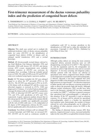
Sensitivity may also be affected by the timing of the DV
measurement, as DV flow seems to normalize towards
14 weeks, even when CHD are present25 (Figure 4).
When considering DV measurements as a screen for
CHD, aspects such as learning curve26
and repeatability
of waveforms9
should be considered. Intra- and inter-
observer reproducibility in studies of DV measurements
show results that vary from substantial to acceptable27–29
.
We observed a mixed a-wave in 15% of the fetuses with
enlarged NT and in 6% of the fetuses with normal NT. In
the case of a-wave discrepancy, the cases were classified
as normal for the analysis, whereas for the DV-PIV the
average of three different recordings was used.
Such variation in DV waveforms is also reported in
second- and third-trimester fetuses30,31
, but a patho-
physiologic explanation for this intrinsic variability is
still lacking. Ultrasonographers should be aware of this,
regardless of whether it is the result of true biologi-
cal variation or a purely methodological problem (e.g.
contamination of the nearby vessel), especially when an
abnormal waveform is found in an otherwise normal fetus.
Repetition of the investigation to confirm consistency of
the results is warranted.
A possible way of implementing DV investigation
in a clinical setting is by using a two-step contin-
gent screening approach32. This can reduce false-positive
rates in fetuses at increased risk after the combined
test33
and can be used to select chromosomally normal
Copyright 2010 ISUOG. Published by John Wiley & Sons, Ltd. Ultrasound Obstet Gynecol 2010; 36: 668–675.
6. DV-PIV to predict CHD 673
1.0
0.8
0.6
0.4
0.2
0.0
Sensitivity
1.0
0.8
0.6
0.4
0.2
0.0
Sensitivity
0.0 0.2 0.4 0.6 0.8 1.0
1 – Specificity
0.0 0.2 0.4 0.6 0.8 1.0
1 – Specificity
(a)
(b)
Figure 2 Receiver–operating characteristics (ROC) curves for the
prediction of all cardiac defects (a) and major cardiac defects (b)
using our logistic regression model including nuchal translucency
multiples of the median (NT-MoM) and ductus venosus pulsatility
index for veins multiples of the median (DV-PIV-MoM) in fetuses
with an enlarged NT. Area under both ROC curves was 0.72.
fetuses to be referred for detailed scans, including early
echocardiography9,11,15,17,28,32,34–38
. Such an approach
would also minimize interpretation problems by restrict-
ing DV studies to a few specialist centers. Moreover,
analysis of DV-PIV as a continuous variable can further
refine the individual risk assessment for CHD in these
fetuses.
The pathophysiologic mechanism linking NT, CHD
and abnormal DV flow is still obscure3,39
. In second-
trimester fetuses an abnormal DV flow is usually
associated with cardiac failure40. It has therefore been
tempting to extrapolate this also to the first-trimester
1.0
0.9
0.8
0.7
0.6
0.5
0.4
0.3
0.2
0.1
0
Observedprobability
0.1 0.2 0.3 0.4 0.5 0.6 0.7 0.8 0.9 1.0
Predicted probability
Figure 3 Calibration plot of the prediction model for congenital
heart defects (CHDs) (mean predicted and observed CHDs in 10
decile groups of the study population).
Figure 4 Ductus venosus flow patterns in a Down syndrome fetus
with an atrioventricular septal defect and tetralogy of Fallot. (a) At
13 weeks, the crown–rump length was 72 mm and the ductus
venosus pulsatility index for veins (DV-PIV) was 2.8 with reversed
a-wave. (b) At 17 + 4 weeks, the DV-PIV was 0.89 with positive
a-wave; normalization of abnormal DV flow with advancing
gestation was observed.
fetus with enlarged NT9,12,14,41. Abnormal DV flow
may reflect impaired atrial contraction and reduced
myocardial compliance with fluid accumulation akin
to that seen during right ventricular failure. Tricuspid
valve regurgitation, commonly observed in first-trimester
fetuses with CHD, seems to point in the same direction42
,
as does the preponderance of atrioventricular septal
defects (AVSDs) and hypoplastic left heart syndrome
in our cohort. Increased right atrial pressure can be
Copyright 2010 ISUOG. Published by John Wiley & Sons, Ltd. Ultrasound Obstet Gynecol 2010; 36: 668–675.
7. 674 Timmerman et al.
expected in these cases, as a result of atrioventricular valve
regurgitation or left-to-right shunting over the foramen
ovale.
A large study on diastolic cardiac function measured
at 11–14 weeks in chromosomally normal and abnormal
fetuses failed to show differences among fetuses with
normal and abnormal hearts43
. Recently, our group found
a correlation between tricuspid valve E/A-wave velocities,
E/TVI (E-wave velocity corrected for stroke volume) and
NT thickness in chromosomally normal fetuses with
normal hearts at 11–14 weeks’ gestation44
. Hence, the
DV anomaly may reflect changes in right heart function.
Enlarged NT, CHD and DV abnormality may have
as common denominator an insult39
, such as hypoxia44
,
that affects the neural crest cells migrating to the neck,
the conotruncal region of the heart and the DV45,46
.
Another proposed mechanism for DV flow abnormality
is abnormal innervation or endothelial thickening of the
DV, analogous to that observed in trisomy 16 mouse
fetuses47
.
In conclusion, in chromosomally normal fetuses with
enlarged NT, an increased DV-PIV is a marker for CHD.
An abnormal DV-PIV is present in almost three-quarters
of CHD cases. DV-PIV can be used as a continuous
variable to increase specificity. Further research is needed
to investigate the advantage of DV-PIV, used as a
continuous variable, above a-wave assessment, in the
prediction of CHD in fetuses with an enlarged NT and
a normal karyotype. This study suggests that DV-PIV
measurement could lead to a substantial improvement in
individual risk assessment for CHD. The pathophysiology
of abnormal DV flow in first-trimester fetuses with
enlarged NT and CHD is still a challenging mystery.
REFERENCES
1. Tegnander E, Williams W, Johansen OJ, Blaas HG, Eik-
Nes SH. Prenatal detection of heart defects in a non-selected
population of 30149 fetuses – detection rates and outcome.
Ultrasound Obstet.Gynecol 2006; 27: 252–265.
2. McBrien A, Sands A, Craig B, Dornan J, Casey F. Major con-
genital heart disease: antenatal detection, patient characteris-
tics and outcomes. J Matern Fetal Neonatal Med 2009; 22:
101–105.
3. Clur SA, Ottenkamp J, Bilardo CM. The nuchal translucency
and the fetal heart: a literature review. Prenat Diagn 2009; 29:
739–748.
4. Becker R, Wegner RD. Detailed screening for fetal anomalies
and cardiac defects at the 11-13-week scan. Ultrasound Obstet
Gynecol 2006; 27: 613–618.
5. Carvalho JS. Fetal heart scanning in the first trimester. Prenat
Diagn 2004; 24: 1060–1067.
6. Hyett J. Does nuchal translucency have a role in fetal cardiac
screening? Prenat Diagn 2004; 24: 1130–1135.
7. Ghi T, Huggon IC, Zosmer N, Nicolaides KH. Incidence of
major structural cardiac defects associated with increased nuchal
translucency but normal karyotype. Ultrasound Obstet Gynecol
2001; 18: 610–614.
8. Makrydimas G, Sotiriadis A, Huggon IC, Simpson J, Shar-
land G, Carvalho JS, Daubeney PE, Ioannidis JP. Nuchal
translucency and fetal cardiac defects: a pooled analysis of
major fetal echocardiography centers. Am J Obstet Gynecol
2005; 192: 89–95.
9. Bilardo CM, Muller MA, Zikulnig L, Schipper M, Hecher K.
Ductus venosus studies in fetuses at high risk for chromosomal
or heart abnormalities: relationship with nuchal translucency
measurement and fetal outcome. Ultrasound Obstet Gynecol
2001; 17: 288–294.
10. Oh C, Harman C, Baschat AA. Abnormal first-trimester ductus
venosus blood flow: a risk factor for adverse outcome in fetuses
with normal nuchal translucency. Ultrasound Obstet Gynecol
2007; 30: 192–196.
11. Maiz N, Valencia C, Emmanuel EE, Staboulidou I, Nico-
laides KH. Screening for adverse pregnancy outcome by ductus
venosus Doppler at 11–13 + 6 weeks of gestation. Obstet
Gynecol 2008; 112: 598–605.
12. Matias A, Huggon I, Areias JC, Montenegro N, Nicolaides KH.
Cardiac defects in chromosomally normal fetuses with abnormal
ductus venosus blood flow at 10–14 weeks. Ultrasound Obstet
Gynecol 1999; 14: 307–310.
13. Montenegro N, Matias A, Areias JC, Barros H. Ductus venosus
revisited: a Doppler blood flow evaluation in the first trimester
of pregnancy. Ultrasound Med Biol 1997; 23: 171–176.
14. Favre R, Cherif Y, Kohler M, Kohler A, Hunsinger MC, Bouf-
fet N, Tanghe M, Cancellier M, Nisand I. The role of
fetal nuchal translucency and ductus venosus Doppler at
11–14 weeks of gestation in the detection of major congenital
heart defects. Ultrasound Obstet Gynecol 2003; 21: 239–243.
15. Maiz N, Plasencia W, Dagklis T, Faros E, Nicolaides K. Ductus
venosus Doppler in fetuses with cardiac defects and increased
nuchal translucency thickness. Ultrasound Obstet Gynecol
2008; 31: 256–260.
16. Haak MC, Twisk JW, Bartelings MM, Gittenberger-de Groot
AC, van Vugt JM. Ductus venosus flow velocities in relation to
the cardiac defects in first-trimester fetuses with enlarged nuchal
translucency. Am J Obstet Gynecol 2003; 188: 727–733.
17. Borrell A. The ductus venosus in early pregnancy and congenital
anomalies. Prenat Diagn 2004; 24: 688–692.
18. The Fetal Medicine Foundation. http://www.fetalmedicine.com/
fmf/FMF-English.pdf [Accessed 27 October 2009].
19. Teixeira LS, Leite J, Viegas MJ, Faria MM, Chaves AS, Teix-
eira RC, Pires MC, Pettersen H. Ductus venosus Doppler
velocimetry in the first trimester: a new finding. Ultrasound
Obstet Gynecol 2008; 31: 261–265.
20. Altman DG, Royston P. What do we mean by validating a
prognostic model? Stat Med 2000; 19: 453–473.
21. Vogel M, Sharland GK, McElhinney DB, Zidere V, Simp-
son JM, Miller OI, Allan LD. Prevalence of increased nuchal
translucency in fetuses with congenital cardiac disease and a
normal karyotype. Cardiol Young 2009; 19: 441–445.
22. Berg C, Kremer C, Geipel A, Kohl T, Germer U, Gembruch U.
Ductus venosus blood flow alterations in fetuses with
obstructive lesions of the right heart. Ultrasound Obstet
Gynecol 2006; 28: 137–142.
23. Hung JH, Fu CY, Lu JH, Hung CY. Ductus venosus blood flow
resistance and congenital heart defects in the second trimester.
J Clin Ultrasound 2008; 36: 72–78.
24. Mart´ınez JM, Comas M, Borrell A, Bennasar M, G´omez O,
Puerto B, Gratac´os E. Abnormal first trimester ductus venosus:
a marker of cardiac defects in fetuses with normal karyotype
and nuchal translucency. Ultrasound Obstet Gynecol 2010; 35:
267–272.
25. Huisman TW, Bilardo CM. Transient increase in nuchal
translucency thickness and reversed end-diastolic ductus
venosus flow in a fetus with trisomy 18. Ultrasound Obstet
Gynecol 1997; 10: 397–399.
26. Maiz N, Kagan KO, Milovanovic Z, Celik E, Nicolaides KH.
Learning curve for Doppler assessment of ductus venosus flow at
11 + 0 to 13 + 6 weeks’ gestation. Ultrasound Obstet Gynecol
2008; 31: 503–506.
27. Borrell A, Perez M, Figueras F, Meler E, Gonce A, Grata-
cos E. Reliability analysis on ductus venosus assessment at
11–14 weeks’ gestation in a high-risk population. Prenat Diagn
2007; 27: 442–446.
Copyright 2010 ISUOG. Published by John Wiley & Sons, Ltd. Ultrasound Obstet Gynecol 2010; 36: 668–675.
8. DV-PIV to predict CHD 675
28. Mavrides E, Holden D, Bland JM, Tekay A, Thilaganathan B.
Intraobserver and interobserver variability of transabdominal
Doppler velocimetry measurements of the fetal ductus venosus
between 10 and 14 weeks of gestation. Ultrasound Obstet
Gynecol 2001; 17: 306–310.
29. Prefumo F, Risso D, Venturini PL, De Biasio P. Reference values
for ductus venosus Doppler flow measurements at 10–14 weeks
of gestation. Ultrasound Obstet Gynecol 2002; 20: 42–46.
30. Kiserud T, Eik-Nes SH, Hellevik LR, Blaas H-G. Ductus veno-
sus – A longitudinal doppler velocimetric study of the human
fetus. J Matern Fetal Invest 1992; 2: 5–11.
31. Diemert A, Hecher K. Severely abnormal flow patterns in the
ductus venosus in the presence of otherwise normal fetal
Doppler parameters in two cases of severe intrauterine growth
restriction. Ultrasound Obstet Gynecol 2009; 34: 605–607.
32. Nicolaides KH, Spencer K, Avgidou K, Faiola S, Falcon O.
Multicenter study of first- trimester screening for trisomy 21
in 75 821 pregnancies: results and estimation of the potential
impact of individual risk-orientated two-stage first-trimester
screening. Ultrasound Obstet Gynecol 2005; 25: 221–226.
33. Timmerman E, Oude Rengerink K, Pajkrt E, Opmeer BC, Van
der Post JAM, Bilardo CM. Ductus venosus pulsatility index
measurement reduces the false-positive rate in first-trimester
screening. Ultrasound Obstet Gynecol 2010; 36: 661–667.
34. Antolin E, Comas C, Torrents M, Munoz A, Figueras F,
Echevarria M, Cararach M, Carrera JM. The role of ductus
venosus blood flow assessment in screening for chromosomal
abnormalities at 10–16 weeks of gestation. Ultrasound Obstet
Gynecol 2001; 17: 295–300.
35. Hecher K. Assessment of ductus venosus flow during the first
and early second trimesters: what can we expect? Ultrasound
Obstet Gynecol 2001; 17: 285–287.
36. Matias A, Montenegro N. Ductus venosus blood flow in
chromosomally abnormal fetuses at 11 to 14 weeks of gestation.
Semin Perinatol 2001; 25: 32–37.
37. Toyama JM, Brizot ML, Liao AW, Lopes LM, Nomura RM,
Saldanha FA, Zugaib M. Ductus venosus blood flow assessment
at 11 to 14 weeks of gestation and fetal outcome. Ultrasound
Obstet Gynecol 2004; 23: 341–345.
38. Bilardo CM, Timmerman E, Pajkrt E, Van Maarle MC.
Increased nuchal translucency in euploid fetuses – what should
we be telling the parents? Prenat Diagn 2010; 30: 93–102.
39. Allan LD. The mystery of nuchal translucency. Cardiol Young
2006; 16: 11–17.
40. Gembruch U, Meise C, Germer U, Berg C, Geipel A. Venous
Doppler ultrasound in 146 fetuses with congenital heart disease.
Ultrasound Obstet Gynecol 2003; 22: 345–350.
41. Murta CG, Moron AF, Avila MA, Weiner CP. Application of
ductus venosus Doppler velocimetry for the detection of fetal
aneuploidy in the first trimester of pregnancy. Fetal Diagn Ther
2002; 17: 308–314.
42. Faiola S, Tsoi E, Huggon IC, Allan LD, Nicolaides KH. Likeli-
hood ratio for trisomy 21 in fetuses with tricuspid regurgitation
at the 11 to 13 + 6-week scan. Ultrasound Obstet Gynecol
2005; 26: 22–27.
43. Huggon IC, Turan O, Allan LD. Doppler assessment of cardiac
function at 11–14 weeks’ gestation in fetuses with normal
and increased nuchal translucency. Ultrasound Obstet Gynecol
2004; 24: 390–398.
44. Clur SA, Oude Rengerink K, Mol BWJ, Ottencamp J, Bilardo CM.
Fetal cardiac function between 11 and 35 weeks’ gestation
and nuchal translucency thickness. Ultrasound Obstet Gynecol
2010; DOI:10.1002/uog.8807.
45. Coceani F, Adeagbo AS, Cutz E, Olley PM. Autonomic mecha-
nisms in the ductus venosus of the lamb. Am J Physiol 1984;
247: H17–H24.
46. Nakamura T, Gulick J, Colbert MC, Robbins J. Protein tyro-
sine phosphatase activity in the neural crest is essential for
normal heart and skull development. Proc Natl Acad Sci USA
2009; 106: 11270–11275.
47. Bekker MN, Arkesteijn JB, Van Den Akker NM, Hoffman S,
Webb S, Van Vugt JM, Gittenberger-De Groot AC. Increased
NCAM expression and vascular development in Trisomy 16
mouse embryos: relationship with nuchal translucency. Pediatr
Res 2005; 58: 1222–1227.
SUPPORTING INFORMATION ON THE INTERNET
The following supporting information may be found in the online version of this article:
Table S1 Cardiac defects, nuchal translucency (NT), ductus venosus pulsatility index for veins (DV-PIV) and
outcome in chromosomally normal fetuses with an enlarged NT
Copyright 2010 ISUOG. Published by John Wiley & Sons, Ltd. Ultrasound Obstet Gynecol 2010; 36: 668–675.